. 2024 Apr;244(4):557-593.
doi: 10.1111/joa.13983. Epub 2023 Dec 1.

Reconstruction of the pelvic girdle and hindlimb musculature of the early tetanurans Piatnitzkysauridae (Theropoda, Megalosauroidea)

Affiliations

Reconstruction of the pelvic girdle and hindlimb musculature of the early tetanurans Piatnitzkysauridae (Theropoda, Megalosauroidea)

Mauro B S Lacerda et al. J Anat. 2024 Apr.

Abstract

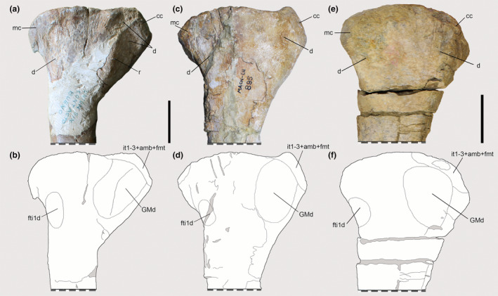
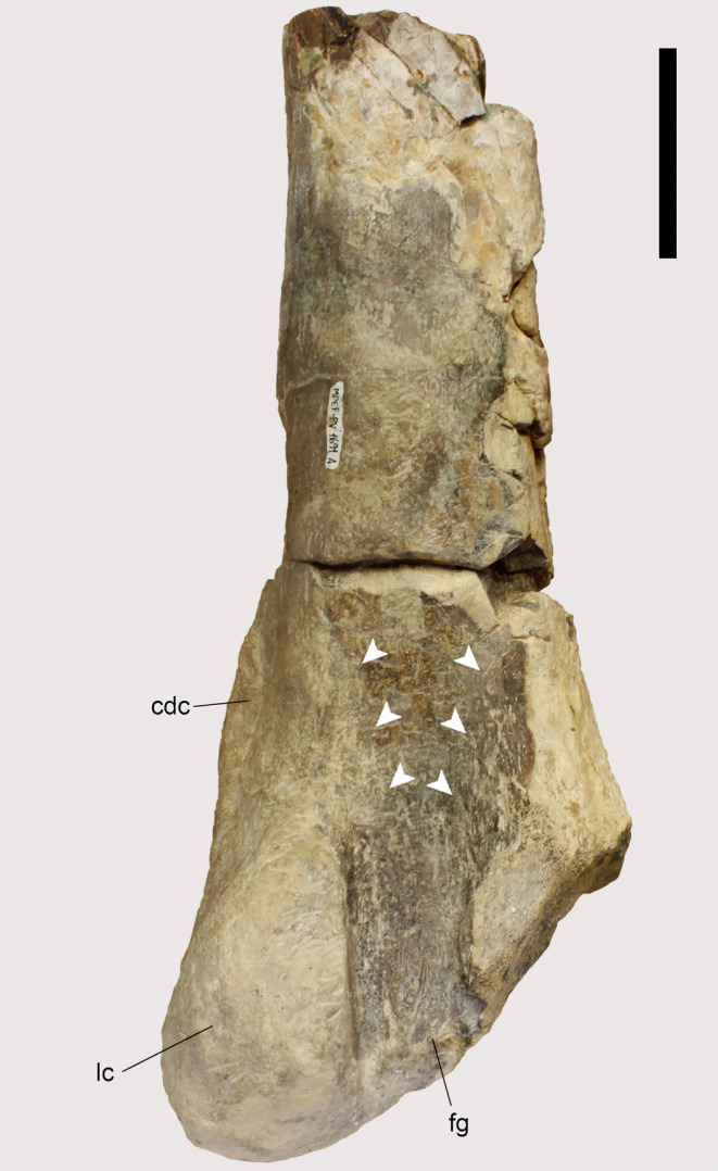
Piatnitzkysauridae were Jurassic theropods that represented the earliest diverging branch of Megalosauroidea, being one of the earliest lineages to have evolved moderate body size. This clade's typical body size and some unusual anatomical features raise questions about locomotor function and specializations to aid in body support; and other palaeobiological issues. Biomechanical models and simulations can illuminate how extinct animals may have moved, but require anatomical data as inputs. With a phylogenetic context, osteological evidence, and neontological data on anatomy, it is possible to infer the musculature of extinct taxa. Here, we reconstructed the hindlimb musculature of Piatnitzkysauridae (Condorraptor, Marshosaurus, and Piatnitzkysaurus). We chose this clade for future usage in biomechanics, for comparisons with myological reconstructions of other theropods, and for the resulting evolutionary implications of our reconstructions; differential preservation affects these inferences, so we discuss these issues as well. We considered 32 muscles in total: for Piatnitzkysaurus, the attachments of 29 muscles could be inferred based on the osteological correlates; meanwhile, in Condorraptor and Marshosaurus, we respectively inferred 21 and 12 muscles. We found great anatomical similarity within Piatnitzkysauridae, but differences such as the origin of M. ambiens and size of M. caudofemoralis brevis are present. Similarities were evident with Aves, such as the division of the M. iliofemoralis externus and M. iliotrochantericus caudalis and a broad depression for the M. gastrocnemius pars medialis origin on the cnemial crest. Nevertheless, we infer plesiomorphic features such as the origins of M. puboischiofemoralis internus 1 around the "cuppedicus" fossa and M. ischiotrochantericus medially on the ischium. As the first attempt to reconstruct muscles in early tetanurans, our study allows a more complete understanding of myological evolution in theropod pelvic appendages.

Keywords: Dinosauria; Jurassic; extant phylogenetic bracket; functional morphology; soft-tissue.


Figures

FIGURE 1
FIGURE 1
(a) Simplified example of the Extant Phylogenetic Bracket (EPB) application in Theropoda. (b), Theropod phylogeny (up to Coelurosauria on the right side of the phylogeny) highlighting the phylogenetic position of Piatnitzkysauridae. (a), adapted from Grillo & Azevedo, ; (b), adapted from Carrano et al., . M, muscle; O, osteological correlate. Silhouettes are from phylopic.org; see Acknowledgements.
FIGURE 2
FIGURE 2
Osteological correlates observed in the ilia of Piatnitzkysauridae (left ilia, lateral view). (a, b) Piatnitzkysaurus (MACN‐Pv‐CH 895). (c, d) Condorraptor (MPEF‐PV 1687). (e, f) Marshosaurus (UMNH VP 6372). The M. flexor tibialis externus is not marked in the line drawings. Anatomical/muscular abbreviations: bf, brevis fossa; cf, “cuppedicus” fossa; CFBf, M. caudofemoralis brevis origin fossa; IFEf, M. iliofemoralis externus origin fossa; ILFBf, M. iliofibularis origin fossa; ip, ischiadic peduncle; IT1–3, Mm. iliotibiales 13 origin scars; PIFI1, M. puboischiofemoralis internus 1 origin fossa; poap, postacetabular process; pp, pubic peduncle; prap, preacetabular process; sac, supraacetabular crest; vr, vertical ridge. Arrows indicate potential subdivision of IT heads. Dark grey represents broken areas of bones. Scale bar = 100 mm.
FIGURE 3
FIGURE 3
Osteological correlates of M. iliotibiales 1–3 observed on the ilia of Piatnitzkysauridae (left ilia, lateral view). (a), Piatnitzkysaurus (MACN‐Pv‐CH 895). (b), Marshosaurus (UMNH VP 6372). Anatomical/muscular abbreviations: cdi, crista dorsolateralis ilii; IT1–3s, Mm. iliotibiales scars; IT1–2l, M. iliotibialis 1 and 2 limits. Arrows indicate muscle scars. Scale bar = 20 mm.
FIGURE 4
FIGURE 4
Osteological correlates of the triceps femoris insertion and origins of lower leg muscles from the tibiae of Piatnitzkysauridae (left tibiae, lateral view). (a, b) Piatnitzkysaurus (PVL 4073). (c, d) Condorraptor (MPEF‐PV 1672). Anatomical/muscular abbreviations: cc, cnemial crest; EDLs, M. extensor digitorum longus scar; fc, fibular crest; it1–3 + amb + fmt, insertion of the tendon from the iliotibiales + ambiens + femorotibiales muscles; lc, lateral condyle; mc, medial condyle; pas, proximal articular surface; si, sulcus intercnemialis; TAd, M. tibialis anterior depression. Scale bar = 50 mm.
FIGURE 5
FIGURE 5
Osteological correlates observed on the pubes of Piatnitzkysauridae (right pubes, lateral and anterior views). (a–d) Piatnitzkysaurus (MACN‐Pv‐CH 895). (e–h) Condorraptor (MPEF‐PV 1696). (i, j) Marshosaurus (UMNH VP 6387). Anatomical/muscular abbreviations: ac, acetabulum; AMBt, M. ambiens tubercle; ap, apron; ilc, iliac peduncle; ip, ischial peduncle; of, obturator foramen; pb, pubic boot; PIFE1s, M. puboischiofemoralis externus 1 scar; PIFE2s, M. puboischiofemoralis externus 2 scar; pt, pubic tubercle. (a, b, e, f, i, j) in lateral view; (c, d, g, h) in anterior view. Scale bar = 50 mm.
FIGURE 6
FIGURE 6
Osteological correlates observed on the femur of Piatnitzkysaurus (right femur, PVL 4073). (a, b) lateral view; (c, d) anterior view; (e, f) medial view; (g, h) posterior view. Anatomical/muscular abbreviations: add1 + 2 s, Mm. adductores femores insertion scar; at, acessory trochanter; cdc, craniomedial distal crest; cfbs, M. caudofemoralis brevis insertion scar; cfls, M. caudofemoralis longus insertion scar; eg, extensor groove; fh, femoral head; fg, flexor groove; fmtes, M. femorotibialis externus scar; fmtis, M. femorotibialis internus scar; fn, femoral neck; ft, fourth trochanter; GLd, M. gastrocnemius pars lateralis depression; gt, greater trochanter; ifes, M. iliofemoralis externus insertion scar; istrs, M. ischiotrochantericus insertion scar; itcs, M. iliotrochantericus caudalis insertion scar; la, linea aspera; lc, lateral condyle; lia, linea intermuscularis cranialis; lip, linea intermuscularis caudalis; lt, lesser trochanter; mc, medial condyle; pifes, Mm. puboischiofemorales externi insertion scar; pifi1s, M. puboischiofemoralis internus 1 insertion scar; pifi2s, M. puboischiofemoralis internus 2 insertion scar; TA?, M. tibialis anterior?, origin?; tfc, tibiofibular crest; ts, trochanteric shelf. Scale bar = 100 mm.
FIGURE 7
FIGURE 7
Osteological correlates observed on the femur of Condorraptor (left femur, MPEF‐PV 1690). (a, b) lateral view; (c, d) anterior view; (e, f) medial view; (g, h) posterior view. Anatomical/muscular abbreviations: add1 + 2 s, Mm. adductores femores insertion scar; cdc, craniomedial distal crest; cfb + cfls, Mm. caudofemorales insertion scar; cfbs, M. caudofemoralis brevis insertion scar; eg, extensor groove; fg, flexor groove; fmtes, M. femorotibialis externus scar; fmtis, M. femorotibialis internus scar; ft, fourth trochanter; GLd, M. gastrocnemius pars lateralis depression; ifes?, M. iliofemoralis externus insertion scar?; itcs?, M. iliotrochantericus caudalis insertion scar?; la, linea aspera; lc, lateral condyle; lia, linea intermuscularis cranialis; lip, linea intermuscularis caudalis; lt, lesser trochanter; TA?, M. tibialis anterior?, origin?; tfc, tibiofibular crest; ts?, trochanteric shelf? Scale bar = 100 mm.
FIGURE 8
FIGURE 8
Osteological correlate observed on the left fibula of Piatnitzkysaurus (PVL 4073). (a, b) lateral view. Anatomical/muscular abbreviations: ift, iliofibularis (fibular) tubercle; ilfbs, M. iliofibularis insertion scar. Scale bar = 50 mm.
FIGURE 9
FIGURE 9
Osteological correlates observed on the femur of Piatnitzkysaurus (right femur, PVL 4073). (a, b) lateral view. Anatomical/muscular abbreviations: at, acessory trochanter; cfbs, M. caudofemoralis brevis insertion scar; ft, fourth trochanter; gt, greater trochanter; ifes, M. iliofemoralis externus insertion scar; istrs, M. ischiotrochantericus insertion scar; itcs, M. iliotrochantericus caudalis insertion scar; lt, lesser trochanter; pifes, Mm. puboischiofemorales externi insertion scar; pifi2, M. puboischiofemoralis internus 2 insertion scars; ts, trochanteric shelf. Scale bar = 20 mm.
FIGURE 10
FIGURE 10
Osteological correlates observed on the vertebrae of Piatnitzkysaurus (PVL 4073) in lateral view. (a) 19th dorsal vertebra. (b) 20th dorsal vertebra. Anatomical/muscular abbreviations: ns, neural spine; poz, postzygapophysis; prz, prezygapophysis; vb, vertebral body. Arrows indicate the fossa. Scale bar = 20 mm.
FIGURE 11
FIGURE 11
Osteological correlates observed on the ischia of Piatnitzkysauridae. (a, b) Piatnitzkysaurus (right ischium, MACN‐Pv‐CH 895). (c, d) Condorraptor (left ischium, MPEF‐PV 1696). (d–f) Marshosaurus (left ischium, UMNH VP 6387) in lateral view. Anatomical/muscular abbreviations: ac, acetabulum; ADD1s, M. adductor femoris 1 scar; ADD2s, M. adductor femoris 2 scars; de, distal expansion; FTI1t, M. flexor tibialis internus 1 tubercle; FTI3t, M. flexor tibialis internus 3 tubercle; ip, iliac peduncle; it, ischial tuberosity; op, obturator process; pp, pubic peduncle; PIFE3, M. puboischiofemoralis externus 3; ti, ischiadic tubercle. Scale bar = 50 mm.
FIGURE 12
FIGURE 12
Osteological correlates observed on the tibiae of Piatnitzkysauridae (left tibiae, medial view). (a, b) Piatnitzkysaurus (PVL 4073). (c, d) Piatnitzkysaurus (MACN‐Pv‐CH 895). (e, f) Condorraptor (MPEF‐PV 1672). Anatomical/muscular abbreviations: cc, cnemial crest; d, depression; fti1d, M. flexor tibialis internus 1 depression; GMd, M. gastrocnemius pars medialis depression; it1–3 + amb + fmt, insertion of the tendons of the iliotibiales + ambiens + femorotibiales muscles; mc, medial condyle; r, ridge. Scale bar = 50 mm.
FIGURE 13
FIGURE 13
Possible Mm. adductores femores division on the Condorraptor distal right femur (MPEF‐PV 1691, posterior view). Anatomical abbreviations: cdc, craniomedial distal crest, fg, flexor groove; lc, lateral condyle. Arrows indicate these scars that may or may not pertain to ADD 1 + 2 (see text). Scale bar = 50 mm.
FIGURE 14
FIGURE 14
Ventral view of the brevis fossa in Piatnitzkysauridae. (a) Piatnitzkysaurus (PVL 4073). (b) Marshosaurus (UMNH VP 6372). Anatomical/muscular abbreviations: CFBf, M. caudofemoralis brevis fossa. Not to scale.
FIGURE 15
FIGURE 15
Proximal tibiae of Piatnitzkysaurus (left tibiae, lateral view). (a) PVL 4073 specimen. (b) MACN‐Pv‐CH 895 specimen. Anatomical/muscular abbreviations: cc, cnemial crest; fc, fibular crest; lc, lateral condyle, mc, medial condyle. Arrows indicate muscle scar. Scale bar = 50 mm.
FIGURE 16
FIGURE 16
Metatarsal IV of Piatnitzkysauridae (left, posterior view). (a, b) Piatnitzkysaurus (MACN‐Pv‐CH 895). (c, d) Condorraptor (MPEF‐PV 1692). Anatomical/muscular abbreviations: das, distal articular surface; gl + gms, Mm. gastrocnemii insertion scar; pas, proximal articular surface; r, ridge. Scale bar = 50 mm.
FIGURE 17
FIGURE 17
Pelvic and hindlimb ‘muscle map’ inferred for Piatnitzkysauridae (left lateral view). (a) Piatnitzkysaurus floresi. (b) Condorraptor currumili. (c) Marshosaurus bicentesimus. Note that some muscles are not shown here, and some bones have been mirrored to illustrate the reconstructions of the three piatnitzkysaurid species. Muscle abbreviations are provided in Table 1; see text for inference levels and other comparisons. CFL and PIFI2 origins are much smaller than expected; simply shown for relative positions. Medial muscle origins (e.g., FMTI) and insertions (e.g., PIFI1) are not shown. Mm. gastrocnemii are labelled as “Flexores” due to their action around the knee; lower leg (FB, FL, etc.) as “Flexores” for ankle dorsiflexion. Not to scale.
FIGURE 18
FIGURE 18
Restoration of pelvic and hindlimb muscles in Piatnitzkysaurus floresi (left lateral view). Artwork by Júlia d'Oliveira.

References

    1. Allen, V.R. , Kilbourne, B.M. & Hutchinson, J.R. (2021) The evolution of pelvic limb muscle moment arms in bird‐line archosaurs. Science Advances, 7(12), eabe2778. - PMC - PubMed
    1. Barbi, M. , Bell, P.R. , Fanti, F. , Dynes, J.J. , Kolsceke, A. , Buttigieg, J. et al. (2019) Integumentary structure and composition in an exceptionally well‐preserved hadrosaur (Dinosauria: Ornithischia). PeerJ, 7, e7875. - PMC - PubMed
    1. Bates, K.T. , Benson, R.B.J. & Falkingham, P.L. (2012) A computational analysis of locomotor anatomy and body mass evolution in Allosauroidea (Dinosauria, Theropoda). Paleobiology, 38(3), 486–507.
    1. Bates, K.T. & Falkingham, P.L. (2018) The importance of muscle architecture in biomechanical reconstructions of extinct animals: a case study using Tyrannosaurus rex . Journal of Anatomy, 233(5), 625–635. - PMC - PubMed
    1. Bates, K.T. , Maidment, S.C. , Allen, V. & Barrett, P.M. (2012) Computational modelling of locomotor muscle moment arms in the basal dinosaur Lesothosaurus diagnosticus: assessing convergence between birds and basal ornithischians. Journal of Anatomy, 220(3), 212–232. - PMC - PubMed

Publication types