. 2024 May 3;147(5):1837-1855.
doi: 10.1093/brain/awad403.

Gain-of-function and loss-of-function variants in GRIA3 lead to distinct neurodevelopmental phenotypes

Berardo Rinaldi  1 Allan Bayat  2   3   4 Linda G Zachariassen  2 Jia-Hui Sun  5   6 Yu-Han Ge  5   7 Dan Zhao  2 Kristine Bonde  2 Laura H Madsen  2 Ilham Abdimunim Ali Awad  2 Duygu Bagiran  2 Amal Sbeih  2 Syeda Maidah Shah  2 Shaymaa El-Sayed  2 Signe M Lyngby  2 Miriam G Pedersen  2 Charlotte Stenum-Berg  2 Louise Claudia Walker  8 Ilona Krey  9 Andrée Delahaye-Duriez  10   11   12 Lisa T Emrick  13   14 Krystal Sully  13 Chaya N Murali  14 Lindsay C Burrage  14 Julie Ana Plaud Gonzalez  13 Mered Parnes  13   15 Jennifer Friedman  16   17   18 Bertrand Isidor  19 Jérémie Lefranc  20 Sylvia Redon  21   22 Delphine Heron  23   24 Cyril Mignot  23   24 Boris Keren  25 Mélanie Fradin  26 Christele Dubourg  27   28 Sandra Mercier  19   29 Thomas Besnard  19   29 Benjamin Cogne  19   29 Wallid Deb  19   29 Clotilde Rivier  30 Donatella Milani  31 Maria Francesca Bedeschi  1 Claudia Di Napoli  1 Federico Grilli  1 Paola Marchisio  32   33 Suzanna Koudijs  34 Danielle Veenma  35 Emanuela Argilli  36   37 Sally Ann Lynch  38 Ping Yee Billie Au  39 Fernando Eduardo Ayala Valenzuela  40 Carolyn Brown  41 Diane Masser-Frye  42 Marilyn Jones  42 Leslie Patron Romero  43 Wenhui Laura Li  44 Erin Thorpe  41 Laura Hecher  45 Jessika Johannsen  45 Jonas Denecke  45 Vanda McNiven  46   47 Anna Szuto  46   48 Emma Wakeling  49 Vincent Cruz  50 Valerie Sency  50 Heng Wang  50 Juliette Piard  51   52 Fanny Kortüm  53 Theresia Herget  53 Tatjana Bierhals  53 Angelo Condell  54 Bruria Ben-Zeev  55   56 Simranpreet Kaur  54   57 John Christodoulou  54   57   58   59 Amelie Piton  60 Christiane Zweier  61   62 Cornelia Kraus  61 Alessia Micalizzi  63 Marina Trivisano  64 Nicola Specchio  64 Gaetan Lesca  65   66 Rikke S Møller  3   4 Zeynep Tümer  67   68 Maria Musgaard  8 Benedicte Gerard  69 Johannes R Lemke  70 Yun Stone Shi  5   7   71 Anders S Kristensen  2
Affiliations

Gain-of-function and loss-of-function variants in GRIA3 lead to distinct neurodevelopmental phenotypes

Berardo Rinaldi et al. Brain. .

Abstract

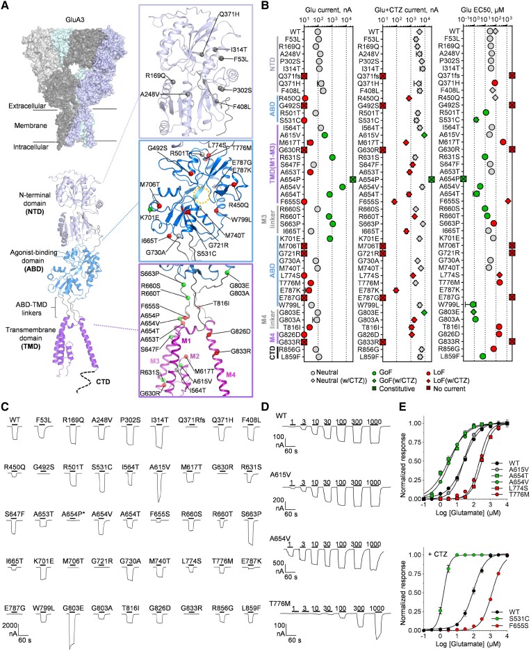
AMPA (α-amino-3-hydroxy-5-methyl-4-isoxazole propionic acid) receptors (AMPARs) mediate fast excitatory neurotransmission in the brain. AMPARs form by homo- or heteromeric assembly of subunits encoded by the GRIA1-GRIA4 genes, of which only GRIA3 is X-chromosomal. Increasing numbers of GRIA3 missense variants are reported in patients with neurodevelopmental disorders (NDD), but only a few have been examined functionally. Here, we evaluated the impact on AMPAR function of one frameshift and 43 rare missense GRIA3 variants identified in patients with NDD by electrophysiological assays. Thirty-one variants alter receptor function and show loss-of-function or gain-of-function properties, whereas 13 appeared neutral. We collected detailed clinical data from 25 patients (from 23 families) harbouring 17 of these variants. All patients had global developmental impairment, mostly moderate (9/25) or severe (12/25). Twelve patients had seizures, including focal motor (6/12), unknown onset motor (4/12), focal impaired awareness (1/12), (atypical) absence (2/12), myoclonic (5/12) and generalized tonic-clonic (1/12) or atonic (1/12) seizures. The epilepsy syndrome was classified as developmental and epileptic encephalopathy in eight patients, developmental encephalopathy without seizures in 13 patients, and intellectual disability with epilepsy in four patients. Limb muscular hypotonia was reported in 13/25, and hypertonia in 10/25. Movement disorders were reported in 14/25, with hyperekplexia or non-epileptic erratic myoclonus being the most prevalent feature (8/25). Correlating receptor functional phenotype with clinical features revealed clinical features for GRIA3-associated NDDs and distinct NDD phenotypes for loss-of-function and gain-of-function variants. Gain-of-function variants were associated with more severe outcomes: patients were younger at the time of seizure onset (median age: 1 month), hypertonic and more often had movement disorders, including hyperekplexia. Patients with loss-of-function variants were older at the time of seizure onset (median age: 16 months), hypotonic and had sleeping disturbances. Loss-of-function and gain-of-function variants were disease-causing in both sexes but affected males often carried de novo or hemizygous loss-of-function variants inherited from healthy mothers, whereas affected females had mostly de novo heterozygous gain-of-function variants.

Keywords: AMPA receptor; GRIA; GRIA3; clinical biomarker; genotype-phenotype.


Conflict of interest statement

The authors report no competing interests.

Figures

Figure 1
Figure 1
Location of GRIA3 variants in the GluA3 receptor and effect on glutamate-gated channel function. (A) Structural model of homomeric GluA3 receptor encoded by the GRIA3 gene built from structures of the GluA2 receptor (Supplementary material, ‘Materials and methods’ section). The top left panel shows a surface representation of the tetrameric receptor complex with the four identical subunits. The bottom panel shows a cartoon representation of a single GluA3 subunit with the N-terminal domain (NTD), the agonist-binding domain (ABD), and the transmembrane domain (TMD). Zoomed views of the NTD, ABD and TMD show the position of genetic variants caused by GRIA3 missense variants highlighted by different colours according to the apparent effect on homomeric GluA3 function as neutral, loss-of-function (LoF) and gain-of-function (GoF). The stippled circle indicates the position of the Glu binding site in the ABD. (B) Summary of desensitized (Glu) and non-desensitized (Glu + CTZ) current amplitudes and Glu EC50 for homomeric GluA3 receptors containing genetic variants encoded by the GRIA3 variants evaluated in this study. Values, number of measurements, and statistical parameters are given in Supplementary Tables 2 and 3. Individual data-points are colour-coded according to the effect on currents or EC50 (LoF effect) or increase (GoF effect). For the EC50 panel, data-points shown as squares represent EC50 values determined with cyclothiazide (CTZ). (C) Representative current responses from two-electrode voltage-clamp (TEVC) recordings of Xenopus laevis oocytes (XOs) (VHOLD −40 mV) expressing wild-type (WT) or GRIA3 variant-containing GluA3 receptors in response to Glu application (300 µM, black bar) in the presence of CTZ (100 µM) to block desensitization. (D) Representative current recordings from TEVC Glu concentration-response experiments of wild-type GluA3 and selected variants exemplifying neutral [p.(Ala615Val)], increasing [p.(Ala654Val)] or decreasing [p.(Thr776Met)] effect on receptor responsiveness to Glu. (E) Composite concentration-response curves for wild-type and selected GRIA3 variant-containing GluA3 receptors. Data-points represent the mean of 6–12 oocytes. Error bars are the standard error of the mean (SEM) and are shown when larger than the symbol size. The current responses are normalized to the maximal response evoked by Glu. In all panels, variants are labelled with single-letter amino acid codes.
Figure 2
Figure 2
Variant effects on receptor desensitization and activation properties. (A) Representative currents evoked by sequential 10–20 s applications of Glu (1 mM, black bar) alone and in the presence of cyclothiazide (CTZ) (100 μM, grey bar) from oocytes expressing wild-type (WT) GluA3 and GluA3 carrying selected GRIA3 missense variants. The p.(Pro302Ser) variant shows no change in the size of the desensitized current relative to the non-desensitized Glu current compared to wild-type, the p.(Ala654Val) variant shows increased desensitized current, and the p.(Thr816Ile) variant show decreased desensitized current. (B) Representative currents evoked by sequential 10–20 s applications of Glu (1 mM, black bar) and kainic acid (KA) (300 µM; blue bars) in the presence of CTZ (100 μM, grey bar) from oocytes expressing wild-type GluA3 and GluA3 containing selected variants exemplifying different types of variant effects on KA/GLU response ratio. For wild-type GluA3 and the p.(Pro302Ser) variant, the KA-evoked current has an amplitude of 16% of the Glu current amplitude. In contrast, the p.(Ala654Val) variant has a relative KA current of 41%, indicating an increase in activation properties, and p.(Ala653Thr) variant has decreased relative KA response amplitude of 3.5%, indicating decreased activation properties. The holding potential was −40 mV in all shown recordings. (C) Representative currents illustrating 1-naphthyl acetyl spermine (NASPM) (1 µM, red bar) inhibition of Glu-evoked currents for wild-type GluA3 and GluA3 containing the variants p.(Arg631Ser) and p.(Ala654Pro). (D) Summary of the ratio of desensitized and non-desensitized current amplitude (IGLU/IGLU+CTZ), non-desensitized Glu and KA (IKA+CTZ/IGLU+CTZ) current amplitudes and NASPM inhibition of Glu-evoked current for homomeric GluA3 receptors containing genetic variants encoded by the GRIA3 variants evaluated in this study. Values, number of measurements and statistical parameters are given in Supplementary Table 2. Individual data-points are colour-coded according to the effect on currents or EC50 [loss-of-function (LoF) effect; red] or increase [gain-of-function (GoF) effect; green]. (EG) Summary of phenotype and domain location of variants with overall GoF (E), LoF (F) and neutral (G) effect on homomeric GluA3 receptor function. Inverted triangle = decrease; triangle = increase; filled circle = no change; dash = not determined. Colour-coding indicates a predicted LoF (red) or GoF (green) effect of change on overall receptor function. (H) Missense tolerance ratio (MTR) of GRIA3 variants analysed with a 31 amino acid window calculated using the MTR-viewer online tool (https://biosig.lab.uq.edu.au/mtr-viewer/). A line graph displays the MTR distribution for GRIA3 (gene transcript NM_000828) with regions in orange indicating observed variation differs significantly from neutrality. Dashed lines on the plot denote gene-specific MTRs: green = fifth percentile; purple = 25th percentile; black = 50th percentile. Above the MTR distribution is shown the domain structure of the GluA3 subunit. Variant positions are shown as circles on the MTR line graph and coloured according to functional effect as: neutral (grey), GoF (green) and LoF (red). Orange line segments indicate regions where the observed variation differs significantly from neutrality. In all panels, variants are labelled with single-letter amino acid codes. ABD = agonist binding domain; CTD = carboxy-terminal domain; NTD = N-terminal domain; TMD = transmembrane domain.
Figure 3
Figure 3
Variant effects in heteromeric GluA2/A3 receptors. (A) Representative currents evoked by sequential 10–20 s applications of Glu (1 mM) alone and in the presence of cyclothiazide (CTZ) (100 μM) from oocytes expressing wild-type (WT) GluA2, wild-type GluA3 and wild-type GluA2 with GluA3 carrying selected GRIA3 missense variants illustrating increased [p.(Ala654Val), middle trace] and decreased [p.(Leu774Ser); lower trace] desensitized current. (B) Representative currents evoked by sequential 10–20 s applications of Glu (1 mM) and kainic acid (KA) (300 µM) the presence of CTZ (100 μM) from oocytes expressing wild-type GluA2, wild-type GluA3 and wild-type GluA2 with GluA3 carrying selected GRIA3 missense variants illustrating increased [p.(Ala615Val), middle trace] and decreased [p.(Ala653Thr); lower trace] current response to KA relative to Glu. (C) Representative current recordings from two-electrode voltage-clamp (TEVC) Glu concentration-response experiments of wild-type and selected variants in heteromeric GluA2/A3 receptors with corresponding fitted dose-response curves for homomeric (A3) and heteromeric (A2/A3) receptors. The p.(Trp799Leu) exemplifies a variant changing the EC50 in both homomeric and heteromeric receptors, whereas p.(Thr776Met) exemplifies a variant affecting only homomeric receptors. (D) Overview and summary of the effects on heteromeric GluA2/A3 receptor parameters (squares) of GRIA3 variants with gain-of-function (GoF) and loss-of-function (LoF) effects. Data-points represent the mean and 95% confidence interval (CI) values (Supplementary Tables 2 and 3). (E) Current-voltage (IV) relationships of Glu-evoked currents from oocytes expressing homomeric wild-type and variant-containing GluA3 alone and with wild-type GluA2R. The current amplitude at the different holding potentials is normalized to the current at −40 mV. Data-points represent the mean from 6 to 10 oocytes. Error bars indicate the standard error of the mean (SEM) and are shown when larger than the symbol size. In all panels, variants are labelled with single-letter amino acid codes.
Figure 4
Figure 4
Characterization of variant effect on fast receptor kinetics. (A) Representative whole-cell currents evoked by a 500 ms application of glutamate (Glu) (10 mM, black bar) from homomeric GluA3 (left) and heteromeric GluA2/A3 receptors carrying the indicated GRIA3 variants subunits expressed in HEK293 cells. The holding potential was −70 mV in all recordings. Note that scale bars for current amplitude differ between recordings. (B) The time constant (τdes) and level (Iss) of current desensitization determined from the fitting of the current decay (insert) during 500 ms applications of Glu (10 mM, black bars) fitted to two-exponential decay functions weighted by proportional contributions for wild-type (WT) and variant homomeric GluA3 (left) and heteromeric GluA2/A3 (right) receptors. (C) Summary of the τdes and Iss values. Bars represent the mean with standard error of the mean (SEM) error. Values not determined due to low or no current are labelled ‘nd’. (D) Deactivation rates (τdeact) determined from the fitting of the current decay (insert) following 1 ms application of Glu (10 mM, black bars) fitted to a mono-exponential decay function (inserts) for wild-type and variant homomeric GluA3 (left) and heteromeric GluA2/A3 (right) receptors. (E) Summary of τdeact values. Bars represent the mean with SEM error. Values not determined due to low or no current are labelled ‘nd’. (F) Summary of effects of patient variants on current kinetics and location in GluA3 subunit. Variants with loss-of-function (LoF) effects are shown in red and gain-of-function (GoF) in green. Inverted triangle = decrease; triangle = increase; filled circle = no change; dash = not determined. In all panels, variants are labelled with single-letter amino acid codes.
Figure 5
Figure 5
Variant classification and phenotype correlations for Patients M1–M14 and F1–F11. (A) Schematic overview of the classification of receptor phenotype for Patients M1–M14 and F1–F11 into severe and mild gain-of-function (GoF; green) and loss-of-function (LoF; red) categories based on variant effect patterns on GluA3-containing receptor function together with an overview of the number of patients and prevalence of key patient symptoms for each category. (B) Summary of key and supporting features for the clinical phenotypes associated with LoF and GoF variants. The diagram summarizes several clinical findings that can help predict if a GRIA3 variant leads to LoF and GoF. GoF variants manifest with seizures occurring before the first year of life (with a median age of 1 month) and are characterized by supporting features such as hypertonia, hyperekplexia/excessive startle reflex, and the absence of sleep disturbances. LoF variants manifest with key features such as seizure onset after the first year of life (with a median age of 16 months) and supporting features including hypotonia, sleep disturbances and the absence of hyperekplexia/excessive startle reflex. If a patient’s phenotypical presentation displays a combination of these features, functional testing of the variant is required to determine whether a GRIA3 variant displays LoF or GoF characteristics. a–dP-values for comparing proportions of clinical indicators between the LoF or GoF patients. aAge of seizure onset 12 months; P = 0.004. bHypertonia versus hypotonia; P = 0.0004. cHyperekplexia/startle versus no hyperekplexia/startle; P = 0.003. dSleep disturbance versus no sleep disturbance; P = 0.018.

References

    1. Hansen KB, Wollmuth LP, Bowie D, et al. Structure, function, and pharmacology of glutamate receptor ion channels. Pharmacol Rev. 2021;73:298–487. - PMC - PubMed
    1. Raman IM, Trussell LO. The kinetics of the response to glutamate and kainate in neurons of the avian cochlear nucleus. Neuron. 1992;9:173–186. - PubMed
    1. Sah P, Hestrin S, Nicoll RA. Properties of excitatory postsynaptic currents recorded in vitro from rat hippocampal interneurones. J Physiol. 1990;430:605–616. - PMC - PubMed
    1. Silver RA, Traynelis SF, Cull-Candy SG. Rapid-time-course miniature and evoked excitatory currents at cerebellar synapses in situ. Nature. 1992;355:163–166. - PubMed
    1. Hayashi Y, Shi SH, Esteban JA, Piccini A, Poncer JC, Malinow R. Driving AMPA receptors into synapses by LTP and CaMKII: Requirement for GluR1 and PDZ domain interaction. Science. 2000;287:2262 LP–2262267. - PubMed

Publication types

Substances