. 2023 Dec 12;17(23):23772-23783.
doi: 10.1021/acsnano.3c07706. Epub 2023 Dec 1.

Cyclophospholipids Enable a Protocellular Life Cycle

Affiliations

Cyclophospholipids Enable a Protocellular Life Cycle

Ö Duhan Toparlak et al. ACS Nano. .

Abstract

There is currently no plausible path for the emergence of a self-replicating protocell, because prevalent formulations of model protocells are built with fatty acid vesicles that cannot withstand the concentrations of Mg2+ needed for the function and replication of nucleic acids. Although prebiotic chelates increase the survivability of fatty acid vesicles, the resulting model protocells are incapable of growth and division. Here, we show that protocells made of mixtures of cyclophospholipids and fatty acids can grow and divide in the presence of Mg2+-citrate. Importantly, these protocells retain encapsulated nucleic acids during growth and division, can acquire nucleotides from their surroundings, and are compatible with the nonenzymatic extension of an RNA oligonucleotide, chemistry needed for the replication of a primitive genome. Our work shows that prebiotically plausible mixtures of lipids form protocells that are active under the conditions necessary for the emergence of Darwinian evolution.

Keywords: Darwinian evolution; artificial cells; cyclophospholipids; prebiotic chemistry; protocells.


Conflict of interest statement

The authors declare no competing financial interest.

Figures

Figure 1
Figure 1
Schematic of a protocellular life cycle. Cyclophospholipid vesicles grow through lipid exchange, acquire activated nucleotides (pink) from the environment, copy RNA, bud, and divide, all in the presence of citrate-chelated Mg2+ (gold-salmon).
Figure 2
Figure 2
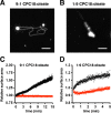
Growth of cyclophospholipid vesicles in the presence of Mg2+-citrate. (A, C) Growth of 9:1 CPC18:oleate MLVs fed with 50 equiv (A) or 25 equiv (C) of 1:9 CPC18:oleate LUVs. (B, D) Growth of 1:9 CPC18:oleate MLVs fed with 5 equiv of pure CPC18 (B) or 25 equiv of 9:1 CPC18:oleate (D) LUVs. Reactions were monitored by epifluorescence microscopy with receiver vesicles labeled with 0.15 mol % LR-DHPE (A, B) and fluorescence spectrophotometry with receiver vesicles containing lipids labeled with FRET donor and acceptor fluorophores (C, D). Red data points represent receiver and donor vesicles of the same lipid composition and black indicates feeding with LUVs of different composition, i.e., 1:9 CPC18:oleate (C) and 9:1 CPC18:oleate (D). Reactions were in 25 mM Mg2+-citrate, 0.2 M Na+-HEPES, pH 8.0. Scale bars are 20 μm.
Figure 3
Figure 3
Division of cyclophospholipid vesicles in the presence of Mg2+-citrate. (A) Time-lapse images of vesicle division of cyclophospholipid-rich MLVs (9:1 CPC18:oleyl alcohol MLVs) fed with 50 equiv of 1:1 CPC18:oleate LUVs. (B) The bar graph represents the number of vesicles observed when 9:1 CPC18:oleate MLVs were fed with either 9:1 or 1:9 CPC18:oleate LUVs. (C) Time-lapse imaging of vesicle division of 1:9 CPC18:oleate MLVs fed with 5 equiv. pure CPC18 LUVs. (D) The bar graph represents the number of vesicles observed when 1:9 CPC18:oleate MLVs were fed with either 1:9 or 9:1 CPC18:oleate LUVs. Conditions were 25 mM Mg2+-citrate, 0.2 M HEPES, pH 8.0. Error bars indicate the ± SD of the mean from n ≥ 4 fields in n ≥ 2 independent experiments. Scale bars are 10 μm. Vesicles were labeled with 0.15 mol % LR-DHPE.
Figure 4
Figure 4
Membrane heterogeneity and monomer kinetics drive the growth and division of protocells. (A) Determination of the lipid exchange rates between vesicles containing oleate and CPC18 with FRET-labeled POPC vesicles. Data were linearly fit with a R2 = 0.8. (B) Flip-flop rates of oleate and CPC18. Representative stopped-flow kinetic data showing the decay of a pH gradient across the membranes of oleate and CPC18 LUVs. Data were fit to a one-phase exponential decay with R2 > 0.82. (C) Determination of the fluidity of membranes with varying ratios of oleate and CPC18. Data are of the fluorescence anisotropy of the hydrophobic reporter molecule 1,6-diphenyl-1,3,5-hexatriene (DPH). Data were fit to a one-phase exponential decay with R2 = 0.99. (D) Acidification of POPC vesicles upon mixing with vesicles containing different ratios of oleate and CPC18. Data were linearly fit with R2 = 0.96. Experiments were performed in 0.2 M Na+-HEPES, pH 8.0.
Figure 5
Figure 5
Cyclophospholipid protocells retain oligonucleotides during growth and division in the presence of Mg2+-citrate. (A) Stability of protocells and leakage of molecules across the bilayers of 6:3:1, 1:1:1, and 1:3:6 CPC10:dodecanol:decanoate vesicles. (B) Stability of multilamellar vesicular protocells with membrane composition of (left) 6:3:1 CPC10:dodecanol:decanoate (CPC10-rich) and (right) 1:3:6 CPC10:dodecanol:decanoate (decanoate-rich). The protocells were fed with 5 equiv. LUVs. All data was collected in 25 mM Mg2+-citrate, 0.2 M Na+-HEPES, pH 8.0. The protocells contained 10 μM entrapped 6-FAM-labeled 10-mer DNA.
Figure 6
Figure 6
Template-directed, nonenzymatic RNA primer extension within cyclophospholipid protocells. (A) A schematic of the experiment illustrating diffusion of 2-AI activated nucleotide and primer extension. (B) The RNA template and labeled primer encapsulated within the protocell. 2-AImpG was added externally to (C) cyclophospholipid-deficient (2:3:5 CPC10:dodecanol:decanoate) and (D) cyclophospholipid-rich (6:3:1 CPC10:dodecanol:decanoate) protocells. The protocells contained an entrapped fluorescein-labeled RNA primer and an RNA template. Aliquots were removed and purified 0, 2, 4, 7, 24, and 48 h after the addition of the nucleotide and run on a denaturing polyacrylamide gel at approximately the same concentration of RNA. Time zero shows a band corresponding to the primer (P).

References

    1. Horning D. P.; Joyce G. F. Amplification of RNA by an RNA polymerase ribozyme. Proc. Natl. Acad. Sci. U. S. A. 2016, 113, 9786–9791. 10.1073/pnas.1610103113. - DOI - PMC - PubMed
    1. Unrau P. J.; Bartel D. P. RNA-catalysed nucleotide synthesis. Nature 1998, 395, 260–263. 10.1038/26193. - DOI - PubMed
    1. Doudna J. A.; Szostak J. W. RNA-catalysed synthesis of complementary-strand RNA. Nature 1989, 339, 519–522. 10.1038/339519a0. - DOI - PubMed
    1. Attwater J.; Raguram A.; Morgunov A. S.; Gianni E.; Holliger P. Ribozyme-catalysed RNA synthesis using triplet building blocks. eLife 2018, 7, e35255.10.7554/eLife.35255. - DOI - PMC - PubMed
    1. Li L.; et al. Enhanced nonenzymatic RNA copying with 2-aminoimidazole activated nucleotides. J. Am. Chem. Soc. 2017, 139, 1810–1813. 10.1021/jacs.6b13148. - DOI - PMC - PubMed

Publication types