. 2023 Dec 19;4(12):101305.
doi: 10.1016/j.xcrm.2023.101305. Epub 2023 Nov 30.

A broadly reactive antibody targeting the N-terminal domain of SARS-CoV-2 spike confers Fc-mediated protection

Affiliations

A broadly reactive antibody targeting the N-terminal domain of SARS-CoV-2 spike confers Fc-mediated protection

Lucas J Adams et al. Cell Rep Med. .

Abstract

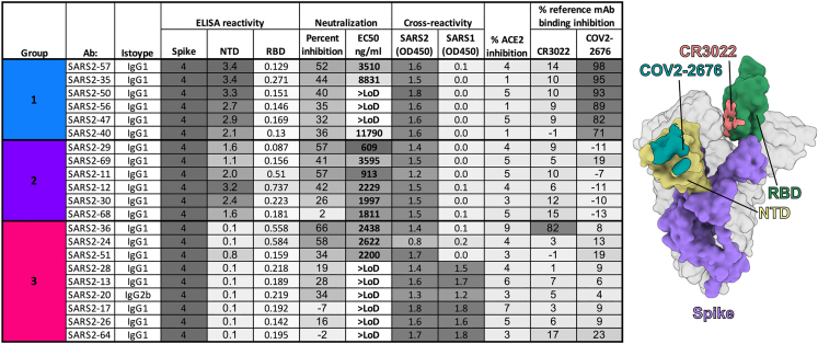
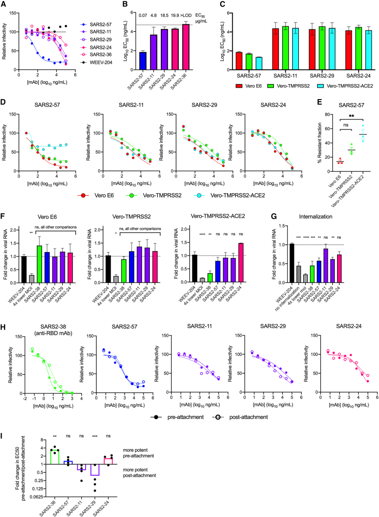
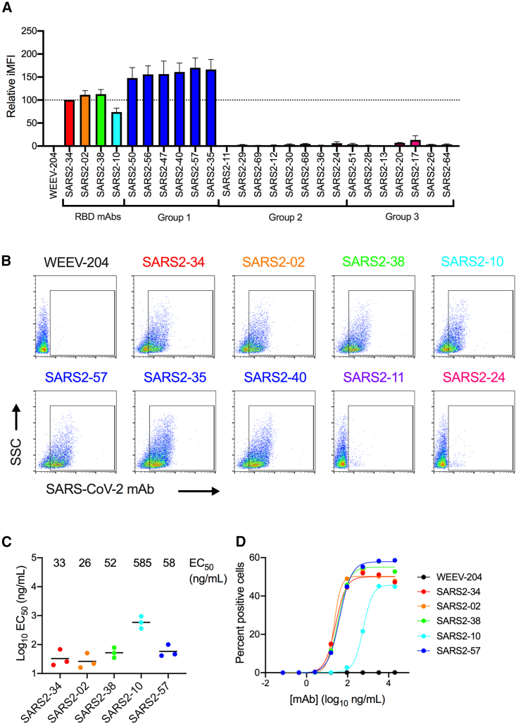
Most neutralizing anti-SARS-CoV-2 monoclonal antibodies (mAbs) target the receptor binding domain (RBD) of the spike (S) protein. Here, we characterize a panel of mAbs targeting the N-terminal domain (NTD) or other non-RBD epitopes of S. A subset of NTD mAbs inhibits SARS-CoV-2 entry at a post-attachment step and avidly binds the surface of infected cells. One neutralizing NTD mAb, SARS2-57, protects K18-hACE2 mice against SARS-CoV-2 infection in an Fc-dependent manner. Structural analysis demonstrates that SARS2-57 engages an antigenic supersite that is remodeled by deletions common to emerging variants. In neutralization escape studies with SARS2-57, this NTD site accumulates mutations, including a similar deletion, but the addition of an anti-RBD mAb prevents such escape. Thus, our study highlights a common strategy of immune evasion by SARS-CoV-2 variants and how targeting spatially distinct epitopes, including those in the NTD, may limit such escape.

Keywords: B cell epitope mapping; SARS-CoV-2; cryo-EM; neutralizing antibodies; variants of concern.


Conflict of interest statement

Declaration of interests M.S.D. is a consultant for Inbios, Vir Biotechnology, Ocugen, Topspin Therapeutics, GlaxoSmithKline, Moderna, and Immunome. The Diamond laboratory has received unrelated funding support in sponsored research agreements from Vir Biotechnology, Emergent BioSolutions, and Moderna. Some of the mAbs described in this study have been licensed by Washington University to Bio X Cell. D.H.F. is a founder of Courier Therapeutics and has received funding support in a sponsored research agreement from Emergent BioSolutions. J.E.C. has served as a consultant for Eli Lilly and Luna Biologics, is a member of the Scientific Advisory Boards of CompuVax and Meissa Vaccines, and is the founder of IDBiologics. The Crowe laboratory at Vanderbilt University Medical Center has received sponsored research agreements from AstraZeneca and IDBiologics.

Figures

None
Graphical abstract
Figure 1
Figure 1
Panel of anti-SARS-CoV-2 mAbs Hybridoma supernatants from the panel of anti-SARS-CoV-2 murine mAbs were assayed for inhibition of SARS-CoV-2 by focus-reduction neutralization test (FRNT), cross-reactivity to SARS-CoV-1 S protein, and ability to inhibit SARS-CoV-2 S protein binding to hACE2 or a panel of reference human mAbs through competition ELISA. MAbs are grouped by reference mAb competition properties. EC50 values reflect neutralization experiments performed with purified mAbs. Data represent the mean (or geometric mean for EC50 values) from two to four experiments. Beside the table, an approximate epitope is illustrated for each reference mAb, with that of CR3022 (PDB 7LOP) in pink and COV2-2676 (EMDB: EMD-23155) in blue.
Figure 2
Figure 2
Neutralization by anti-SARS-CoV-2 mAbs (A–E) Anti-SARS-CoV-2 mAbs were assayed for neutralization by FRNT against SARS-CoV-2 using Vero E6 cells (A and B) or comparing Vero E6 cells with Vero-TMPRSS2 and Vero-hACE2-TMPRSS2 cells (C–E). (A and D) Representative dose-response curves are shown; error bars represent the range from two technical replicates. Data are from three to four experiments. (B and C) Geometric mean EC50 values are shown. (E) Neutralization-resistant fraction of SARS2-57 dose-response curves. (F) Anti-SARS-CoV-2 mAbs were assayed for attachment inhibition of SARS-CoV-2 to Vero E6, Vero-TMPRSS2, or Vero-hACE2-TMPRSS2 cells. Data are from three experiments. (G) Anti-SARS-CoV-2 mAbs were assayed for inhibition of virus internalization in Vero E6 cells. Data are from four experiments. (H and I) Anti-SARS-CoV-2 mAbs were assayed for pre- or post-attachment neutralization of SARS-CoV-2 using Vero E6 cells. An anti-RBD mAb, SARS2-38, is included as a control. (H) Representative dose-response curves are shown. Error bars represent the range from two technical replicates. (I) Fold change in EC50 values for pre-attachment over post-attachment neutralization. Error bars represent standard error of the mean (SEM) from four experiments. (I) ANOVA with Sidak’s post-test comparing pre- versus post-attachment EC50 values for each mAb; (F) and (G) one-way ANOVA with Dunnett’s post-test compared mAb treatment with isotype control mAb treatment. ns, not significant; ∗p < 0.05; ∗∗p < 0.01; ∗∗∗p < 0.001, ∗∗∗∗p < 0.0001.
Figure 3
Figure 3
Staining of S protein on the surface of infected cells by anti-SARS-CoV-2 mAbs (A and B) Vero cells infected with SARS2-CoV-2 were stained with the indicated anti-SARS-CoV-2 mAbs (10 μg/mL) and analyzed by flow cytometry. Positively stained cells were gated, and their integrated mean fluorescence intensity (iMFI) was determined (MFI ✕ percent positive cells). (A) The mean relative iMFI from three experiments. Relative iMFI for each experiment was determined by normalizing iMFI values to a representative anti-RBD mAb, SARS2-34. (B) Representative flow cytometry plots with positive gates are shown. (C and D) SARS-CoV-2-infected cells were stained with serial dilutions of anti-SARS-CoV-2 mAbs, and the EC50 values for the percent of positively stained cells were determined. (C) Mean EC50 values for each mAb. Error bars represent SEM from three experiments. (D) Representative dose-response curves are shown.
Figure 4
Figure 4
MAbs protect against SARS-CoV-2 infection in vivo (A–D) K18-hACE2 transgenic mice were passively administered 100 μg (∼5 mg/kg) of the indicated mAb by intraperitoneal injection 24 h prior to intranasal inoculation with 103 ffu of SARS-CoV-2 WA1/2020. (A) Mice were monitored for weight change for 7 days following viral infection. Mean weight change is shown. Error bars represent SEM. (B and C) At 7 dpi, nasal washes (B) and lungs (C) were collected, and viral RNA levels were determined. Median levels are shown; dotted line represents the limit of detection (LOD) of the assay. (D) A subset of the lungs from (C) were assessed for infectious virus by plaque assay. Median PFU/mL is shown. Dotted line indicates the LOD. (A–D) Data for each mAb are from two experiments; WEEV-204 (isotype control): n = 12; all other mAbs: n = 6 per group. (E) SARS2-57 chimeric mouse Fv/human IgG1 Fc mAb was assayed for neutralization by FRNT against SARS-CoV-2 WA/2020. m, mouse hybridoma-derived mAb; h, recombinant chimeric mAb. Representative dose-response curves are shown from one of three experiments. (F) hSARS2-57 N297Q mAb was assayed for binding to murine Fcγ receptors FcγRI and FcγRIV relative to intact hSARS2-57 by ELISA, using an anti-human capture mAb as a control. Shown is the mean relative absorbance obtained for N297Q over intact/WT mAb for each FcγR or capture mAb. Error bars represent SEM from three experiments. (G and H) K18-hACE2 transgenic mice were administered 100 μg (∼5 mg/kg) of the indicated chimeric mAb by intraperitoneal injection 24 h prior to intranasal inoculation with 103 ffu of SARS-CoV-2 WA1/2020. Data are from two experiments; hE16 (isotype control): n = 8 per group; hSARS2-57 and hSARS2-57 N297Q: n = 7 per group. (I and J) K18-hACE2 transgenic mice were administered 200 μg (10 mg/kg) of the indicated mAb by intraperitoneal injection 24 h after intranasal inoculation with 103 ffu of SARS-CoV-2 WA1/2020. Data are from two experiments; hE16 (isotype control) and hSARS2-57: n = 6; hSARS2-57 N297Q: n = 5 per group. (K and L) K18-hACE2 transgenic mice were administered 100 μg (∼5 mg/kg) of the indicated mAb by intraperitoneal injection 24 h prior to intranasal inoculation with 103 ffu of SARS-CoV-2 WA1/2020 with sequence-confirmed wild-type furin cleavage site (FCS) sequence. Data are from two experiments; n = 12 per group. (G, I, and K) Mean weight change is shown. Error bars represent SEM. (H, J, and L) At 7 dpi, lung, nasal washes, heart, and brain were collected and viral RNA levels were determined. (A, G, and I) One-way ANOVA of area under the curve of 4–7 dpi with Dunnet’s post-test; (K) t test of area under the curve of 4–7 dpi; (B, C, H, and J) Kruskal-Wallis with Dunn’s post-test; (D and L) Mann-Whitney test (ns, not significant, ∗p < 0.05, ∗∗p < 0.01, ∗∗∗p < 0.001, ∗∗∗∗p < 0.0001).
Figure 5
Figure 5
SARS2-57 binds NTD loops N3 and N5 (A) Biolayer interferometry signal (left) and steady-state analysis (right) of SARS2-57 Fab interacting with immobilized SARS-CoV-2 spike. Kinetic values were fitted to a global 1:1 binding model. Plots are representative of three technical replicates. (B) Density map of SARS2-57 Fab bound to trimeric SARS-CoV-2 S with all RBDs in the down position. The NTD bound by SARS2-57 is shown in yellow, with the RBD of the same monomer colored green and the rest of the monomer shown in purple. The S trimer is otherwise colored gray. The SARS2-57 heavy and light chains are shown in royal blue and cyan, respectively. (C) Magnified region from black box in (B). Focused density map of the Fv/NTD complex encompassing a refined atomic model. The NTD is shown in yellow, with loops N3 and N5 colored green. The SARS2-57 heavy and light chains are colored royal blue and cyan, respectively. (D) Magnified regions from blue and purple boxes in (C), as indicated. A ribbon diagram of the NTD abutting a surface rendering of the SARS2-57 Fv. Loops N3 and N5 are colored mint and forest green, respectively, with glycan N149 colored pink. The SARS2-57 heavy and light chains are colored royal blue and cyan, respectively. PISA contact residues are shown as sticks. (E) A ribbon diagram of the SARS2-57 complementarity-determining regions (CDR) overlying a surface rendering of the NTD. Loops N3 and N5 are colored mint and forest green, respectively, with glycan N149 colored pink and the rest of the NTD shown in yellow. CDRs of the SARS2-57 heavy and light chain are colored royal blue and cyan, respectively, with PISA paratope residues shown as sticks.
Figure 6
Figure 6
Identification of SARS2-57 escape mutations (A and B) Neutralization escape mutants were isolated by passaging a VSV-eGFP-SARS-CoV-2-S chimeric virus (S from D614G strain) in the presence of SARS2-57. Wild-type and escape mutant viruses were tested for neutralization sensitivity to SARS2-57 by plaque assay. (A) Representative plaque assay images are shown. Data are representative of two experiments. (B) The locations of escape mutants are highlighted on the structure of the SARS-CoV-2 S protein (PDB: 7C2L). Spike is colored tan, the NTD is colored blue, and the RBD is colored green. Mutations on NTD loop N3 are highlighted in red, while mutations on NTD loop N5 are highlighted in orange. (C) SARS-CoV-2 variants and their mutations in spike. (D) Vero-TMPRSS2 cells were inoculated with the indicated SARS-CoV-2 strains. At 48 h after infection, cells were stained with mAb SARS2-38 (upper) or SARS2-57 (lower) at a concentration of 10, 1, or 0.1 μg/mL prior to analysis by flow cytometry. The percent of positively stained cells was determined after gating on staining with WEEV-204 (isotype control). Shown is the relative percent of positively stained cells (normalized to SARS2-38 staining at 10 μg/mL for each variant or a polyclonal mixture for Omicron subvariants) from two experiments. Error bars represent SEM.
Figure 7
Figure 7
The SARS2-57 epitope may be remodeled by deletions in emerging variants (A) Density map and fitted model of SARS2-57 Fv bound to trimeric SARS-CoV-2 S with all RBDs in the down position. The NTD bound by SARS2-57 is shown in yellow, with the RBD of the same monomer colored green and the rest of the monomer shown in purple. The S trimer is otherwise colored gray. The SARS2-57 heavy and light chains are shown in royal blue and cyan, respectively. (B) Multiple sequence alignment of the NTD loops N3 and N5 from WA1/2020 and SARS-CoV-2 VOCs/VOIs with the PISA binding footprint of SARS2-57 boxed in blue. VOC/VOI mutations are highlighted in red, and SARS2-57 escape mutations are designated with purple triangles (substitutions) or a dotted black line (deletion). Red triangles indicate variants that escape SARS2-57 recognition. Secondary structure annotation is displayed above the alignment in yellow with loops N3 and N5 noted in green. The PISA binding footprints of previously described human anti-NTD mAbs are compared below the alignment as blue lines. Antibodies 2–51, 4–18, 5–24, and 4–8 also contact loop N1 (not pictured). (C) Magnified region from the black box in (A) displaying only residues included in (B). NTD loops are displayed as round ribbons with residues of interest shown as sticks. Residues are color coded to indicate epitope residues identified by PISA (blue, left), VOC/VOI substitutions or deletions (red or gray, middle), and escape substitutions or deletions (purple or gray, right). VOCs represented include those listed in (B) (i.e., Wash-B.1.351, B.1.630, B.1.621, B.1.1.7, B.1.1.298, Wash-B.1.1.28, B.1.429, B.1.617.1, B.1.617.2, B.1.526, BA.1, BA.1.1, and BA.2).

References

    1. Baden L.R., El Sahly H.M., Essink B., Kotloff K., Frey S., Novak R., Diemert D., Spector S.A., Rouphael N., Creech C.B., et al. Efficacy and Safety of the mRNA-1273 SARS-CoV-2 Vaccine. N. Engl. J. Med. 2021;384:403–416. - PMC - PubMed
    1. Cathcart A.L., Havenar-Daughton C., Lempp F.A., Ma D., Schmid M.A., Agostini M.L., Guarino B., Iulio J.D., Rosen L.E., Tucker H., et al. The dual function monoclonal antibodies VIR-7831 and VIR-7832 demonstrate potent in Vitro and in Vivo activity against SARS-CoV-2. bioRxiv. 2021 Preprint at. 2021.03.09.434607.
    1. Chen P., Nirula A., Heller B., Gottlieb R.L., Boscia J., Morris J., Huhn G., Cardona J., Mocherla B., Stosor V., et al. SARS-CoV-2 Neutralizing Antibody LY-CoV555 in Outpatients with Covid-19. N. Engl. J. Med. 2021;384:229–237. - PMC - PubMed
    1. Keech C., Albert G., Cho I., Robertson A., Reed P., Neal S., Plested J.S., Zhu M., Cloney-Clark S., Zhou H., et al. Phase 1-2 Trial of a SARS-CoV-2 Recombinant Spike Protein Nanoparticle Vaccine. N. Engl. J. Med. 2020;383:2320–2332. - PMC - PubMed
    1. Polack F.P., Thomas S.J., Kitchin N., Absalon J., Gurtman A., Lockhart S., Perez J.L., Pérez Marc G., Moreira E.D., Zerbini C., et al. Safety and Efficacy of the BNT162b2 mRNA Covid-19 Vaccine. N. Engl. J. Med. 2020;383:2603–2615. - PMC - PubMed

Publication types

Supplementary concepts