. 2024 Feb:506:85-94.
doi: 10.1016/j.ydbio.2023.11.008. Epub 2023 Nov 29.

Fgf signalling is required for gill slit formation in the skate, Leucoraja erinacea

Affiliations

Fgf signalling is required for gill slit formation in the skate, Leucoraja erinacea

Jenaid M Rees et al. Dev Biol. 2024 Feb.

Abstract

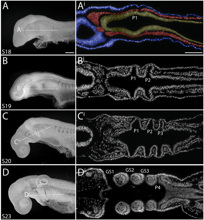
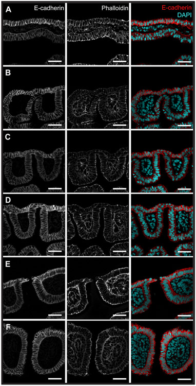
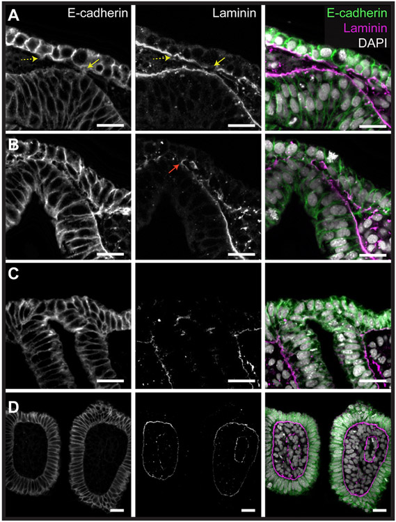
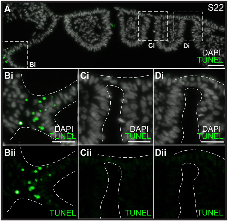
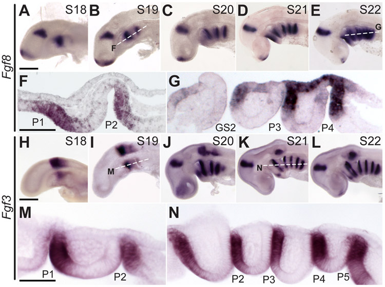
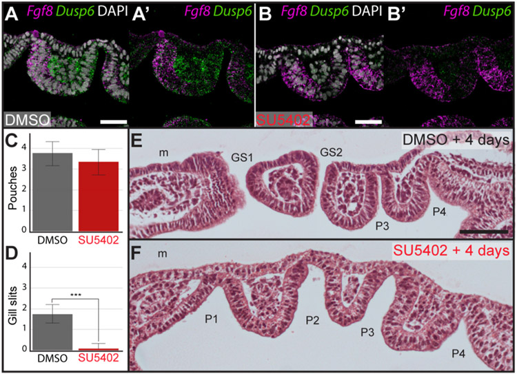
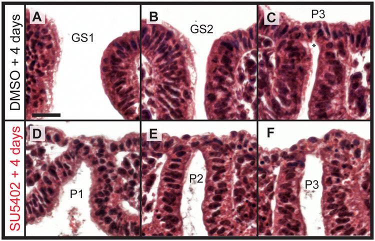
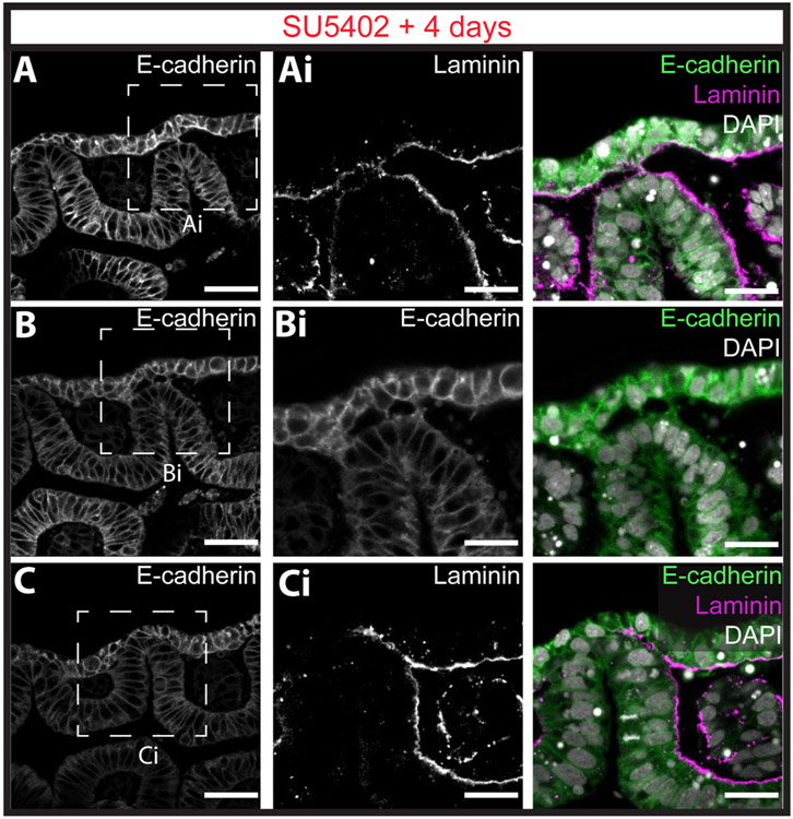
The gill slits of fishes develop from an iterative series of pharyngeal endodermal pouches that contact and fuse with surface ectoderm on either side of the embryonic head. We find in the skate (Leucoraja erinacea) that all gill slits form via a stereotypical sequence of epithelial interactions: 1) endodermal pouches approach overlying surface ectoderm, with 2) focal degradation of ectodermal basement membranes preceding endoderm-ectoderm contact; 3) endodermal pouches contact and intercalate with overlying surface ectoderm, and finally 4) perforation of a gill slit occurs by epithelial remodelling, without programmed cell death, at the site of endoderm-ectoderm intercalation. Skate embryos express Fgf8 and Fgf3 within developing pharyngeal epithelia during gill slit formation. When we inhibit Fgf signalling by treating skate embryos with the Fgf receptor inhibitor SU5402 we find that endodermal pouch formation, basement membrane degradation and endodermal-ectodermal intercalation are unaffected, but that epithelial remodelling and gill slit perforation fail to occur. These findings point to a role for Fgf signalling in epithelial remodelling during gill slit formation in the skate and, more broadly, to an ancestral role for Fgf signalling during pharyngeal pouch epithelial morphogenesis in vertebrate embryos.

Keywords: Ectoderm; Endoderm; Fgf3; Fgf8; Gill slit; Pharyngeal pouch; Skate.


Figures

Fig. 1.
Fig. 1.. Overview of pharyngeal arch development in the little skate.
Skate embryos at stages (S)18, 19, 20 and 23 in lateral view (anterior to the left), and corresponding horizontal sections through the pharynx, stained with DAPI. (A–Ai) The first pharyngeal pouch begins to form as an outpocket from the pharyngeal endoderm at S18. Additional pouches will follow in an anterior-to-posterior sequence. By (B–Bi) S19 and (C-Ci) 20, two or three pharyngeal pouches have formed, and these pouches are in contact with surface ectoderm. (D–Di) By S23, the first, second and third pharyngeal pouches have fused with surface ectoderm to form gill slits, while the fourth pouch has contacted the surface ectoderm, but has not yet fused. In Ai, endoderm is false-coloured yellow, mesoderm is false-coloured red, and ectoderm is false-coloured blue. GS1-GS3, gill slits 1–3; P1P4, pharyngeal pouches 1–4. Scale bars: A,B,C,D = 100 μm; Ai,Bi,Ci,Di = 100 μm.
Fig. 2.
Fig. 2.. Sequence of epithelial interactions during gill slit development in the little skate.
Immunofluorescence for E-cadherin and phalloidin staining of developing pharyngeal pouches and gill slits in the skate. (A) Endodermal pouches initially bulge out toward the overlying surface ectoderm, displacing the mesoderm layer that sits between the endoderm and ectoderm. (B) The endoderm then contacts the overlying ectoderm and (C) begins to push against the surface ectoderm. (D) Endoderm and ectoderm form a single epithelium. (E) The epithelium perforates at the site of endoderm–ectoderm contact, forming a gill slit opening. (F) The columns of tissue that are delineated by adjacent gill slits are called pharyngeal arches. All scale bars = 50 μm.
Fig. 3.
Fig. 3.. Basement membrane degradation and epithelial protrusions precede endoderm-ectoderm intercalation and gill slit formation.
Immunofluorescence for E-cadherin and laminin reveals that (A) degradation of the ectodermal basement membrane precedes endoderm-ectoderm contact, with small cytoskeletal protrusions (dashed yellow arrow) forming bridges (yellow arrow) between the ectoderm and the endodermal basement membrane. (B) These bridges continue to form and are associated with further basement membrane degradation in the ectoderm and endoderm. The endodermal and ectodermal basement membranes flanking the point of endoderm-ectoderm contact join to form a new continuous basement membrane (red arrow) (C) The basement membrane fully degrades at the site of endoderm-ectoderm contact, leading to intercalation of the endoderm and ectoderm. At this stage, endoderm and ectoderm are no longer recognisable as distinct epithelia. (D) After perforation of the gill slit is complete, the new continuous endodermal-ectodermal basement membrane flanking the gill slit forms persists within the pharyngeal arch. All scale bar = 25 μm.
Fig. 4.
Fig. 4.. Cell death does not contribute to gill slit perforation in the skate.
(A) Horizontal section through a S22 skate embryo stained with TUNEL and DAPI. (Bi-Bii) There is a clear region of positive TUNEL staining indicating cell death between the rostral pharyngeal epithelium and the neural tube. However, (Ci/Cii-Di/Dii) no cell death was detected at any stage during endoderm–ectoderm intercalation or gill slit perforation. All sections were stained and imaged for n = 4 S22 skate embryos. Scale bars: A = 50 μm, Bi-Di = 25 μm.
Fig. 5.
Fig. 5.. Fgf8 and Fgf3 are expressed in the skate endoderm and ectoderm during pharyngeal pouch formation and fusion.
(AE) Fgf8 is expressed sequentially with the formation of pharyngeal endodermal pouches, as shown by wholemount mRNA ISH at S18–22. Horizontal sections of (F) S19 and (G) S22 embryos indicate that Fgf8 is expressed in the posterior endodermal epithelium of each pharyngeal pouch and in the overlying ectodermal epithelium during endoderm-ectoderm intercalation. (HL) Fgf3 is expressed sequentially with the formation of pharyngeal endodermal pouches, as shown by wholemount mRNA ISH at S18–22. Horizontal sections of (M) S19 and (N) S22 embryos indicate that Fgf3 is expressed in the posterior endodermal epithelium of each developing pharyngeal pouch. GS2, gill slit 2; P1P5, pharyngeal pouches 1–5. Scale bars; A–E and H–L = 500 μm; F,G,M,N = 100 μm.
Fig. 6.
Fig. 6.. Fgf signalling is required for gill slit perforation in the skate.
Expression of Dusp6 offers a transcriptional readout of Fgf signalling in epithelium and mesenchyme of developing pharyngeal pouches. To validate the effectiveness of our Fgf signalling inhibition strategy, we compared expression of Dusp6 expression around developing pharyngeal pouches using mRNA ISH by chain reaction (HCR) 48 h after in ovo injection of (A) DMSO (control) or (B) SU5402 at S19/20. With HCR and imaging performed in parallel, under identical conditions, we observed a striking reduction in expression of Dusp6 in pharyngeal epithelium and mesenchyme with SU5402 treatment. (C) There is no significant reduction in the number of pharyngeal pouches following four days of in ovo exposure to the Fgf receptor antagonist SU5402, relative to control. However, (D) there is a significant reduction in the number of gill slits following four days of in ovo exposure to SU5402, relative to control. (E) Representative histological section through an embryo after four days of exposure to DMSO. This embryo possesses two gill slits and two additional pharyngeal pouches that have not yet formed gill slits. (F) Representative histological section through an embryo after four days of exposure to SU5402. This embryo possesses four pharyngeal pouches, but no gill slits. GS1-GS2, gill slits 1–2; P1P4, pharyngeal pouches 1–4; m, mandibular arch. Scale bars: A, B = 25 μm; E,F = 100 μm.
Fig. 7.
Fig. 7.. Fgf signalling is dispensable for epithelial intercalation but is required for gill slit perforation in the skate.
In a representative skate embryo treated with DMSO in ovo for 4 days from S19/20, (A) gill slit 1 and (B) gill slit 2 have perforated, and (C) pouch 3 has intercalated with the surface ectoderm and perforation is initiating (*). By contrast, in a representative skate embryo treated with SU5402 in ovo for 4 days from S19/20, (D) pouch 1, (E) pouch 2 and (F) pouch 3 have all intercalated with the surface ectoderm, but gill slits have perforated. GS1-GS2, gill slits 1–2; P1P3: pharyngeal pouches 1–3. All scale bar = 30 μm.
Fig. 8.
Fig. 8.. Fgf signalling is dispensable for basement membrane degradation and endoderm–ectoderm intercalation but is required for gill slit perforation in the skate.
Immunofluorescence for E-cadherin and laminin reveals that with SU5402 treatment (A) ectodermal basement membrane degradation, (B) ectodermal cytoskeletal protrusions toward the endoderm, (C) endoderm basement membrane degradation and endoderm–ectoderm intercalation all occur normally. However, Fgf signalling is required for remodelling of intercalated epithelium and gill slit perforation. All pouches were imaged for n = 3 SU5402-treated embryos, and representative images are shown here. Scale bars: A,B,C = 50 μm; Ai,Bi,Ci = 25 μm.

Similar articles

Cited by

References

    1. Abu-Issa R, Smyth G, Smoak I, Yamamura K-I, Meyers EN, 2002. Fgf8 is required for pharyngeal arch and cardiovascular development in the mouse. Development 129, 4613–4625. - PubMed
    1. Ballard WW, Mellinger J, Lechenault H, 1993. A series of normal stages for development of Scyliorhinus canicula, the lesser spotted dogfish (Chondrichthyes: Scyliorhinidae). J. Exp. Zool 267, 318–336.
    1. Bertrand S, Camasses A, Somorjai I, Belgacem MR, Chabrol O, Escande ML, Pontarotti P, Escriva H, 2011. Amphioxus FGF signaling predicts the acquisition of vertebrate morphological traits. Proc. Natl. Acad. Sci. U.S.A 108, 9160–9165. - PMC - PubMed
    1. Chen J, Jacox LA, Saldanha F, Sive H, 2017. Mouth Development, vol. 6. Wiley Interdiscip Rev Dev Biol. - PMC - PubMed
    1. Choe CP, Collazo A, Trinh Le A, Pan L, Moens CB, Crump JG, 2013. Wnt-dependent epithelial transitions drive pharyngeal pouch formation. Dev. Cell 24, 296–309. - PMC - PubMed

Publication types

LinkOut - more resources