. 2023 Dec 1;11(12):e007415.
doi: 10.1136/jitc-2023-007415.

Hsa-LINC02418/mmu-4930573I07Rik regulated by METTL3 dictates anti-PD-L1 immunotherapeutic efficacy via enhancement of Trim21-mediated PD-L1 ubiquitination

Affiliations

Hsa-LINC02418/mmu-4930573I07Rik regulated by METTL3 dictates anti-PD-L1 immunotherapeutic efficacy via enhancement of Trim21-mediated PD-L1 ubiquitination

Zhijia Sun et al. J Immunother Cancer. .

Abstract

Background: Limited response to programmed death ligand-1 (PD-L1)/programmed death 1 (PD-1) immunotherapy is a major hindrance of checkpoint immunotherapy in non-small cell lung cancer (NSCLC). The abundance of PD-L1 on the tumor cell surface is crucial for the responsiveness of PD-1/PD-L1 immunotherapy. However, the negative control of PD-L1 expression and the physiological significance of the PD-L1 inhibition in NSCLC immunotherapy remain obscure.

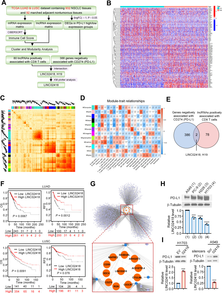
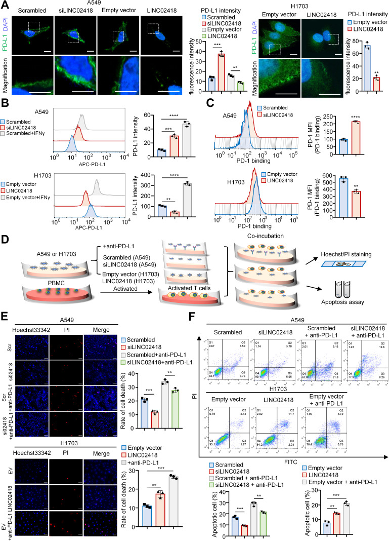
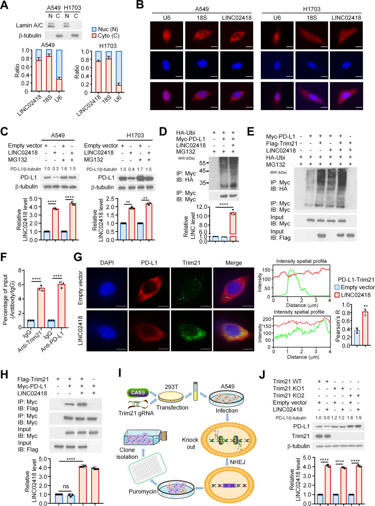
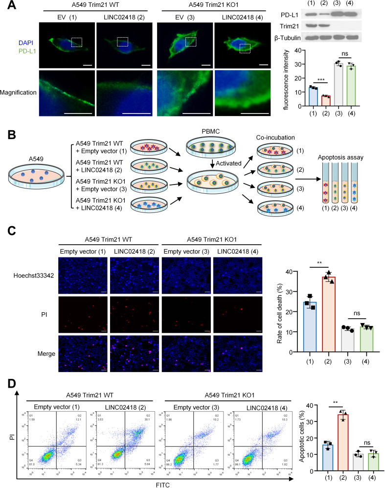
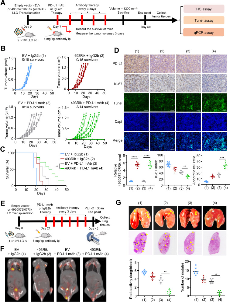
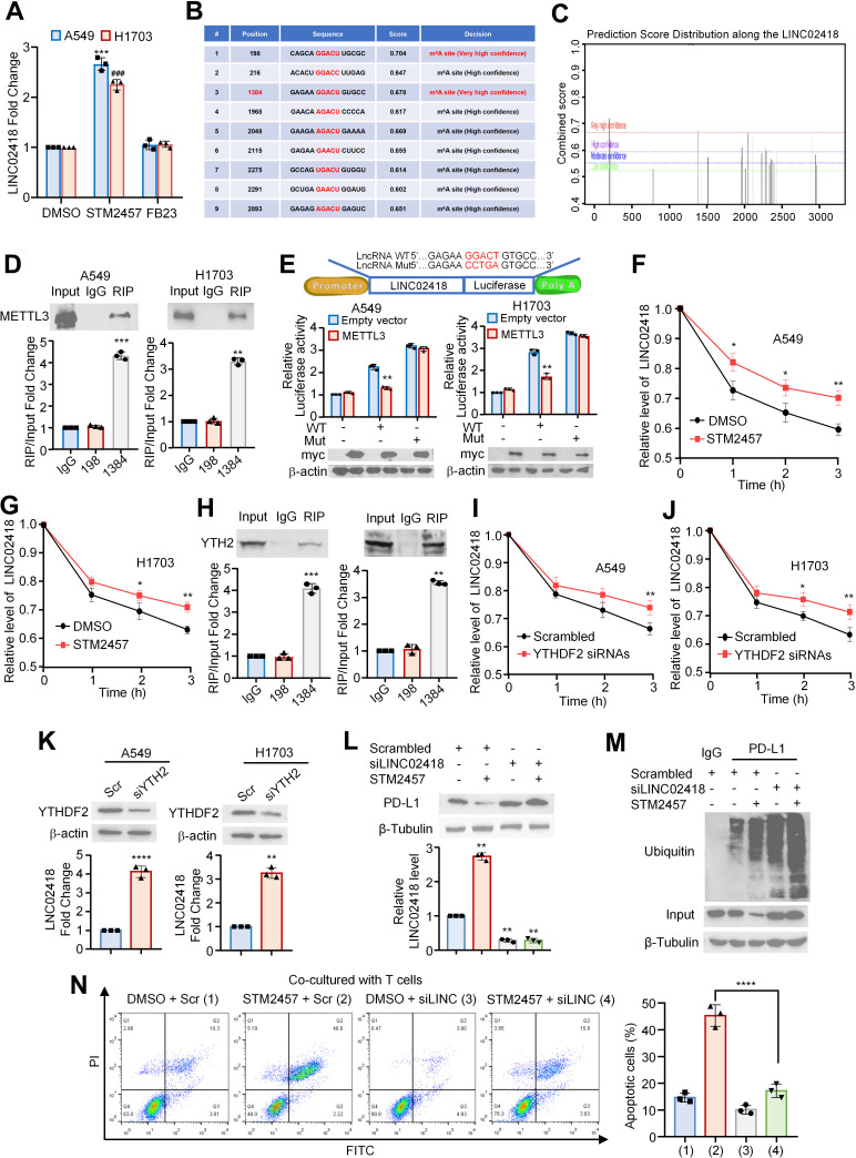
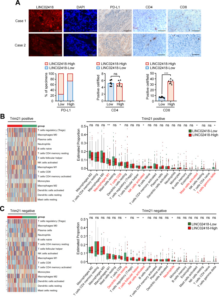
Methods: Bioinformatics analysis was performed to profile and investigate the long non-coding RNAs that negatively correlated with PD-L1 expression and positively correlated with CD8+T cell infiltration in NSCLC. Immunofluorescence, in vitro PD-1 binding assay, T cell-induced apoptosis assays and in vivo syngeneic mouse models were used to investigate the functional roles of LINC02418 and mmu-4930573I07Rik in regulating anti-PD-L1 therapeutic efficacy in NSCLC. The molecular mechanism of LINC02418-enhanced PD-L1 downregulation was explored by immunoprecipitation, RNA immunoprecipitation (RIP), and ubiquitination assays. RIP, luciferase reporter, and messenger RNA degradation assays were used to investigate the m6A modification of LINC02418 or mmu-4930573I07Rik expression. Bioinformatics analysis and immunohistochemistry (IHC) verification were performed to determine the significance of LINC02418, PD-L1 expression and CD8+T cell infiltration.

Results: LINC02418 is a negative regulator of PD-L1 expression that positively correlated with CD8+T cell infiltration, predicting favorable clinical outcomes for patients with NSCLC. LINC02418 downregulates PD-L1 expression by enhancing PD-L1 ubiquitination mediated by E3 ligase Trim21. Both hsa-LINC02418 and mmu-4930573I07Rik (its homologous RNA in mice) regulate PD-L1 therapeutic efficacy in NSCLC via Trim21, inducing T cell-induced apoptosis in vitro and in vivo. Furthermore, METTL3 inhibition via N6-methyladenosine (m6A) modification mediated by YTHDF2 reader upregulates hsa-LINC02418 and mmu-4930573I07Rik. In patients with NSCLC, LINC02418 expression is inversely correlated with PD-L1 expression and positively correlated with CD8+T infiltration.

Conclusion: LINC02418 functions as a negative regulator of PD-L1 expression in NSCLC cells by promoting the degradation of PD-L1 through the ubiquitin-proteasome pathway. The expression of LINC02418 is regulated by METTL3/YTHDF2-mediated m6A modification. This study illuminates the underlying mechanisms of PD-L1 negative regulation and presents a promising target for improving the effectiveness of anti-PD-L1 therapy in NSCLC.

Keywords: immune checkpoint inhibitors; lung neoplasms; non-small cell lung cancer; programmed cell death 1 receptor.


Conflict of interest statement

Competing interests: None declared.

Figures

Figure 1
Figure 1
Identification and characterization of a novel lncRNA positively correlates with CD8+T cell infiltration and predicts a good prognosis in NSCLC. (A) Schematic diagram of the screening process. (B) Heatmap of the top 100 dysregulated lncRNAs in the high-CD274 and low-CD274 (PD-L1) expression groups. (C) The TOM heatmap shows the module genes obtained by WGCNA analysis combining the lncRNA expression matrix and the immune cell score. (D) The heatmap shows the correlation between the module genes of lncRNA and immune cells, with the upper number in each module represents the Pearson coefficient and the lower number represents the p value. (E) Venn diagram shows the intersected genes that are negatively associated with CD274 and positively associated with CD8+T cells. (F) Kaplan-Meier analysis of the overall survival (OS) rate and recurrence-free survival (RFS) rate of patients with NSCLC with low or high expression of LINC02418 (http://kmplot.com/analysis). Low or high LINC02418 expression was grouped by choosing the auto select best cut-off option. (G) Network analysis of the lncRNA (LINC02418), mRNAs, and miRNAs intersection in (A). The orange circles represent LINC02418 and the top 10 mRNAs with the highest correlation coefficient, the light blue circles represent the other mRNAs exhibiting a smaller correlation coefficient, and the dark blue circles represent miRNAs. (H) Protein levels of PD-L1 and expression levels of LINC02418 in four NSCLC cell lines. (I) Expression levels of LINC02418 and PD-L1 in H1703 or A549 cells following transfection of LINC02418 vector or LINC02418 smart pool of silencers. ***p<0.001, ****p<0.0001 versus the corresponding controls. DEG, differentially expressed gene; FC, fold change; IncRNA, long non-coding RNA; LUAD, lung adenocarcinoma; LUSC, lung squamous cell carcinoma; mRNA, messenger RNA; NSCLC, non-small cell lung cancer; PD-L1, programmed cell death ligand 1. TCGA, the cancer genome atlas; WGCNA, weighted correlation network analysis.
Figure 2
Figure 2
LINC02418 dampens PD-1 binding ability and enhances T cell-dependent toxicity in NSCLC. (A) Immunofluorescence analysis of PD-L1 expression in A549 cells treated with the LINC02418 smart pool of silencers (siLINC02418) or scrambled siRNA (scrambled), as well as in A549 cells or H1703 cells treated with either empty vector or LINC02418. Scale bars represent 10 µm in all panels. (B) FCM was used to detect PD-L1 expression on the surface of NSCLC cells with different conditions. (C) FCM was used to detect the intensity of fluorescence binding to PD-1 of NSCLC cells with different conditions. The PD-1 mean fluorescence intensity (MFI) graph was plotted. (D) Schematic diagram of the effect of LINC02418 on tumor cells killing by PBMC. (E) Hoechst 33342/PI staining assay was used to determine the effect of LINC02418 on PBMC cytotoxicity, with histograms showing the rate of cell death. (F) Cell apoptosis assay was used to determine the effect of LINC02418 on PBMC cytotoxicity, with histograms showing the proportion of apoptotic cells. All experiments were conducted three times, and the results were similar. All values were presented as mean±SD, and statistical differences were calculated using two-sided Student’s t-test. **p<0.01, ***p<0.001, ****p<0.0001 versus the corresponding control. APC, allophycocyanin; DAPI, 4’,6-Diamidine-2’-phenylindole dihydrochloride; FCM, flow cytometric; FITC, fluorescein Isothiocyanate; IFN, interferon; PBMC, peripheral blood mononuclear cell; PD-1, programmed death 1; PD-L1, programmed cell death ligand 1; PI, propidium iodide; NSCLC, non-small cell lung cancer.
Figure 3
Figure 3
LINC02418 regulates PD-L1 protein stability through enhancing the Trim21-mediated ubiquitin-proteasome pathway. (A) LINC02418 expression in H1703 and A549 cells was detected using qRT-PCR. The separated nucleus and cytoplasm fractions were assessed using western blot assays, with lamin A/C and β-tubulin selected as markers, respectively. (B) Subcellular localization of LINC02418 (red), U6 (nuclear marker, red), and 18S (cytoplasmic marker, red) in H1703 and A549 cells was observed using fluorescence in situ hybridization. The nuclei were stained blue by 4’,6-Diamidine-2’-phenylindole dihydrochloride (DAPI). Scale bar, 10 µm. (C) H1703 and A549 cells were transfected with empty vector or LINC02418 and treated with the proteasome inhibitor MG132 (10 μM). PD-L1 expression was detected by Western blot. Histograms showed corresponding expression levels of LINC02418 measured by qRT-PCR. (D) Effects of LINC02418 on the ubiquitination of PD-L1 was analyzed by immunoprecipitation treated with MG132 and incubated with indicated antibodies. (E) Effects of LINC02418 on Trim21-mediated PD-L1 ubiquitination. (F) The interaction between LINC02418 with Trim21 and PD-L1 were assessed by an RNA immunoprecipitation assay. (G) Immunofluorescence showed the co-localization of PD-L1/Trim21 under empty vector and LINC02418 vector transfection in A549 cells. The intensity distribution of PD-L1/Trim21 was plotted in the middle panel, and the statistical quantification of colocalization (Pearson’s R value) was shown on the right panel. Values were presented as mean ± SD from three independent experiments, with comparison using a two-sided Student’s t-test. Scale bar, 10 µm. (H) Co-immunoprecipitation was performed using lysates of A549 cells expressing Flag-Trim21 and Myc-PD-L1 with or without overexpression of LINC02418. (I) Schematic diagram of the construction of Trim21 knockout (KO) A549 cell lines. (J) Immunoblot analysis of PD-L1 in A549 cells with Trim21 KO cells (Trim21 KO1, Trim21 KO2) stably transfected with empty vector or LINC02418 vector. **p<0.01, ***p<0.001, ****p<0.0001 versus the corresponding control, ns means no significance.IP, immunoprecipitation; IB, immunoblotting; PD-L1, programmed cell death ligand 1; qRT-PCR, quantitative real-time polymerase chain reaction; WT, wildtype.
Figure 4
Figure 4
LINC02418 downregulates PD-L1 expression and enhances T cell-induced apoptosis dependent on Trim21 in NSCLC. (A) A549 cells with either Trim21 WT or knockout (Trim21 KO1) were transfected with either empty vector (EV) or LINC02418, and immunofluorescence was used to analyze PD-L1 expression. The histogram displayed the fluorescence intensity on the cell surfaces. (B) Schematic diagram showed the verification of the effect of LINC02418 on T cell-dependent toxicity via Trim21. (C) Hoechst33342/PI staining assay was used to verify whether the effect of LIINC02418 on T cell-induced apoptosis required Trim21 participation. The histogram showed the apoptotic rate of the indicated groups. (D) Apoptosis test was used to verify the effect of LINC02418 on T cell-induced apoptosis in one representative clone of the A549 Trim21 knockout cell lines (Trim21 KO1). The histogram showed the apoptosis rate of the indicated groups. **p<0.01, ***p<0.001 versus the corresponding control, ns means no significance. DAPI, 4’,6-Diamidine-2’-phenylindole dihydrochloride; FITC, fluorescein Isothiocyanate; PBMC, peripheral blood mononuclear cell; PD-L1, programmed cell death ligand 1; PI, propidium iodide; WT, wildtype.
Figure 5
Figure 5
Mmu-4930573I07Rik (493Rik), the mouse homologous RNA of hsa-LINC02418, enhances the efficacy of PD-L1 antibody therapy in vivo. (A) The experimental scheme of the in vivo study. (B) Volumes of LLC tumors carrying empty vector (treated with IgG2b, blue lines, n=15; treated with anti-PD-L1 mAb, gray lines, n=14), or 493Rik vector (treated with IgG2b, red lines, n=15; treated with anti-PD-L1 mAb, green lines, n=14) were plotted individually. (C) Kaplan-Meier survival curves for each group. The p value was calculated using a two-sided Gehan-Breslow-Wilcoxon test. (D) The top two rows were representative IHC staining of PD-L1 and Ki67 of the collected tumors. The histograms showed the 493Rik levels determined by qRT-PCR and Ki-67 index determined by proportion of positive cell count. The lower three rows were representative Tunel staining of the collected tumors. The histogram showed the apoptotic cell ratio. Scale bar, 50 µm. (E) The experimental scheme of the in vivo study of lung metastasis. (F) The representative PET-CT scan images of the mice in each group. (G) Representative lung tissue anatomy and H&E staining pictures of the mice. The histogram showed the number of tumor nodules observed under the microscope. *p<0.05, **p<0.01, ***p<0.001, ****p<0.0001 versus the corresponding control, ns means no significance. IHC, immunohistochemistry; LLC, Lewis lung carcinoma cell; mAb, monoclonal Antibody; PD-L1, programmed cell death ligand 1; PET-CT, positron emission tomography-CT; qRT-PCR, quantitative real-time polymerase chain reaction.
Figure 6
Figure 6
Hsa-LINC02418 is m6A-modificated by METTL3 and degraded in a manner dependent on the m6A reader protein YTHDF2. (A) The expression level of LINC02418 in A549 and H1703 cells treated with DMSO, STM2457 or FB23, respectively. (B) The prediction score distribution along the LINC02418 sequence. (C) The specific possible position of m6A sites in the LINC02418 sequence. (D) RIP-RT-PCR assays for detection of the two m6A sites in A549 and H1703 cell lysates immunoprecipitated with METTL3 antibody. (E) The upper panel showed the sequence scheme of the wild-type (WT) and mutant (Mut) luciferase reporter plasmids. The lower panel showed the luciferase activity after co-transfection of LINC02418 WT or LINC02418 Mut plasmid with myc tagged METTL3 plasmid or empty vector. (F) The mRNA levels of LINC02418 were detected at different time points after treated with STM2457 or DMSO in A549 cells. (G) The mRNA levels of LINC02418 were detected at different time points after treated with STM2457 or DMSO in H1703 cells. (H) Two m6A sites with very high confidence were detected by qRT-PCR assay after immunoprecipitated with YTHDF2 antibody. The mRNA level of LINC02418 were detected at different time points after treated with YTHDF2 siRNAs or scrambled in (I) A549 cells and (J) H1703 cells, respectively. (K) The fold change of LINC02418 in A549 and H1703 cells under YTHDF2 gene was knocked down with specific siRNAs. (L) The mRNA level and protein expression level of PD-L1 in A549 cells which were treated by LINC02418 siRNAs or scrambled siRNAs with or without STM2457. (M) Immunoprecipitation and immunoblotting analysis of A549 cells treated by LINC02418 siRNAs or scrambled siRNAs with or without STM2457. (N) Apoptosis assay tested T-cell cytotoxic effect to A549 cells treated by LINC02418 siRNAs or scrambled siRNAs with or without STM2457. The histogram showed the percentage of apoptosis cells. Data are presented as mean±SD, *p<0.05, **p<0.01, ***p<0.001, ****p<0.0001 versus the corresponding control. DMSO, Dimethyl sulfoxidem; FITC, fluorescein Isothiocyanate; mRNA, messenger RNA; PD-L1, programmed cell death ligand 1; RIP, RNA immunoprecipitation; qRT-PCR, quantitative real-time polymerase chain reaction.
Figure 7
Figure 7
Correlation between the expression of LINC02418, PD-L1 and T immune cell infiltration in human NSCLC. (A) The representative fluorescence in situ hybridization staining of LINC02418 and IHC staining of PD-L1, CD4 and CD8 in tumor sections of clinical NSCLC cases. Scale bar, 50 µm. The lower three histograms showed the percentage of sections with low or high PD-L1 displaying low or high LINC02418 levels in NSCLC specimens, the positive CD4 cell counts and the positive CD8 cell counts. (B, C) In the TCGA database, patients with NSCLC were divided into Trim21 positive and Trim21 negative group. The proportions of immune cells were compared under LINC02418 low or high expression status. *p<0.05, **p<0.01 versus the corresponding control, ns means no significance. DAPI, 4’,6-Diamidine-2’-phenylindole dihydrochloride; IHC, immunohistochemistry; NK, natural killer; NSCLC, non-small cell lung cancer; PD-L1, programmed cell death ligand 1; TCGA, the cancer genome atlas.

References

    1. Bade BC, Dela Cruz CS. Lung cancer 2020: epidemiology, etiology, and prevention. Clin Chest Med 2020;41:1–24. 10.1016/j.ccm.2019.10.001 - DOI - PubMed
    1. Pardoll DM. The blockade of immune checkpoints in cancer immunotherapy. Nat Rev Cancer 2012;12:252–64. 10.1038/nrc3239 - DOI - PMC - PubMed
    1. Chen Y, Pei Y, Luo J, et al. . Looking for the optimal PD-1/PD-L1 inhibitor in cancer treatment: a comparison in basic structure, function, and clinical practice. Front Immunol 2020;11:1088. 10.3389/fimmu.2020.01088 - DOI - PMC - PubMed
    1. Huang M-Y, Jiang X-M, Wang B-L, et al. . Combination therapy with PD-1/PD-L1 blockade in non-small cell lung cancer: strategies and mechanisms. Pharmacol Ther 2021;219:107694. 10.1016/j.pharmthera.2020.107694 - DOI - PubMed
    1. Yang Y, Yu Y, Lu S. Effectiveness of PD-1/PD-L1 inhibitors in the treatment of lung cancer: brightness and challenge. Sci China Life Sci 2020;63:1499–514. 10.1007/s11427-019-1622-5 - DOI - PubMed

Publication types