

MiR-1246b, a novel miRNA molecule of extracellular vesicles in bronchoalveolar lavage fluid, promotes nodule growth through FGF14 in patients with lung cancer
- PMID: 38040694
- PMCID: PMC10692082
- DOI: 10.1038/s41419-023-06218-9
MiR-1246b, a novel miRNA molecule of extracellular vesicles in bronchoalveolar lavage fluid, promotes nodule growth through FGF14 in patients with lung cancer
Abstract
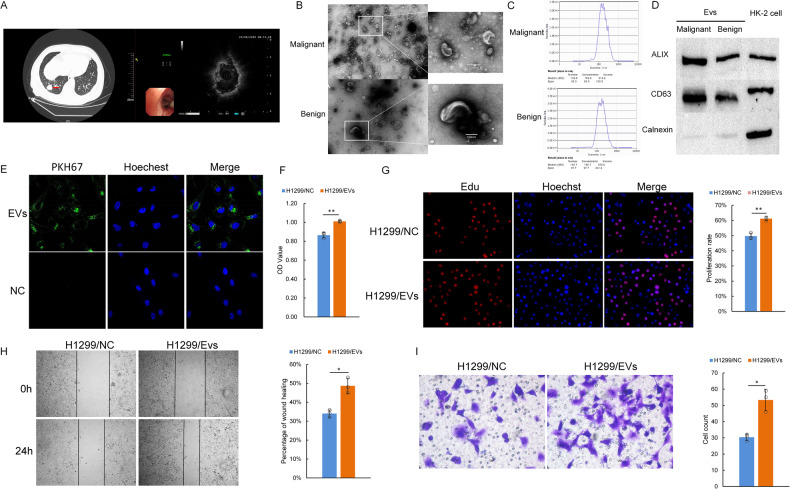
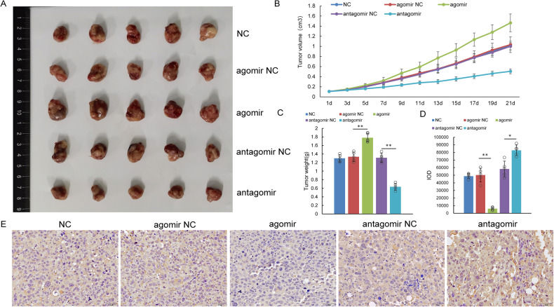
With the widespread development of chest computed tomography (CT), the detection rate of pulmonary nodules has increased; therefore, the classification of benign vs. malignant nodules has become a common problem in the clinic. MicroRNA, a potential tool, is expected to become a good choice for diagnosing and studying the occurrence and development of diseases through the vector of bronchoalveolar lavage fluid extracellular vesicles (BALF-EVs). In this study, radial endobronchial ultrasound (R-EBUS) was used to locate pulmonary nodules in patients. BALF was obtained, EVs were isolated, and small RNA sequencing was performed to screen differentially expressed miRNAs between benign and malignant pulmonary nodules. The binding targets and underlying mechanisms of the differentially expressed miRNAs were verified by in vitro and in vivo experiments. R-EBUS localization and sampling was used to obtain BALF, and EVs were successfully isolated and characterized. Differentially expressed miRNAs in BALF-EVs of patients with benign vs. malignant pulmonary nodules were screened by high-throughput small RNA sequencing. A new miRNA, miR-1246b, was identified. We found that FGF14 was the binding target of miR-1246b by luciferase assay. Subsequent mechanistic studies showed that miR-1246b inhibited the expression of FGF14 in lung cancer cells, further leading to ERK phosphorylation and epithelial-to-mesenchymal transition (EMT), which ultimately contributed to lung cancer cell proliferation, migration and invasion. In summary, our study demonstrates that the detection of miRNAs in BALF-EVs, a means of liquid biopsy, could assist in distinguishing malignant nodules from benign nodules. miR-1246b, which was extracted from BALF-EVs, targets FGF14 to promote lung cancer cell proliferation, migration and invasion.
© 2023. The Author(s).
Conflict of interest statement
The authors declare no competing interests.
Figures


Similar articles
-
Diagnostic Potential of microRNAs in Extracellular Vesicles Derived from Bronchoalveolar Lavage Fluid for Pneumonia-A Preliminary Report.Cells. 2022 Sep 22;11(19):2961. doi: 10.3390/cells11192961. Cells. 2022. PMID: 36230923 Free PMC article.
-
Acute respiratory distress vs healthy lung environments differently affect mesenchymal stromal cell extracellular vesicle miRNAs.Cytotherapy. 2025 May;27(5):581-596. doi: 10.1016/j.jcyt.2025.01.006. Epub 2025 Jan 20. Cytotherapy. 2025. PMID: 39945694
-
Identification of miRNA-rich vesicles in bronchoalveolar lavage fluid: Insights into the function and heterogeneity of extracellular vesicles.J Control Release. 2019 Jan 28;294:43-52. doi: 10.1016/j.jconrel.2018.12.008. Epub 2018 Dec 7. J Control Release. 2019. PMID: 30529727 Free PMC article.
-
EVs from BALF-Mediators of Inflammation and Potential Biomarkers in Lung Diseases.Int J Mol Sci. 2021 Apr 1;22(7):3651. doi: 10.3390/ijms22073651. Int J Mol Sci. 2021. PMID: 33915715 Free PMC article. Review.
-
Extracellular Vesicle MicroRNA Transfer in Lung Diseases.Front Physiol. 2017 Dec 12;8:1028. doi: 10.3389/fphys.2017.01028. eCollection 2017. Front Physiol. 2017. PMID: 29311962 Free PMC article. Review.
Cited by
-
DNA Molecular Computing with Weighted Signal Amplification for Cancer miRNA Biomarker Diagnostics.Adv Sci (Weinh). 2025 Jun;12(22):e2416490. doi: 10.1002/advs.202416490. Epub 2025 Apr 11. Adv Sci (Weinh). 2025. PMID: 40213938 Free PMC article.
-
Current trends in theranostic applications of extracellular vesicles in cancer.Front Oncol. 2025 Jun 3;15:1592006. doi: 10.3389/fonc.2025.1592006. eCollection 2025. Front Oncol. 2025. PMID: 40530018 Free PMC article. Review.
-
Integrated multi-omics landscape of non-small cell lung cancer with distant metastasis.Front Immunol. 2025 Mar 17;16:1560724. doi: 10.3389/fimmu.2025.1560724. eCollection 2025. Front Immunol. 2025. PMID: 40165954 Free PMC article.
-
Harnessing EVs-ncRNA for Lung Cancer: From Oncogenic Pathways to Novel Diagnostic and Therapeutic Strategies.Int J Nanomedicine. 2025 Jul 14;20:9031-9054. doi: 10.2147/IJN.S528115. eCollection 2025. Int J Nanomedicine. 2025. PMID: 40689018 Free PMC article. Review.
-
Recent advances to address challenges in extracellular vesicle-based applications for lung cancer.Acta Pharm Sin B. 2024 Sep;14(9):3855-3875. doi: 10.1016/j.apsb.2024.06.010. Epub 2024 Jun 20. Acta Pharm Sin B. 2024. PMID: 39309489 Free PMC article. Review.
References
-
- Haidong H, Yunye N, Wei Z, Zarogoulidis P, Hohenforst-Schmidt W, Man YG, et al. Multiple guided technologies based on radial probe endobronchial ultrasound for the diagnosis of solitary peripheral pulmonary lesions: a single-center study. J Cancer. 2017;8:3514–21. doi: 10.7150/jca.20035. - DOI - PMC - PubMed
-
- Halpenny D, Das K, Ziv E, Plodkowski A, Zheng J, Capanu M, et al. Percutaneous computed tomography guided biopsy of sub-solid pulmonary nodules: differentiating solid from ground glass components at the time of biopsy. Clin Imaging. 2021;69:332–8. doi: 10.1016/j.clinimag.2020.07.011. - DOI - PMC - PubMed
Publication types
MeSH terms
Substances
LinkOut - more resources
Full Text Sources
Medical
Miscellaneous
