. 2023 Dec 1;14(1):7762.
doi: 10.1038/s41467-023-43498-3.

SMARCB1 loss activates patient-specific distal oncogenic enhancers in malignant rhabdoid tumors

Affiliations

SMARCB1 loss activates patient-specific distal oncogenic enhancers in malignant rhabdoid tumors

Ning Qing Liu et al. Nat Commun. .

Abstract

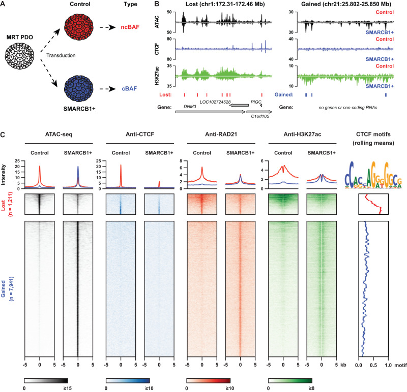
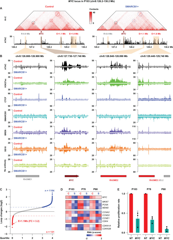
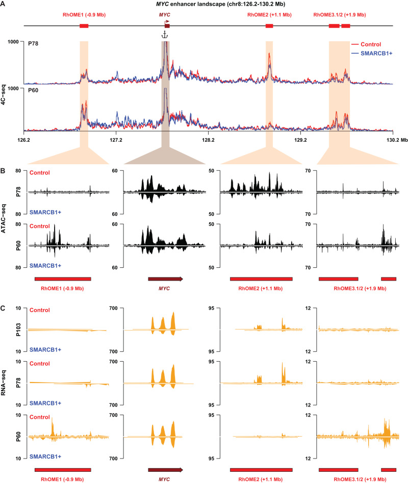
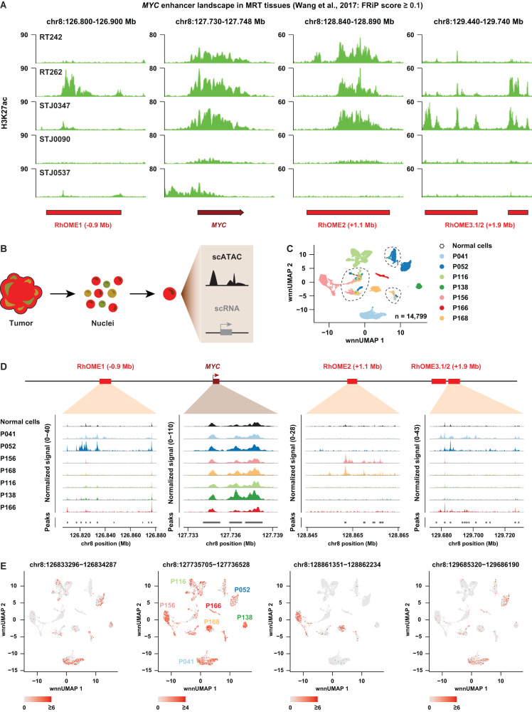
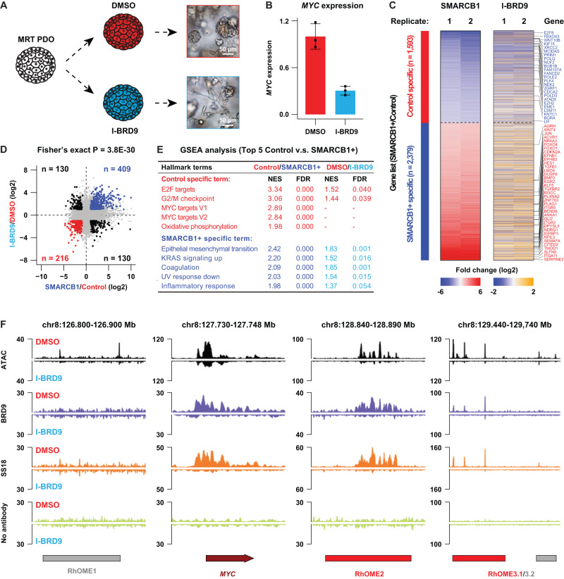
Malignant rhabdoid tumor (MRT) is a highly malignant and often lethal childhood cancer. MRTs are genetically defined by bi-allelic inactivating mutations in SMARCB1, a member of the BRG1/BRM-associated factors (BAF) chromatin remodeling complex. Mutations in BAF complex members are common in human cancer, yet their contribution to tumorigenesis remains in many cases poorly understood. Here, we study derailed regulatory landscapes as a consequence of SMARCB1 loss in the context of MRT. Our multi-omics approach on patient-derived MRT organoids reveals a dramatic reshaping of the regulatory landscape upon SMARCB1 reconstitution. Chromosome conformation capture experiments subsequently reveal patient-specific looping of distal enhancer regions with the promoter of the MYC oncogene. This intertumoral heterogeneity in MYC enhancer utilization is also present in patient MRT tissues as shown by combined single-cell RNA-seq and ATAC-seq. We show that loss of SMARCB1 activates patient-specific epigenetic reprogramming underlying MRT tumorigenesis.


Conflict of interest statement

The authors declare no competing interests.

Figures

Fig. 1
Fig. 1. SMARCB1 reconstitution reshapes open chromatin landscape in the MRT PDO model.
A Schematic overview of the experimental set-up of lentiviral transduction (adapted from Custers et al.); B Representative loci that lose (left) or gain (right) chromatin accessibility after SMARCB1 reconstitution (ATAC-seq: n = 3, an average of the 3 replicates; ChIP-seq of CTCF and H3K27Ac: n = 1). Called lost and gained peaks are indicated below the tracks (FDR cut-off <0.05, FC difference in accessibility > 2); C Tornado-plot showing that the lost (control-specific) open chromatin regions are enriched for the binding of CTCF, RAD21 and H3K27ac, while the gained (SMARCB1 + -specific) open chromatin regions are only enriched for RAD21 and H3K27ac (CTCF ChIP n = 1, RAD21 ChIP n = 1, H3K27Ac ChIP n = 1).
Fig. 2
Fig. 2. SMARCB1 reconstitution affects distal enhancers of the MYC oncogene in MRT PDOs.
A Hi-C contact maps depicting the MYC locus including the MYC promoter and three distal regulatory regions in P103 (E-0.9 Mb; E + 1.1 Mb; E + 1.8 Mb). Left panel depicts chromatin contact in control and the right panel contacts after SMARCB1 reconstitution. Circles indicate established chromatin loops and dashed circles indicate reduced chromatin loop formation. B ATAC-seq, ChIP-seq and CUT&RUN of the indicated proteins in MRT PDO P103 with (SMARCB1 + , lower tracks) and without (Control, upper tracks) SMARCB1 expression at the MYC locus. No antibody was included as negative control for CUT&RUN tracks (BRD9 and SS18). C Waterfall plot summarizes changes of the identified chromatin loops before and after SMARCB1 reconstitution (n = 1). The E + 1.1 Mb enhancer (RhOME2) is one of the most affected loops. D Z-scores calculated for expression of MYC, cell proliferation markers and cell cycle markers in the Control and SMARCB1+ samples of the three PDOs using bulk RNA-seq. E CellTiter-Glo assay measuring cell viability of MRT PDOs 7 days after transduction with a non-targeting shRNA (NT) or an shRNA targeting MYC, showing that knockdown of MYC leads to a decreased proliferation of the tumor PDOs. Data is represented as means ± SD (n = 3 independent experiments) (n = 3). Source data are provided as a source data file.
Fig. 3
Fig. 3. Identification of patients-specific MYC enhancer-ome in different MRT PDOs.
A Contact map of 4C-seq data identifies chromatin interactions between the MYC promoter and three Rhabdoid Oncogenic MYC Enhancers (RhOMEs) in two additional MRT PDO models (P60 and P78; n = 2). B Chromatin accessibility and (C) enhancer RNA (eRNA) expression at the three RhOMEs and the MYC mRNA expression in the indicated MRT PDOs with (SMARCB1 + ) and without (Control) SMARCB1 expression.
Fig. 4
Fig. 4. Patient-specific super enhancers form TAD boundaries in the SMARCB1-null tumor PDOs that are independent of CTCF.
A A K-means clustering analysis on the control-specific (lost) OCRs, based on their chromatin features in these PDOs (ATAC n = 3, CTCF ChIP n = 1, RAD21 ChIP n = 1, H3K27Ac ChIP n = 1). B Contact map of P103 Hi-C data depicting an example of a super-enhancer forming a TAD boundary (left panel), that is diminished after SMARCB1 reconstitution (right panel). Supporting tracks of ATAC-seq and ChIP-seq (CTCF, H3K27Ac) are displayed above. C Aggregate Region Analysis (ARA) showing the insulation borders in each separate K-mean cluster. The K3 cluster is comprised of chromatin loops with SMARCB1-sensitive insulation borders in P103.
Fig. 5
Fig. 5. Analyses of MRT tissues reveal inter- and intra-tumor heterogeneity of MYC enhancer landscape.
A Genomic tracks depicting H3K27Ac signal at the MYC locus from publicly available H3K27Ac ChIP-seq data. Data was filtered based on an average FRiP score of higher than 0.1. B Acquisition of single-cell Multiome data (gene expression + ATAC-seq) from MRT tissues using Chromium 10x Genomics platform. C UMAP discriminating tumor cells, but not normal cells, from the MRT tissues derived from different patients (n = 1 per patient). Tumor cells cluster separately while normal cells are depicted as a mixed population from different patients. D A pseudo-bulk view of scATAC-seq reads at the MYC locus for 7 MRT patients. E UMAP depicting RhOME specific peak signal within each single cell (n = 1 per patient).
Fig. 6
Fig. 6. A therapeutic strategy for MRT patients via targeting a ncBAF subunit named BRD9 using a pharmacological inhibitor.
A Schematic representation of the experiment and brightfield images showing a differentiation phenotype upon BRD9 inhibition in MRT PDOs (adapted from Custers et al.). B RT-qPCR for MYC in P103 treated with DMSO or I-BRD9 (10 μM). Data is represented as means ± SD (n = 3 independent experiments). Source data are provided as a source data file. C Heatmaps showing the transcriptomic changes of differential expressed genes between SMARCB1+ and Control in either SMARCB1 reconstitution or BRD9 inhibition experiments (n = 2). D Correlation of transcriptomic changes of SMARCB1 reconstitution with BRD9 inhibition. Upper right panel show all genes significantly upregulated in both experiments (blue dots) (n = 2). Lower left panel represent all genes specific for control condition in both experiments (red dots). Statistical significance was calculated by a fisher’s exact test. E GSEA analyses showing top five hallmark gene sets identified in the SMARCB1 reconstitution and the corresponding differential expression analysis after BRD9 inhibition. F Chromatin occupancy at the MYC locus 5 days after I-BRD9 treatment depicting ATAC-seq and CUT&RUN for BRD9 and SS18. No antibody was included as the negative control.

References

    1. Mashtalir N, et al. Modular Organization and Assembly of SWI/SNF Family Chromatin Remodeling Complexes. Cell. 2018;175:1272–1288.e20. doi: 10.1016/j.cell.2018.09.032. - DOI - PMC - PubMed
    1. Shain AH, Pollack JR. The spectrum of SWI/SNF mutations, ubiquitous in human cancers. PLoS One. 2013;8:e55119. doi: 10.1371/journal.pone.0055119. - DOI - PMC - PubMed
    1. Kadoch C, et al. Proteomic and bioinformatic analysis of mammalian SWI/SNF complexes identifies extensive roles in human malignancy. Nat. Genet. 2013;45:592–601. doi: 10.1038/ng.2628. - DOI - PMC - PubMed
    1. Plass C, et al. Mutations in regulators of the epigenome and their connections to global chromatin patterns in cancer. Nat. Rev. Genet. 2013;14:765–780. doi: 10.1038/nrg3554. - DOI - PubMed
    1. Masliah-Planchon J, Bièche I, Guinebretière JM, Bourdeaut F, Delattre O. SWI/SNF chromatin remodeling and human malignancies. Annu Rev. Pathol. 2015;10:145–171. doi: 10.1146/annurev-pathol-012414-040445. - DOI - PubMed

Publication types

Associated data