. 2024 Feb:69:102975.
doi: 10.1016/j.redox.2023.102975. Epub 2023 Nov 29.

Epigenetically upregulated NSUN2 confers ferroptosis resistance in endometrial cancer via m5C modification of SLC7A11 mRNA

Affiliations

Epigenetically upregulated NSUN2 confers ferroptosis resistance in endometrial cancer via m5C modification of SLC7A11 mRNA

Shuai-Jun Chen et al. Redox Biol. 2024 Feb.

Abstract

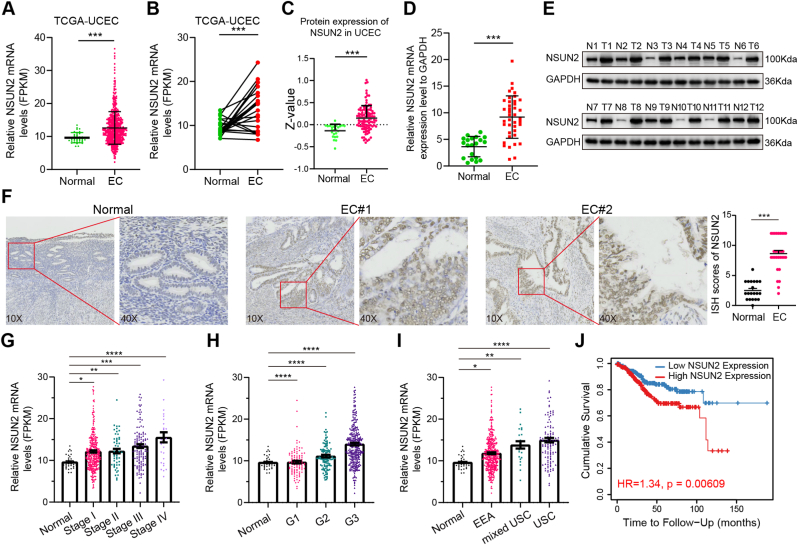
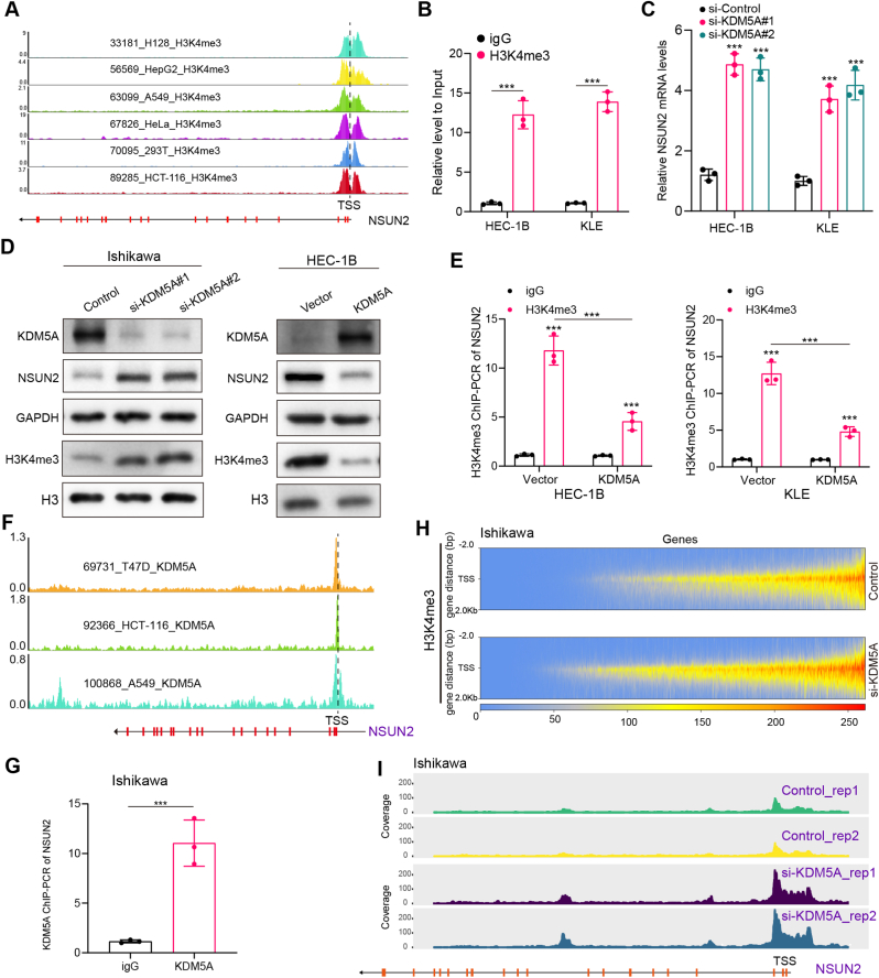
Endometrial cancer (EC) is a prevalent gynecological malignancy worldwide, and 5-methylcytosine (m5C) modification of mRNA is a crucial epigenetic modification associated with the development and occurrence of several cancers. However, the precise function of m5C modification in EC remains elusive. This study aimed to investigate the expression and clinical significance of the primary m5C modification writer, NSUN2, in EC. Our findings indicated that NSUN2 exhibited a substantial up-regulation in EC as a result of an epigenetic augmentation in H3K4me3 levels within the promoter region, which was triggered by the down-regulation of KDM5A. Moreover, gain- and loss-of-function experiments revealed the role of NSUN2 in enhancing m5C modification of mRNA, thereby promoting EC cell proliferation. RNA bisulfite sequencing and transcriptomic sequencing were employed to elucidate the involvement of NSUN2 in the regulation of ferroptosis. Subsequent in vitro experiments confirmed that the knockdown of NSUN2 significantly up-regulated the levels of lipid peroxides and lipid ROS in EC cells, thereby augmenting the susceptibility of EC to ferroptosis. Mechanistically, NSUN2 stimulated the m5C modification of SLC7A11 mRNA, and the m5C reader YBX1 exhibited direct recognition and binding to the m5C sites on SLC7A11 mRNA via its internal cold shock domain (CSD), leading to an increase in SLC7A11 mRNA stability and elevated levels of SLC7A11. Additionally, rescue experiments showed that NSUN2 functioned as a suppressor of ferroptosis, which was dependent on SLC7A11. Overall, targeting the NSUN2/SLC7A11 axis inhibited tumor growth by increasing lipid peroxidation and ferroptosis of EC cells both in vitro and in vivo. Therefore, our study provides new insight into the role of NSUN2, suggesting that NSUN2 may serve as a prognostic biomarker and therapeutic target in patients with EC.

Keywords: Endometrial cancer; Ferroptosis; NSUN2; SLC7A11; m(5)C.


Conflict of interest statement

Declaration of competing interest The authors declare that they have no known competing financial interests or personal relationships that could have appeared to influence the work reported in this paper.

Figures

Fig. 1
Fig. 1
NSUN2 overexpression predicts poor prognosis in endometrial cancer. (A) The mRNA levels of NSUN2 were compared between EC and normal tissues in TCGA UCEC dataset. (B) Different mRNA expression of NSUN2 between EC and the paired normal tissues. (C) The protein expression levels of NSUN2 based on the CPTAC database. (D) RT-qPCR analysis displaying the mRNA levels of NSUN2 in surgical EC and normal samples. (E) Western blot was used to detect the protein levels of NSUN2 in EC and paired normal tissues. (F) Representative images of immunohistochemical staining for NSUN2 and the comparison of the ISH scores in EC and normal tissues. The expression levels of NSUN2 were presented in different FIGO stages (G), histological grade (H), and pathological type (I). (J) Kaplan-Meier (KM) curve for OS of patients with UCEC in TCGA and patients were stratified into low and high expression based on the median NSUN2 expression. *P < 0.05; **P < 0.01; ***P < 0.001.
Fig. 2
Fig. 2
NSUN2 upregulation is regulated by the epigenetic alteration of H3K4me3. (A) ChIP-seq data for mapping H3K4me3 modifications near the NSUN2 promoter in the different cell lines were obtained from the Cistrome Data in the WashU Epigenome Browser. (B) ChIP-PCR analysis of H3K4me3 modification in the promoter of NSUN2. (C) The mRNA levels of NSUN2 were detected by qPCR after the knockdown of KDM5A. (D) Protein expression in HEC-1B or Ishikawa cells with overexpression or silencing of KDM5A was assessed using Western blot; GAPDH and H3 were used as the loading control. (E) ChIP-PCR analysis of H3K4me3 modification of the NSUN2 promoter in KDM5A overexpression EC cells. (F) KDM5A ChIP-seq of different cell lines at NSUN2 promoter is displayed using the WashU Epigenome Browser. (G) ChIP-PCR analysis of KDM5A in the promoter of NSUN2. (H) Heatmaps illustrating H3K4me3 levels around gene body regions. (I) IGV tracks presenting the enrichments of H3K4me3 by CUT&Tag. ***P < 0.001.
Fig. 3
Fig. 3
NSUN2 is involved in the regulation of m5C levels and the proliferation ability of EC cells. (A, B) The mRNA and protein levels of NSUN2 in normal endometrium and five different EC cell lines. (C) The m5C levels of total RNA in NSUN2 knockdown HEC-1B and KLE cells or NSUN2 overexpression Ishikawa cells were indicated by m5C dot blot. (D–F) CCK-8, colony formation, and EdU assays were carried out to detect the proliferation ability of EC cells with NSUN2 knockdown or overexpression. *P < 0.05; **P < 0.01; ***P < 0.001.
Fig. 4
Fig. 4
Identification of NSUN2-mediated regulation of m5C-modified mRNAs. (A) Flow chart of the bisulfite sequencing. (B) The number of m5C modified sites on different chromosomes in control or NSUN2 knockdown HEC-1B cells. (C) The sequence context of 10 bases upstream and downstream of NSUN2-m5C sites is depicted in the probability pattern. (D) Distribution and methylation level of all m5C sites in different segments of mRNAs. (E) The heatmap showed differentially methylated genes in HEC-1B cells with NSUN2 knockdown. The screening criteria were as follows: methylation difference greater than 0.1, P-value less than 0.05. (F) The number distribution histogram of differentially methylated mRNAs. (G) Pathway enrichment analysis of differentially methylated genes.
Fig. 5
Fig. 5
NSUN2 knockdown promotes ferroptosis in EC. (A, B) CCK-8 assay was performed to determine the cell viability after treatment with different concentrations of erastin in HEC-1B or KLE cells. (C, D) Evaluation of cell viability after treatment with DMSO, erastin, erastin and Fer-1, erastin and Z-VAD, erastin and 3-MA in the indicated cells. (E) Calcein-AM (green)/PI (red) staining was performed to visualize living and dead cells, respectively. (F) Intracellular lipid peroxides MDA levels were measured with an MDA assay kit. (G) Lipid peroxidation was measured by flow cytometry after C11-BODIPY staining in the indicated cells. (H) Representative TEM images of mitochondria in control or NSUN2 knockdown EC cells. ***P < 0.001; ns, not statistically significant. (For interpretation of the references to colour in this figure legend, the reader is referred to the Web version of this article.)
Fig. 6
Fig. 6
NSUN2 regulates m5C modification of SLC7A11 mRNA to promote its mRNA stability. (A) The m5C modification level of ferroptosis-related genes in RNA bisulfite sequencing data. (B) The mRNA levels of different ferroptosis-related genes in transcriptome sequencing results. (C) Quantitative PCR was conducted to detect the mRNA expression of ferroptosis-related genes in NSUN2 knockdown KLE cells. (D) Western blot was used to evaluate the protein levels of SLC7A11 after NSUN2 knockdown or overexpression. (E) The effect of double mutant NSUN2 on SLC7A11 protein level. (F) RIP/qPCR was carried out to evaluate the binding of NSUN2 to SLC7A11 mRNA. (G) MeRIP-PCR was performed using an m5C-specific antibody to evaluate the levels of m5C modification on SLC7A11 mRNA after NSUN2 knockdown or overexpression. (H) The half-life of SLC7A11 mRNA was measured after actinomycin D treatment. (I) Representative images of anti-SLC7A11 immuno-histochemical staining and the comparison of the ISH scores in EC and normal tissues. (J) The mRNA expression of SLC7A11 in TCGA UCEC and normal tissues. (K) The correlation between mRNA expression levels for NSUN2 and SLC7A11 in TCGA UCEC dataset was evaluated by TIMER online database (https://cistrome.shinyapps.io/timer/). *P < 0.05; **P < 0.01; ***P < 0.001; ns, not statistically significant.
Fig. 7
Fig. 7
YBX1 recognizes m5C modification of SLC7A11 and stabilizes SLC7A11 mRNA. (A) RNA pull-down was performed using the biotin-labeled probes in the HEC-1B cell. Bound proteins were analyzed by SDS-PAGE and silver staining. (B) Mass spectrometry assays revealed YBX1 peptides pulled down by m5C-modified SLC7A11 probes. (C) YBX1 immunoblot analysis of biotinylated SLC7A11 or m5C-SLC7A11 RNA pull-downs in HEC-1B and KLE cells. (D, E) RT-qPCR and Western blotting analysis of SLC7A11 expression in YBX1-knockdown EC cells. (F) The RNA stability assays were conducted using RT-qPCR to measure the half-life of SLC7A11 mRNA in YBX1 knockdown EC cells compared to control cells. (G) RIP assays verified the binding between YBX1 and SLC7A11 mRNA. (H) SLC7A11 protein levels in full-length or CSD domain deletion YBX1 overexpressed Ishikawa cells were determined by Western blot. (I) RIP assays evaluate the binding between full-length or CSD domain deletion YBX1 and SLC7A11 mRNA. (J) Representative images of anti-YBX1 immunohistochemical staining and the comparison of the ISH scores in EC and normal tissues. (K) The Spearman correlation scatter plot of ISH scores for YBX1 and SLC7A11.
Fig. 8
Fig. 8
NSUN2 facilitates tumor progression via a SLC7A11-dependent mechanism in EC. (A) Confirmation of NSUN2 knockdown and re-expression of SLC7A11 in EC cell lines. (B)The tumor proliferation capacity was evaluated through the clone formation assay following transfected with indicated vectors. Cell viability was assessed by CCK8 assay (C) and Calcein-AM/PI staining (D). Lipid peroxidation was evaluated by the MDA assay kit (E) and flow cytometry using Bodipy C11 581/591 (F).
Fig. 9
Fig. 9
NSUN2 regulates EC cell ferroptosis through SLC7A11 in vivo. (A) Pictures of the subcutaneous transplanted tumor. (B) The volume of the subcutaneous transplanted tumor was measured using a vernier caliper every four days. (C) Average tumor weight of indicated groups after dissection. (D) Immunohistochemical staining of 4-HNE in tumor from NSUN2 knockdown HEC-1B cells, with or without SLC7A11 re-expression. (E) Schematic of the model for how NSUN2 regulates EC cell ferroptosis through SLC7A11.

References

    1. Morice P., et al. Endometrial cancer. Lancet. 2016;387(10023):1094–1108. - PubMed
    1. McEachron J., et al. Evaluation of survival, recurrence patterns and adjuvant therapy in surgically staged high-grade endometrial cancer with retroperitoneal metastases. Cancers. 2021;13(9):2052. - PMC - PubMed
    1. Rauh-Hain J.A., Del Carmen M.G. Treatment for advanced and recurrent endometrial carcinoma: combined modalities. Oncol. 2010;15(8):852–861. - PMC - PubMed
    1. Roundtree I.A., et al. Dynamic RNA modifications in gene expression regulation. Cell. 2017;169(7):1187–1200. - PMC - PubMed
    1. Squires J.E., et al. Widespread occurrence of 5-methylcytosine in human coding and non-coding RNA. Nucleic Acids Res. 2012;40(11):5023–5033. - PMC - PubMed

LinkOut - more resources