. 2024 Jan;300(1):105519.
doi: 10.1016/j.jbc.2023.105519. Epub 2023 Nov 30.

Position-specific N- and O-glycosylation of the reactive center loop impacts neutrophil elastase-mediated proteolysis of corticosteroid-binding globulin

Affiliations

Position-specific N- and O-glycosylation of the reactive center loop impacts neutrophil elastase-mediated proteolysis of corticosteroid-binding globulin

Anastasia Chernykh et al. J Biol Chem. 2024 Jan.

Abstract

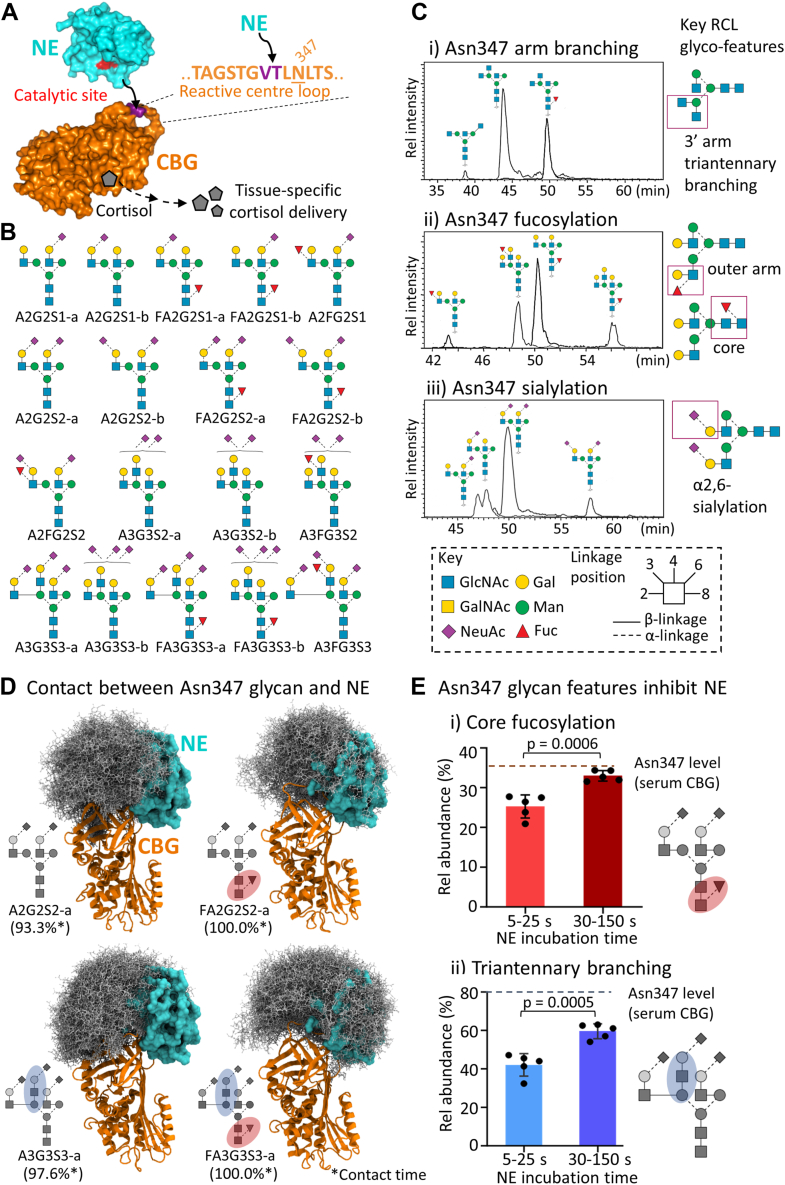
Corticosteroid-binding globulin (CBG) delivers anti-inflammatory cortisol to inflamed tissues through proteolysis of an exposed reactive center loop (RCL) by neutrophil elastase (NE). We previously demonstrated that RCL-localized Asn347-linked N-glycans impact NE proteolysis, but a comprehensive structure-function characterization of the RCL glycosylation is still required to better understand CBG glycobiology. Herein, we first performed RCL-centric glycoprofiling of serum-derived CBG to elucidate the Asn347-glycans and then used molecular dynamics simulations to study their impact on NE proteolysis. Importantly, we also identified O-glycosylation (di/sialyl T) across four RCL sites (Thr338/Thr342/Thr345/Ser350) of serum CBG close to the NE-targeted Val344-Thr345 cleavage site. A restricted N- and O-glycan co-occurrence pattern on the RCL involving exclusively Asn347 and Thr338 glycosylation was experimentally observed and supported in silico by modeling of a CBG-GalNAc-transferase (GalNAc-T) complex with various RCL glycans. GalNAc-T2 and GalNAc-T3 abundantly expressed by liver and gall bladder, respectively, showed in vitro a capacity to transfer GalNAc (Tn) to multiple RCL sites suggesting their involvement in RCL O-glycosylation. Recombinant CBG was then used to determine roles of RCL O-glycosylation through longitudinal NE-centric proteolysis experiments, which demonstrated that both sialoglycans (disialyl T) and asialoglycans (T) decorating Thr345 inhibit NE proteolysis. Synthetic RCL O-glycopeptides expanded on these findings by showing that Thr345-Tn and Thr342-Tn confer strong and moderate protection against NE cleavage, respectively. Molecular dynamics substantiated that short Thr345-linked O-glycans abrogate NE interactions. In conclusion, we report on biologically relevant CBG RCL glycosylation events, which improve our understanding of mechanisms governing cortisol delivery to inflamed tissues.

Keywords: N-glycosylation; O-glycosylation; corticosteroid-binding globulin; cortisol; glycosylation; neutrophil elastase; proteolysis; reactive center loop.


Conflict of interest statement

Conflict of interest The authors declare that they have no conflicts of interest with the contents of this article.

Figures

Figure 1
Figure 1
RCL Asn347-centric glycoprofiling of serum CBG.A, mechanism of cortisol delivery upon NE-mediated cleavage of the RCL and the subsequent structural rearrangement of the cortisol-carrying CBG illustrated using 3D structures of human NE (PDB code: 3Q77, cyan) and uncleaved (intact RCL) human CBG (homology model based on uncleaved human TBG, PDB code: 2CEO, orange). The NE cleavage site (VT) is in purple. B, RCL Asn347-glycans of serum CBG (and their short-hand nomenclature) identified by PGC-LC–MS/MS, see Table S1A for Asn347 site occupancy information. Trace glycoforms have been omitted for simplicity. C, identification of the fine structural features of the Asn347-glycans including (i) the type of antennary branching as determined after sialidase and galactosidase treatment, (ii) the fucose position as determined after sialidase treatment and (iii) the sialyl linkage type. Representative extracted ion chromatograms of key glycans are shown, and prominent Asn347-glycan features are highlighted (right). D, NE contact (steric clash) conferred by four prominent Asn347-glycans (gray, ball/stick representations) as determined in silico by MD simulations of NE (cyan, solid surface) complexed with glycosylated CBG (orange, ribbon structure). In total, 2500 snapshots were extracted from 2.5 μs of cumulative MD simulation time for each of the four CBG glycoforms. Multiple MD-derived snapshots of the Asn347-glycans are shown with a single snapshot of the CBG–NE complex. The proportion of NE contact time (in %) is indicated. E, longitudinal NE-based cleavage of serum CBG demonstrating the impact of (i) core fucosylation and (ii) antennary branching of the Asn347-glycan on the NE cleavage rate replotted, for clarity and support, for six dominating Asn347-glycoforms from our previous study (26). Dashed lines indicate the native modification level for serum CBG. Data plotted as mean ± SD, n = 5, Student's t test. CBG, corticosteroid-binding globulin; MD, molecular dynamics; NE, neutrophil elastase; PDB, Protein Data Bank; RCL, reactive center loop.
Figure 2
Figure 2
Newly discovered RCL O-glycosylation of serum CBG.A, four O-glycosylation sites each carrying one or more O-glycan structures were identified on the RCL of serum CBG at relatively low abundance, see Table S1 for site occupancy. B, (i) Distribution of O-glycans decorating the CBG RCL based on glycopeptide data (mean + SD, n = 3). (ii) CID–MS/MS spectrum of an O-glycan (sialyl T) released from serum CBG (glycomics data). C and D, annotated EThcD–MS/MS spectra of two RCL O-glycopeptides providing evidence of Thr342 and Thr345 RCL O-glycosylation, respectively. These two RCL O-glycopeptides, which were not treated with any exoglycosidases or endoglycosidases, were not found to carry any Asn347-linked N-glycosylation. Insets, precursor (broken box) and key fragment ions (full boxes) documenting the site-specific O-glycosylation events. ∗Interfering fragment of a coisolated precursor ion unrelated to the targeted CBG glycopeptides. See Figure 1 for key to glycan symbols. CBG, corticosteroid-binding globulin; CID, collision-induced dissociatin; EThcD, electron transfer higher-energy collision dissociation; RCL, reactive center loop.
Figure 3
Figure 3
Co-occurrence of RCL N- and O-glycosylation on serum CBG.A, EThcD–MS/MS spectrum of an RCL glycopeptide co-occupied with an O- and N-glycan (Thr338 and Asn347). Inset, precursor (broken) and key fragment ions (full) documenting the site-specific glycosylation events. B, (i) Observed (green) and nondetected (red) RCL O-glycoforms in the absence (−) and presence (+) of Asn347-glycosylation. (ii) Proposed spatiotemporal interplay between RCL N- and O-glycosylation providing a possible explanation for the absence of specific co-occupied RCL glycopeptides (e.g., Thr345><Asn347). C, four in silico models of a complex between nonglycosylated CBG and GalNAc-T2 (see below for rational for choosing GalNAc-T2) produced by aligning the transferase across the four discrete RCL O-glycosylation sites of serum CBG. The local environment around the Asn347 residue (green) was visually assessed for the ability to accommodate a bulky oligomannosidic-type N-glycan (Man5). See Figure 1 for key to glycan symbols. CBG, corticosteroid-binding globulin; EThcD, electron transfer higher-energy collision dissociation; GalNAc-T, GalNAc transferase; RCL, reactive center loop.
Figure 4
Figure 4
GalNAc-T2 and GalNAc-T3 transfer O-glycans to a synthetic RCL peptide in vitro.A, schematic representation of the in vitro glycosylation assay exploring how and where (i) GalNAc-T2 and (ii) GalNAc-T3 transfer GalNAc (Tn) moieties to a synthetic RCL peptide. B, transcript (mRNA) expression profile of GALNTs in liver and gall bladder (the principal tissue origins of CBG) based on RNA consensus tissue gene data from the Human Protein Atlas (4). C, glycopeptide products of the in vitro O-glycosylation reactions by (i) GalNAc-T2 and (ii) GalNAc-T3. The RCL O-glycans sites were identified using EThcD–MS/MS (Figs. S9–S17). See Figure 1 for key to glycan symbols. EThcD, electron transfer higher-energy collision dissociation; nTPM, normalized transcript per million; RCL, reactive center loop.
Figure 5
Figure 5
Protective role of Thr345 O-glycosylation of rCBG against NE-mediated proteolysis.A, RCL glycan occupancies of HEK293-derived rCBG. B, distribution of Thr345 O-glycans of rCBG determined based on glycopeptide data. Data points are plotted as mean + SD, n = 3. C, overview of the RCL peptides before and after NE cleavage with downstream trypsin digestion. D, NE cleavage of rCBG and asialylated rCBG (asialo-rCBG) over time. Distribution of unoccupied uncleaved (dark/light gray) and O-glycosylated (red/pink) uncleaved RCL tryptic peptides of rCBG and asialo-rCBG identified based on glycopeptide data. Data points are plotted as mean + SD, n = 3. E, distribution of nonglycosylated and O-glycosylated C-terminal peptide fragments detected after 480 s of NE incubation. Data points are plotted as mean + SD, n = 3. F, alignment of NE (cyan, PDB code: 7CBK) onto MD-generated shape conformers of uncleaved CBG (orange, PDB code: 4BB2/2CEO) carrying relevant Thr345 O-glycans, that is, (i) disialyl T, (ii) T, and (iii) Tn (3D-SNFG, top) and schematics of their demonstrated or suggested protective roles against NE-mediated RCL cleavage (bottom). The proportion of NE contact time (in %) of the respective CBG O-glycans is provided. See Figure 1 for key to glycan symbols. HEK293, human embryonic kidney 293 cell line; MD, molecular dynamics; NE, neutrophil elastase; rCBG, recombinant corticosteroid-binding globulin; RCL, reactive center loop.
Figure 6
Figure 6
Thr342- and Thr345-GalNAc (Tn) O-glycans impede NE proteolysis of synthetic RCL peptides.A, schematic representation of the synthetic CBG RCL (glyco)peptides and their expected cleavage products upon NE proteolysis. m/z values for both the nonglycosylated and Tn peptides are provided. B, NE-based cleavage of (i) nonglycosylated, (ii) Thr342, and (iii) Thr345 O-glycosylated RCL peptides over time as monitored using MALDI-TOF MS. Intact (uncleaved) nonglycosylated and O-glycosylated RCL peptides are in blue, RCL N-terminal cleavage products in red, and RCL C-terminal cleavage products in green. C, schematics of the observed NE accessibility to (i) unoccupied, (ii) Thr342, and (iii) Thr345 O-glycosylated RCL. See Figure 1 for key to glycan symbols. CBG, corticosteroid-binding globulin; NE, neutrophil elastase; RCL, reactive center loop.
Figure 7
Figure 7
Summary of findings. In this study, we report on newly identified N- and O-glycosylation events modifying the RCL of human CBG, and we have determined how they impact the NE-mediated RCL cleavage process (left side), in turn, leading to an altered cortisol release rate from CBG, and we have established their crosstalk on the RCL (right side). See Figure 1 for key. CBG, corticosteroid-binding globulin; NE, neutrophil elastase; RCL, reactive center loop.

References

    1. Mickelson K.E., Forsthoefel J., Westphal U. Steroid-protein interactions. Human corticosteroid binding globulin: some physicochemical properties and binding specificity. Biochemistry. 1981;20:6211–6218. - PubMed
    1. Lewis J.G., Bagley C.J., Elder P.A., Bachmann A.W., Torpy D.J. Plasma free cortisol fraction reflects levels of functioning corticosteroid-binding globulin. Clin. Chim. Acta. 2005;359:189–194. - PubMed
    1. Hammond G.L., Smith C.L., Goping I.S., Underhill D.A., Harley M.J., Reventos J., et al. Primary structure of human corticosteroid binding globulin, deduced from hepatic and pulmonary cDNAs, exhibits homology with serine protease inhibitors. Proc. Natl. Acad. Sci. U. S. A. 1987;84:5153–5157. - PMC - PubMed
    1. Uhlén M., Fagerberg L., Hallström B.M., Lindskog C., Oksvold P., Mardinoglu A., et al. Tissue-based map of the human proteome. Science. 2015;347 - PubMed
    1. Perogamvros I., Ray D.W., Trainer P.J. Regulation of cortisol bioavailability—effects on hormone measurement and action. Nat. Rev. Endocrinol. 2012;8:717–727. - PubMed

Publication types