. 2023 Dec 2;14(1):7970.
doi: 10.1038/s41467-023-43711-3.

Insights into the ISG15 transfer cascade by the UBE1L activating enzyme

Affiliations

Insights into the ISG15 transfer cascade by the UBE1L activating enzyme

Iona Wallace et al. Nat Commun. .

Abstract

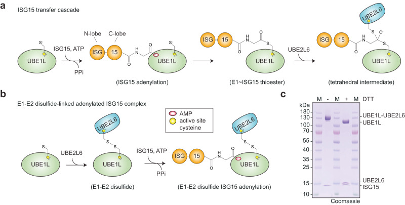
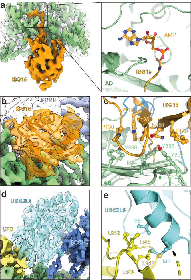
The attachment of the ubiquitin-like protein ISG15 to substrates by specific E1-E2-E3 enzymes is a well-established signalling mechanism of the innate immune response. Here, we present a 3.45 Å cryo-EM structure of a chemically trapped UBE1L-UBE2L6 complex bound to activated ISG15. This structure reveals the details of the first steps of ISG15 recognition and UBE2L6 recruitment by UBE1L (also known as UBA7). Taking advantage of viral effector proteins from severe acute respiratory coronavirus 2 (SARS-CoV-2) and influenza B virus (IBV), we validate the structure and confirm the importance of the ISG15 C-terminal ubiquitin-like domain in the adenylation reaction. Moreover, biochemical characterization of the UBE1L-ISG15 and UBE1L-UBE2L6 interactions enables the design of ISG15 and UBE2L6 mutants with altered selectively for the ISG15 and ubiquitin conjugation pathways. Together, our study helps to define the molecular basis of these interactions and the specificity determinants that ensure the fidelity of ISG15 signalling during the antiviral response.


Conflict of interest statement

B.A.S. is a member of the scientific advisory boards of Interline Therapeutics and BioTheryX. The remaining authors declare no competing interests.

Figures

Fig. 1
Fig. 1. The ISG15 transfer cascade and ISG15 E1-E2 complex formation.
a Schematic of ISG15 adenylation and transfer through the E1 activating enzyme UBE1L to the E2 conjugating enzyme UBE2L6. In an ATP-dependent manner, UBE1L adenylates or ‘activates’ the C-terminus of ISG15. This high-energy ISG15 intermediate reacts with the catalytic cysteine of UBE1L forming a thioester bond ( ~ denotes thioester bond). Subsequently, ISG15 is transferred to UBE2L6 forming a tetrahedral intermediate. b Schematic of ISG15 E1-E2 disulfide-linked complex formation. The active site cysteines of UBE1L and UBE2L6 were initially cross-linked via a disulfide bond (- denotes disulfide bond). Subsequently, this E1-E2 disulfide-linked complex was incubated with ISG15 and Mg-ATP to form a ternary protein complex with adenylated ISG15. c SDS-PAGE of the purified E1-E2 ISG15 adenylated complex. The addition of dithiothreitol (DTT) to the complex reduced the disulfide bond between UBE1L and UBE2L6. Formation of the E1-E2 ISG15 adenylated complex was performed independently in duplicate. Source data are provided in the Source Data file.
Fig. 2
Fig. 2. Cryo-EM structure of ISG15 in complex with UBE1L and UBE2L6.
a Cryo-EM density of UBE1L in complex with UBE2L6 and ISG15. For ISG15, the density surrounding the C-terminal ubiquitin-like fold (C-lobe) is visible. b The UBE1L domains contacting ISG15 are highlighted and include the adenylation domain (AD), first catalytic cysteine half-domain (FCCH), and cross-over loop of the second catalytic cysteine half-domain (SCCH). As expected, ISG15 is positioned within the adenylation catalytic module. c The UBE1L domains contacting UBE2L6 are highlighted and include the UFD (ubiquitin fold domain) and SCCH. UBE2L6 is located at the top of the structure and the E1-E2 catalytic cysteines are positioned for disulfide bond formation. d Cryo-EM density of a subclass of the complex (DeepEMhancer map). The N-terminal ubiquitin-like fold of ISG15 (N-lobe) is visible in the density, however the N-lobe does not make significant contact with UBE1L.
Fig. 3
Fig. 3. Structural analysis of UBE1L specificity.
a Left, cryo-EM density of adenylated ISG15 bound to UBE1L. Right, close-up view of the adenylated ISG15 C-terminus within the adenylation domain (AD) active site (also see Supplementary Fig. 5f). b UBE1L-ISG15 surface contacts. The circled area corresponds to the primary contact site between ISG15 and UBE1L. Amino acid side chain interactions within the interface are shown in c. c ISG15 and AD interactions. The Thr125 patch of ISG15 (Thr125, Phe149, Asn151 – analogous residues to the Ile44 patch of ubiquitin; also see Supplementary Fig. 5g) contact residues within the AD (Tyr885, Tyr892, Ile894). Additional hydrophobic residues of ISG15 (Trp123, Pro130) contact UBE1L (Tyr896). d UBE1L recognition of UBE2L6. Circled area corresponds to the primary contact site between the ubiquitin fold domain (UFD) of UBE1L and UBE2L6. The second catalytic cysteine half-domain (SCCH) of UBE1L also contacts UBE2L6. Amino acid side chain interactions within the UFD-UBE2L6 interface are shown in e. e UBE2L6 and UFD interactions. A hydrophobic surface of the UFD (Ile945, Leu947, Leu952) coordinates UBE2L6 helix-1 residues (Met5, Val8).
Fig. 4
Fig. 4. Biochemical characterization of ISG15 specificity.
a Structure of ISG15 (pdb 1z2m) highlighting residues that contact UBE1L. Residues are shown in ball-and-stick representation under a semi-transparent surface. Interactions are exclusively located in the C-terminal ubiquitin-like fold (ISG15 C-lobe; also see Fig. 3). Analogous residues of ubiquitin are shown in teal. b Diagram representing ISG15 constructs used in cg. From left to right: ISG15C-lobe, full-length ISG15 (ISG15FL), ubiquitin(Ub)-ISG15C-lobe fusion, SUMO1-ISG15C-lobe fusion, ISG15 mutants (ISG15mut(s)). c UBE1L charging reactions with fluorescent ISG15 and ISG15C-lobe. Reactions were separated by SDS-PAGE and visualized with fluorescent imaging. d UBE1L charging reactions with ISG15FL, Ub-ISG15C-lobe, and SUMO1-ISG15C-lobe. Reactions were separated by SDS-PAGE and visualized with Coomassie stain. e Quantification of UBE1L charging reactions with fluorescent ISG15 and ISG15 mutants. ISG15 mutants include: ISG153xmut (T125I/ F149H/N151V), ISG154xmut (T125I/P130Q/F149H/N151V), ISG155xmut (W123R/T125I/P130Q/F149H/N151V). Mutated ISG15 residues were swapped with the analogous residues of ubiquitin (also see Supplementary Fig. 5g). Reactions were performed as in c. Error values represent s.d. from the mean (n = 3 independent experiments). Samples derive from same experiment and gels were processed in parallel. f Quantification of UBA1 charging with ISG15 mutants as in e. Error values represent s.d. from the mean (n = 3 independent experiments). Samples derive from same experiment and gels were processed in parallel. g UBE1L and UBA1 charging reactions with fluorescent ISG15FL and ISG155xmut as in c. Experiments were performed independently in triplicate. Source data are provided in the Source Data file.
Fig. 5
Fig. 5. Accessing ubiquitin E2 enzymes with ubiquitylized ISG15.
a Comparison of UBE1L-mediated UBE2L6 charging with ISG15 and ISG155xmut (W123R/T125I/P130Q/F149H/N151V). Reactions were quenched at the indicated time points, separated by SDS-PAGE, and visualized using fluorescent imaging. b Comparison of UBA1-mediated charging of UBE2L6 and UBE2L3 with ISG155xmut, and charging of UBE2L3 with ubiquitin (Ub). Reactions were visualized as in a. c Comprehensive analysis of UBA1-mediated E2 charging with ISG155xmut. All ubiquitin E2 enzymes tested formed a thioester bond with ISG155xmut, while E2 charging was not observed for SUMO (UBE2I), NEDD8 (UBE2F, UBE2M), and ISG15 (UBE2L6) E2 enzymes (also see Supplementary Fig. 8). Experiments were performed independently in triplicate. Source data are provided in the Source Data file.
Fig. 6
Fig. 6. Accessing the ubiquitin system with ubiquitylised ISG15 in cells.
a Transfection assays with FLAG-tagged full-length ISG15 (ISG15FL), ISG155xmut (W123R/T125I/P130Q/F149H/N151V), and a non-conjugatable version of ISG155xmut (ISG155xmut ΔGG). Cells were treated without or with the proteasomal inhibitor MG132. ISG15 substrate modification was monitored by anti-FLAG western blots. Anti-ubiquitin (Ub) and anti-actin western blots were performed as controls. b Treatment of ISG155xmut transfected cells with a ubiquitin E1 inhibitor. Cells were treated with DMSO, MG132, the UBA1 inhibitor TAK-243, or a combination of both MG132 and TAK-243. ISG15 substrate modification was monitored by anti-FLAG western blots. Anti-Ub and anti-actin western blots were performed as controls. Experiments were performed independently in triplicate. Source data are provided in the Source Data file.
Fig. 7
Fig. 7. The impact of viral proteins on ISG15 E1 charging.
a Structure of non-structural protein 1 from influenza B virus (NS1B) bound to ISG15 (pdb 3sdl) overlaid onto the UBE1L-UBE2L6 ISG15 adenylate complex. NS1B contacts the N-terminal ubiquitin-like fold (N-lobe) of ISG15, but not the C-terminal ubiquitin-like fold (C-lobe). The ISG15 adenylate and UBE1L adenylation active site are highlighted. b Structure of SARS-CoV2 papain-like protease (PLpro) bound to ISG15 (pdb 6yva). PLpro contacts both the N-lobe and C-lobe of ISG15. c Time course analysis of UBE1L charging assays in the presence of viral proteins. The catalytically inactive PLpro mutant Cys111Ala (CA) was used. d Analysis of UBE1L charging in the presence of viral proteins using fluorescence polarization (FP) assays. The indicated concentrations of NS1B, NS1B AA mutant (W36A/Q37A) and PLpro CA were added to fluorescent ISG15, followed by the addition of UBE1L to the sample. e Schematic of the proposed mechanisms to explain data shown in c and d. NS1B binds the N-lobe of ISG15 and remains bound during UBE1L charging, while PLpro CA competes with UBE1L for the C-lobe of ISG15. Experiments were performed independently in triplicate. Source data are provided in the Source Data file.

References

    1. Rathinam VA, Fitzgerald KA. Cytosolic surveillance and antiviral immunity. Curr. Opin. Virol. 2011;1:455–462. doi: 10.1016/j.coviro.2011.11.004. - DOI - PMC - PubMed
    1. Ivashkiv LB, Donlin LT. Regulation of type I interferon responses. Nat. Rev. Immunol. 2014;14:36–49. doi: 10.1038/nri3581. - DOI - PMC - PubMed
    1. Stark GR, Darnell JE. The JAK-STAT pathway at twenty. Immunity. 2012;36:503–514. doi: 10.1016/j.immuni.2012.03.013. - DOI - PMC - PubMed
    1. Schneider WM, Chevillotte MD, Rice CM, Interferon-Stimulated Genes: a complex web of host defenses. Annu. Rev. Immunol. 2014;32:513–545. doi: 10.1146/annurev-immunol-032713-120231. - DOI - PMC - PubMed
    1. Farrell PJ, Broeze RJ, Lengyel P. Accumulation of an mRNA and protein in interferon-treated Ehrlich ascites tumour cells. Nature. 1979;279:523–525. doi: 10.1038/279523a0. - DOI - PubMed

Publication types

MeSH terms