. 2023 Dec 4;13(1):21328.
doi: 10.1038/s41598-023-48940-6.

Relationship between disproportionately enlarged subarachnoid-space hydrocephalus and white matter tract integrity in normal pressure hydrocephalus

Affiliations

Relationship between disproportionately enlarged subarachnoid-space hydrocephalus and white matter tract integrity in normal pressure hydrocephalus

Sunju Lee et al. Sci Rep. .

Abstract

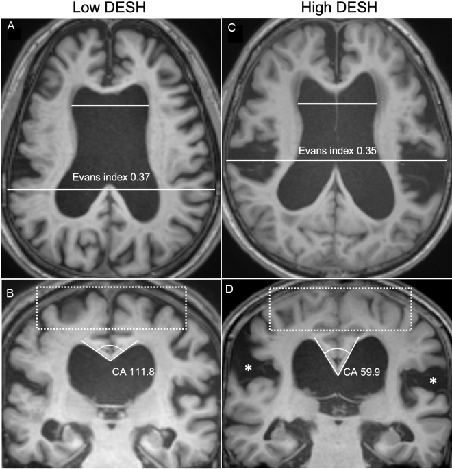
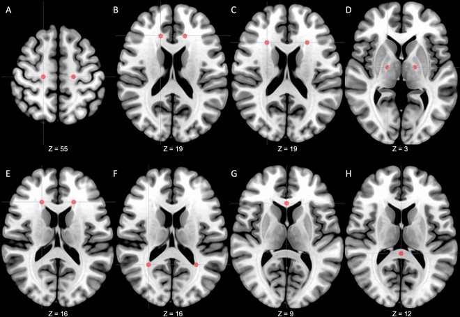
Normal pressure hydrocephalus (NPH) patients had altered white matter tract integrities on diffusion tensor imaging (DTI). Previous studies suggested disproportionately enlarged subarachnoid space hydrocephalus (DESH) as a prognostic sign of NPH. We examined DTI indices in NPH subgroups by DESH severity and clinical symptoms. This retrospective case-control study included 33 NPH patients and 33 age-, sex-, and education-matched controls. The NPH grading scales (0-12) were used to rate neurological symptoms. Patients with NPH were categorized into two subgroups, high-DESH and low-DESH groups, by the average value of the DESH scale. DTI indices, including fractional anisotropy, were compared across 14 regions of interest (ROIs). The high-DESH group had increased axial diffusivity in the lateral side of corona radiata (1.43 ± 0.25 vs. 1.72 ± 0.25, p = 0.04), and showed decreased fractional anisotropy and increased mean, and radial diffusivity in the anterior and lateral sides of corona radiata and the periventricular white matter surrounding the anterior horn of lateral ventricle. In patients with a high NPH grading scale, fractional anisotropy in the white matter surrounding the anterior horn of the lateral ventricle was significantly reduced (0.36 ± 0.08 vs. 0.26 ± 0.06, p = 0.03). These data show that DESH may be a biomarker for DTI-detected microstructural alterations and clinical symptom severity.


Conflict of interest statement

The authors declare no competing interests.

Figures

Figure 1
Figure 1
Representative MR images of the high-DESH and low-DESH group. MR coronal and axial images of representative patients in the low-DESH (a,b) and high-DESH (c,d) groups. In the low-DESH patient, the Evans index is increased to 0.37 (grade 2, panel a), the sylvian fissures are slightly enlarged (grade 1), but no tight high convexity is observed, (white-dotted rectangle) and the callosal angle is 111.8°, which is above 90° (panel b). This patient has a DESH score of 3. In the high-DESH patient, the Evans index is increased to 0.35 (grade 2, panel c), similar to the low-DESH patient, but in addition, the sylvian fissure is markedly dilated bilaterally (grade 2, white asterisks), tight convexity is seen (grade 2, white-dotted rectangle), and the callosal angle is less than 90° at 59.9° (grade 2). This patient also has focal sulcal dilatation (grade 1, not shown), resulting in a DESH score of 9. DESH disproportionately enlarged subarachnoid space hydrocephalus, CA callosal angle.
Figure 2
Figure 2
Predetermined regions-of-interest. A total of 14 regions of interest were predetermined in the centrum semiovale (A), anterior area of corona radiata (B), lateral area of corona radiata (C), posterior limbs of the internal capsule (D), anterior horns of the lateral ventricle (E), posterior horns of the lateral ventricle (F), genu of corpus callosum (G) and splenium of corpus callosum (H). From a radiological perspective, the right side of the figure is the patient's left side. The z coordinate represents the position on the MNI152 standard template. MNI Montreal Neurological Institute.

References

    1. Adams RD, Fisher CM, Hakim S, Ojemann RG, Sweet WH. Symptomatic occult hydrocephalus with “normal” cerebrospinal-fluid pressure. A treatable syndrome. N. Engl. J. Med. 1965;273:117–126. doi: 10.1056/NEJM196507152730301. - DOI - PubMed
    1. Garcia-Armengol R, et al. Comparison of elevated intracranial pressure pulse amplitude and disproportionately enlarged subarachnoid space (DESH) for prediction of surgical results in suspected idiopathic normal pressure hydrocephalus. Acta Neurochir. (Wien) 2016;158:2207–2213. doi: 10.1007/s00701-016-2858-5. - DOI - PubMed
    1. Shinoda N, et al. Utility of MRI-based disproportionately enlarged subarachnoid space hydrocephalus scoring for predicting prognosis after surgery for idiopathic normal pressure hydrocephalus: Clinical research. J. Neurosurg. 2017;127:1436–1442. doi: 10.3171/2016.9.JNS161080. - DOI - PubMed
    1. Kitagaki H, et al. CSF spaces in idiopathic normal pressure hydrocephalus: Morphology and volumetry. AJNR Am. J. Neuroradiol. 1998;19:1277–1284. - PMC - PubMed
    1. Mori E, et al. Guidelines for management of idiopathic normal pressure hydrocephalus: Second edition. Neurol. Med. Chir. Tokyo. 2012;52:775–809. doi: 10.2176/nmc.52.775. - DOI - PubMed

MeSH terms