. 2023 Dec 4;22(1):195.
doi: 10.1186/s12943-023-01887-8.

hsa_circ_0007919 induces LIG1 transcription by binding to FOXA1/TET1 to enhance the DNA damage response and promote gemcitabine resistance in pancreatic ductal adenocarcinoma

Affiliations

hsa_circ_0007919 induces LIG1 transcription by binding to FOXA1/TET1 to enhance the DNA damage response and promote gemcitabine resistance in pancreatic ductal adenocarcinoma

Lei Xu et al. Mol Cancer. .

Erratum in

Abstract

Background: Circular RNAs (circRNAs) play important roles in the occurrence and development of cancer and chemoresistance. DNA damage repair contributes to the proliferation of cancer cells and resistance to chemotherapy-induced apoptosis. However, the role of circRNAs in the regulation of DNA damage repair needs clarification.

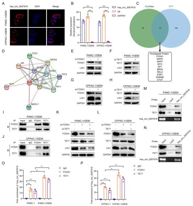
Methods: RNA sequencing analysis was applied to identify the differentially expressed circRNAs. qRT-PCR was conducted to confirm the expression of hsa_circ_0007919, and CCK-8, FCM, single-cell gel electrophoresis and IF assays were used to analyze the proliferation, apoptosis and gemcitabine (GEM) resistance of pancreatic ductal adenocarcinoma (PDAC) cells. Xenograft model and IHC experiments were conducted to confirm the effects of hsa_circ_0007919 on tumor growth and DNA damage in vivo. RNA sequencing and GSEA were applied to confirm the downstream genes and pathways of hsa_circ_0007919. FISH and nuclear-cytoplasmic RNA fractionation experiments were conducted to identify the cellular localization of hsa_circ_0007919. ChIRP, RIP, Co-IP, ChIP, MS-PCR and luciferase reporter assays were conducted to confirm the interaction among hsa_circ_0007919, FOXA1, TET1 and the LIG1 promoter.

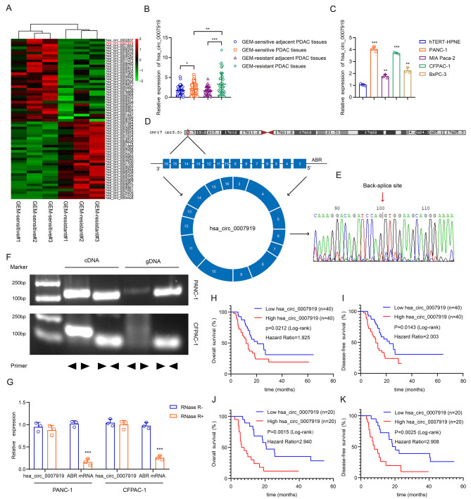
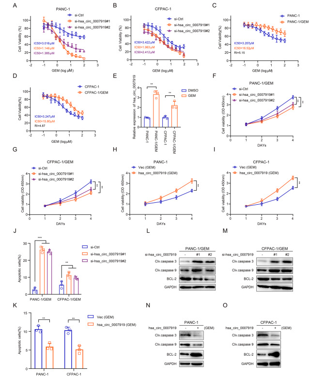
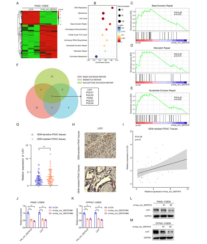
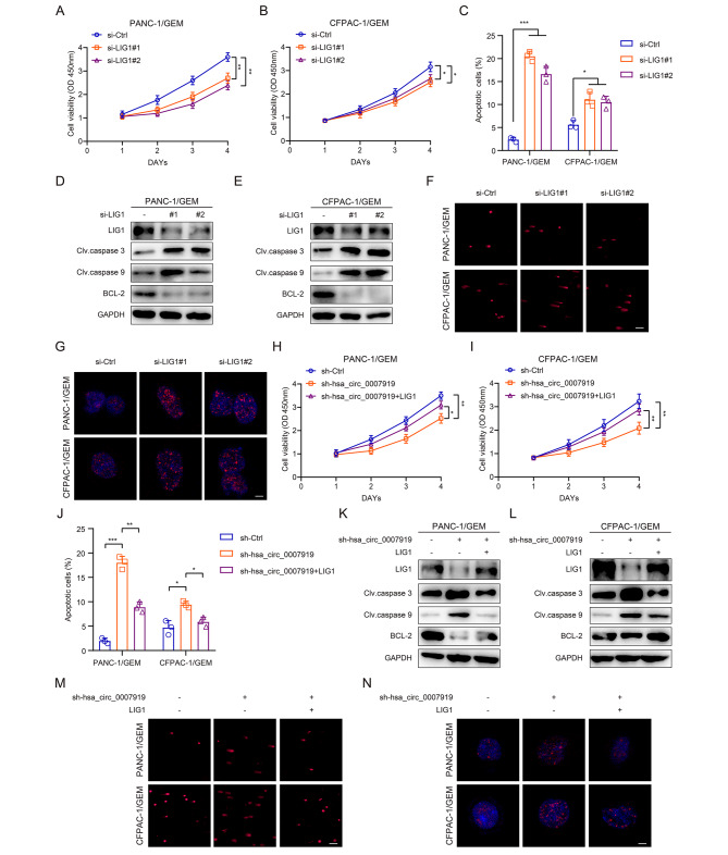
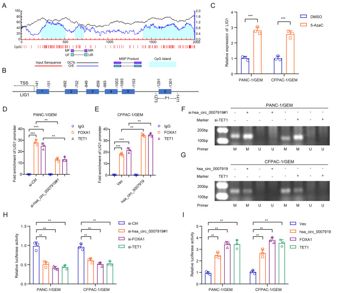
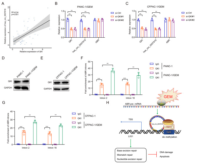
Results: We identified a highly expressed circRNA, hsa_circ_0007919, in GEM-resistant PDAC tissues and cells. High expression of hsa_circ_0007919 correlates with poor overall survival (OS) and disease-free survival (DFS) of PDAC patients. Hsa_circ_0007919 inhibits the DNA damage, accumulation of DNA breaks and apoptosis induced by GEM in a LIG1-dependent manner to maintain cell survival. Mechanistically, hsa_circ_0007919 recruits FOXA1 and TET1 to decrease the methylation of the LIG1 promoter and increase its transcription, further promoting base excision repair, mismatch repair and nucleotide excision repair. At last, we found that GEM enhanced the binding of QKI to the introns of hsa_circ_0007919 pre-mRNA and the splicing and circularization of this pre-mRNA to generate hsa_circ_0007919.

Conclusions: Hsa_circ_0007919 promotes GEM resistance by enhancing DNA damage repair in a LIG1-dependent manner to maintain cell survival. Targeting hsa_circ_0007919 and DNA damage repair pathways could be a therapeutic strategy for PDAC.

Keywords: DNA damage repair; LIG1; Pancreatic ductal adenocarcinoma; QKI; hsa_circ_0007919.


Conflict of interest statement

The authors declare no potential conflicts of interest.

Figures

Fig. 1
Fig. 1
hsa_circ_0007919 is upregulated in GEM-resistant PDAC and predicts poor prognosis (A) Hierarchical clustering showing differentially expressed circRNAs in GEM-sensitive and GEM-resistant PDAC tissues (FC > 1 or <-1, p < 0.05). (B) The relative expression of hsa_circ_0007919 in GEM-sensitive and GEM-resistant PDAC tissues and corresponding adjacent PDAC tissues. (C) The relative expression of hsa_circ_0007919 in PDAC cells and normal pancreatic cells. (D) The genomic location and back-splicing of hsa_circ_0007919. (E) The splicing site of hsa_circ_0007919 validated by Sanger-seq. (F) PCR and agarose gel electrophoresis analysis of the presence of hsa_circ_0007919 and ABR in cDNA and gDNA samples from PDAC cells. (G) Expression of hsa_circ_0007919 and ABR in PDAC cells with or without RNase R treatment. (H-I) Kaplan–Meier analysis of the OS rate and DFS rate in PDAC patients with high or low expression of hsa_circ_0007919. (J-K) Kaplan–Meier analysis of the OS rate and DFS rate in GEM-resistant PDAC patients with high or low expression of hsa_circ_0007919. Data are the means ± SDs (n = 3 independent experiments), * p < 0.05, ** p < 0.01, *** p < 0.001
Fig. 2
Fig. 2
hsa_circ_0007919 inhibits gemcitabine sensitivity and apoptosis of GEM-resistant PDAC cells (A-B) CCK-8 analysis of the sensitivity of PDAC cells with or without hsa_circ_0007919 inhibition under different concentrations of GEM. (C-D) CCK-8 analysis of the sensitivity of normal and GEM-resistant PDAC cells under different concentrations of GEM. (E) Expression of hsa_circ_0007919 in normal and PDAC-resistant PDAC cells. (F-G) CCK-8 analysis of the proliferation of GEM-resistant PDAC cells with or without hsa_circ_0007919 inhibition. (H-I) CCK-8 analysis of the proliferation of PDAC cells with or without hsa_circ_0007919 overexpression under GEM treatment condition. (J) Flow cytometry analysis of the apoptotic rate of PDAC-resistant PDAC cells with or without hsa_circ_0007919 inhibition. (K) Flow cytometry analysis of the apoptotic rate of PDAC PDAC cells with or without hsa_circ_0007919 overexpression under GEM treatment condition. (L-M) Expression of apoptosis-related proteins in GEM-resistant PDAC cells with or without hsa_circ_0007919 inhibition. (N-O) Expression of apoptosis-related proteins in GEM PDAC cells with or without hsa_circ_0007919 overexpression under GEM treatment condition. Data are the means ± SDs (n = 3 independent experiments), ** p < 0.01, *** p < 0.001
Fig. 3
Fig. 3
hsa_circ_0007919 inhibits DNA damage of GEM-resistant PDAC cells and proliferation of tumor in vivo (A) Comet analysis of the DNA damage of GEM-resistant PDAC cells with or without hsa_circ_0007919 inhibition and of the DNA damage of PDAC cells with or without hsa_circ_0007919 overexpression under GEM treatment condition (200×). (B) IF analysis of γ-H2AX accumulation in GEM-resistant PDAC cells with or without hsa_circ_0007919 inhibition and of γ-H2AX accumulation in PDAC cells with or without hsa_circ_0007919 overexpression under GEM treatment condition (1000×). (C) Image of tumors formed by GEM-resistant PDAC cells with or without hsa_circ_0007919 inhibition (n = 5). (D-E) Growth curves of tumors formed by GEM-resistant PDAC cells with or without hsa_circ_0007919 inhibition (n = 5). (F-G) Weights of tumors formed by GEM-resistant PDAC cells with or without hsa_circ_0007919 inhibition (n = 5). (H) IHC analysis of the expression of LIG1, Ki67, caspase 3 and γ-H2AX in tumors formed by GEM-resistant PDAC cells with or without hsa_circ_0007919 inhibition (400×). (I-J) IHC staining score analysis of images from Fig. 3H. Data are the means ± SDs (n = 3 independent experiments), ** p < 0.01
Fig. 4
Fig. 4
hsa_circ_0007919 inhibits DNA damage through LIG1-mediated repair pathways (A) Hierarchical clustering showing differentially expressed genes in GEM-resistant PDAC cells with or without hsa_circ_0007919 inhibition (FC > 1 or < -1, p < 0.05). (B) KEGG enrichment analysis of hsa_circ_0007919-regulated gene expression events. (C-E) GSEA enrichment analysis of hsa_circ_0007919-regulated gene expression events. (F) Venn diagram showing overlapped genes between differentially expressed genes from base excision repair, mismatch repair and nucleotide excision repair pathways. (G-H) Expression of LIG1 in GEM-sensitive and -resistant PDAC tissues at mRNA and protein level (400×). (I) Correlation analysis of hsa_circ_0007919 and LIG1 expression in GEM-resistant tissues. (J-M) Expression of LIG1 in GEM-resistant cells with or without hsa_circ_0007919 inhibition at mRNA and protein level. Data are the means ± SDs (n = 3 independent experiments), ** p < 0.01
Fig. 5
Fig. 5
LIG1 reversed cell proliferation, apoptosis and DNA damage effects of hsa_circ_0007919 (A-B) CCK-8 analysis of the proliferation of GEM-resistant PDAC cells with or without LIG1 inhibition. (C) Flow cytometry analysis of the apoptotic rate of PDAC-resistant PDAC cells with or without LIG1 inhibition. (D-E) Expression of apoptosis-related proteins in GEM-resistant PDAC cells with or without LIG1 inhibition. (F) Comet analysis of the DNA damage of GEM-resistant PDAC cells with or without LIG1 inhibition (200×). (G) IF analysis of γ-H2AX accumulation in GEM-resistant PDAC cells with or without LIG1 inhibition (1000×). (H-I) CCK-8 analysis of the proliferation of hsa_circ_0007919-inhibited GEM-resistant PDAC cells with or without LIG1 overexpression. (J) Flow cytometry analysis of the apoptotic rate of hsa_circ_0007919-inhibited PDAC-resistant PDAC cells with or without LIG1 overexpression. (K-L) Expression of apoptosis-related proteins in hsa_circ_0007919-inhibited GEM-resistant PDAC cells with or without LIG1 overexpression. (M) Comet analysis of the DNA damage of hsa_circ_0007919-inhibited GEM-resistant PDAC cells with or without LIG1 overexpression (200×). (N) IF analysis of γ-H2AX accumulation in hsa_circ_0007919-inhibited GEM-resistant PDAC cells with or without LIG1 overexpression (1000×). Data are the means ± SDs (n = 3 independent experiments), * p < 0.05, ** p < 0.01, *** p < 0.001
Fig. 6
Fig. 6
hsa_circ_0007919 binds FOXA1 and TET1 in GEM-resistant PDAC cells (A) FISH analysis of the cellular localization of hsa_circ_0007919. The hsa_circ_0007919 probes were red while nuclei were stained with DAPI (1000×). (B) Nuclear-cytoplasmic fractionation assay analysis of hsa_circ_0007919 location in GEM-resistant PDAC cells, the U6 and GAPDH were used as nuclear and cytoplasmic controls. (C) Venn diagram showing overlapped genes between interacting with hsa_circ_0007919 and interacting with LIG1 promoter region. (D) Protein-protein interaction network analysis of proteins interact with FOXA1. (E-H) Expression of LIG1 in GEM-resistant PDAC cells with or without FOXA1 or TET1 inhibition. (I-J) IP assay analysis of the interaction between FOXA1 and TET1 in GEM-resistant PDAC cells. (K-L) Expression of LIG1 in FOXA1-silenced GEM-resistant PDAC cells with TET1 inhibition or overexpression. (M-N) ChIRP assay analysis of the interaction between hsa_circ_0007919 and FOXA1 or TET1 in GEM-resistant PDAC cells. (O-P) RIP assay analysis of the interaction between FOXA1 or TET1 and hsa_circ_0007919 in normal or GEM-resistant PDAC cells. Data are the means ± SDs (n = 3 independent experiments), ** p < 0.01, *** p < 0.001
Fig. 7
Fig. 7
hsa_circ_0007919 recruits FOXA1 and TET1 to promote LIG1 transcription (A) DNA methylation analysis of CpG island of LIG1 promoter. (B) Interaction region predicted between FOXA1 and LIG1 promoter. (C) Expression of LIG1 in GEM-resistant PDAC cells with or without 5-AzaC treatment. (D-E) ChIP assay analysis of the interaction between FOXA1 or TET1 and LIG1 promoter. (F-G) MS-PCR analysis of methylation level of LIG1 promoter in GEM-resistant PDAC cells with or without hsa_circ_0007919 or TET1 inhibition and with or without hsa_circ_0007919 or TET1 overexpression. (H-I) Luciferase activity analysis of LIG1 transcriptional activity in GEM-resistant PDAC cells with or without hsa_circ_0007919 or FOXA1 or TET1 inhibition and with or without hsa_circ_0007919 or FOXA1 or TET1 overexpression. Data are the means ± SDs (n = 3 independent experiments), ** p < 0.01, *** p < 0.001
Fig. 8
Fig. 8
Gemcitabine induces hsa_circ_0007919 expression through enhancing QKI-mediated back-splicing (A) Correlation analysis of QKI and hsa_circ_0007919 expression in GEM-resistant PDAC tissues. (B-C) Expression of hsa_circ_0007919 and ABR in GEM-resistant PDAC cells with or without QKI inhibition. (D-E) Expression of QKI in normal and GEM-resistant PDAC cells. (F-G) RIP assay analysis of the interaction between QKI and introns of ABR pre-mRNA in normal and GEM-resistant PDAC cells. (H) Schematic representation showing that GEM enhances interaction between QKI and ABR pre-mRNA and promotes back-splicing and cyclization of hsa_circ_0007919. Highly expressed hsa_circ_0007919 recruits FOXA1 and TET1 to mediate de-methylation and transcription of LIG1 and upregulates the expression of LIG1. Overexpression of LIG1 activates base excision repair, mismatch repair and nucleotide excision repair pathways to inhibit the DNA damage and apoptosis of GEM-resistant PDAC cells. Data are the means ± SDs (n = 3 independent experiments), ** p < 0.01, *** p < 0.001

Similar articles

Cited by

References

    1. Siegel RL, Miller KD, Fuchs HE, Jemal A. Cancer statistics, 2022. Cancer J Clin. 2022;72:7–33. doi: 10.3322/caac.21708. - DOI - PubMed
    1. Kindler HL. A glimmer of Hope for Pancreatic Cancer. N Engl J Med. 2018;379:2463–4. doi: 10.1056/NEJMe1813684. - DOI - PubMed
    1. Wood LD, Canto MI, Jaffee EM, Simeone DM. Pancreatic Cancer: Pathogenesis, screening, diagnosis, and treatment. Gastroenterology. 2022;163:386–402e381. doi: 10.1053/j.gastro.2022.03.056. - DOI - PMC - PubMed
    1. Macheret M, Halazonetis TD. DNA replication stress as a hallmark of cancer. Annu Rev Pathol. 2015;10:425–48. doi: 10.1146/annurev-pathol-012414-040424. - DOI - PubMed
    1. Chatterjee N, Walker GC. Mechanisms of DNA damage, repair, and mutagenesis. Environ Mol Mutagen. 2017;58:235–63. doi: 10.1002/em.22087. - DOI - PMC - PubMed

Publication types

MeSH terms