Review
. 2023 Dec 4:29:e941926.
doi: 10.12659/MSM.941926.

Clinical Application of Adaptive Optics Imaging in Diagnosis, Management, and Monitoring of Ophthalmological Diseases: A Narrative Review

Affiliations
Review

Clinical Application of Adaptive Optics Imaging in Diagnosis, Management, and Monitoring of Ophthalmological Diseases: A Narrative Review

Alina Szewczuk et al. Med Sci Monit. .

Abstract

Visualization of the retinal structure is crucial for understanding the pathophysiology of ophthalmic diseases, as well as for monitoring their course and treatment effects. Until recently, evaluation of the retina at the cellular level was only possible using histological methods, because the available retinal imaging technology had insufficient resolution due to aberrations caused by the optics of the eye. Adaptive optics (AO) technology improved the resolution of optical systems to 2 µm by correcting optical wave-front aberrations, thereby revolutionizing methods for studying eye structures in vivo. Within 25 years of its first application in ophthalmology, AO has been integrated into almost all existing retinal imaging devices, such as the fundus camera (FC), scanning laser ophthalmoscopy (SLO), and optical coherence tomography (OCT). Numerous studies have evaluated individual retinal structures, such as photoreceptors, blood vessels, nerve fibers, ganglion cells, lamina cribrosa, and trabeculum. AO technology has been applied in imaging structures in healthy eyes and in various ocular diseases. This article aims to review the roles of AO imaging in the diagnosis, management, and monitoring of age-related macular degeneration (AMD), diabetic retinopathy (DR), glaucoma, hypertensive retinopathy (HR), central serous chorioretinopathy (CSCR), and inherited retinal diseases (IRDs).


Conflict of interest statement

Conflict of interest: None declared

Figures

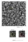
Figure 1
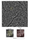
Figure 1. Image of normal cone mosaic
Image of normal cone mosaic in a healthy volunteer obtained with adaptive optics camera 4°×4° degree square (Rtx-1, Imagine Eyes, Orsay, France). The analysis was performed at superior 2° from the fovea (top). The region of interest (ROI) (yellow square in the top image) was used for automated cone segmentation (bottom left) and detection (bottom right) using dedicated software. Red squares correspond to automatically identified cones (bottom right). The image is from the author’s collection.
Figure 2
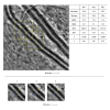
Figure 2. Image of normal retinal arteriole and venule
Evaluation of retinal arteriolar morphology in a healthy volunteer with adaptive optics camera 4°×4° degree square (Rtx-1, Imagine Eyes, Orsay, France) and measurement of morphological parameters using AOdetect software (top). The parameters calculated from the 3 selected regions of interest, for each time landmark (100 μm width and height each) (bottom). The image is from the author’s collection.
Figure 3
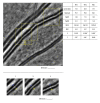
Figure 3. Image of retinal vessels in a patient with diabetes mellitus type 2
Evaluation of retinal arteriolar morphology in a patient with diabetes mellitus type 2 with adaptive optics camera 4°×4° degree square (Rtx-1, Imagine Eyes, Orsay, France) and measurement of morphological parameters using AOdetect software. Retinal artery with focal luminal narrowing (yellow arrow) and increased wall-to-lumen ratio (WLR 0,33; 0,37; 0,29) (top). The parameters calculated from the 3 selected regions of interest, for each time landmark (100 μm width and height each) (bottom). The image is from the author’s collection
Figure 4
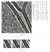
Figure 4. Image of retinal vessels in a patient with hypertensive retinopathy
Evaluation of retinal arteriolar morphology in a patient with hypertensive retinopathy with adaptive optics camera 4°×4° degree square (Rtx-1, Imagine Eyes, Orsay, France) and measurement of morphological parameters using AOdetect software. Retinal artery with increased wall-to-lumen ratio (WLR 0,31; 0,33; 0,32) (top). The parameters calculated from the 3 selected regions of interest, for each time landmark (100 μm width and height each) (bottom). The image is from the author’s collection.
Figure 5
Figure 5. Image of cone mosaic in a patient with cone-rod dystrophy
Image of cone mosaic in a patient with cone-rod dystrophy obtained with adaptive optics camera 4°×4° degree square (Rtx-1, Imagine Eyes, Orsay, France). The analysis was performed at superior 2° from the fovea. Empty spaces at the photoreceptor level and a disrupted mosaic of photoreceptors are demonstrated (top). The region of interest (ROI) (yellow square in the top image) was used for automated cone segmentation (bottom right) and detection (bottom left) using dedicated software. Red squares correspond to automatically identified cones (bottom right). Low density of the cones and the defect in the cones is demonstrated (bottom). The image is from the author’s collection.

References

    1. Burns SA, Elsner AE, Sapoznik KA, et al. Adaptive optics imaging of the human retina. Prog Retin Eye Res. 2019;68:1–30. - PMC - PubMed
    1. Akyol E, Hagag AM, Sivaprasad S, et al. Adaptive optics: Principles and applications in ophthalmology [published correction appears in Eye (Lond). 2021;35(6): 1796] Eye (Lond) 2021;35(1):244–64. - PMC - PubMed
    1. Liang J, Williams DR, Miller DT. Supernormal vision and high-resolution retinal imaging through adaptive optics. J Opt Soc Am A Opt Image Sci Vis. 1997;14(11):2884–92. - PubMed
    1. Lombardo M, Serrao S, Devaney N, et al. Adaptive optics technology for high-resolution retinal imaging. Sensors (Basel) 2012;13(1):334–66. - PMC - PubMed
    1. Hampson KM, Turcotte R, Miller DT, et al. Adaptive optics for high-resolution imaging. Nat Rev Methods Primers. 2021;1:68. - PMC - PubMed

MeSH terms