Review
. 2024 Jan;81(1):75-86.
doi: 10.1161/HYPERTENSIONAHA.123.19943. Epub 2023 Nov 29.

Cerebral Small Vessel Disease, Hypertension, and Vascular Contributions to Cognitive Impairment and Dementia

Affiliations
Review

Cerebral Small Vessel Disease, Hypertension, and Vascular Contributions to Cognitive Impairment and Dementia

Atticus H Hainsworth et al. Hypertension. 2024 Jan.

Abstract

Hypertension-associated cerebral small vessel disease is a common finding in older people. Strongly associated with age and hypertension, small vessel disease is found at autopsy in over 50% of people aged ≥65 years, with a spectrum of clinical manifestations. It is the main cause of lacunar stroke and a major source of vascular contributions to cognitive impairment and dementia. The brain areas affected are subcortical and periventricular white matter and deep gray nuclei. Neuropathological sequelae are diffuse white matter lesions (seen as white matter hyperintensities on T2-weighted magnetic resonance imaging), small ischemic foci (lacunes or microinfarcts), and less commonly, subcortical microhemorrhages. The most common form of cerebral small vessel disease is concentric, fibrotic thickening of small penetrating arteries (up to 300 microns outer diameter) termed arteriolosclerosis. Less common forms are small artery atheroma and lipohyalinosis (the lesions described by C. Miller Fisher adjacent to lacunes). Other microvascular lesions that are not reviewed here include cerebral amyloid angiopathy and venous collagenosis. Here, we review the epidemiology, neuropathology, clinical management, genetics, preclinical models, and pathogenesis of hypertensive small vessel disease. Knowledge gaps include initiating factors, molecular pathogenesis, relationships between arterial pathology and tissue damage, possible reversibility, pharmacological targets, and molecular biomarkers. Progress is anticipated from multicell transcriptomic and proteomic profiling, novel experimental models and further target-finding and interventional clinical studies.

Keywords: arteries; blood pressure; dementia; genetics; neuropathology; stroke; white matter.


Conflict of interest statement

Disclosures A.H. Hainsworth has received honoraria from Eli-Lilly and from National Institute on Aging (NIA). He serves as a consultant for AriBio Co, Ltd. and chairs the DementiasPlatform UK Vascular Experimental Medicine group. J.A. Schneider has received honoraria from Eli Lilly, Alnylam Pharmaceuticals, Inc, Apellis Pharmaceuticals, Inc, and Fondation Alzheimer. She has served as an expert witness for the US National Hockey League. The other author reports no conflicts.

Figures

Figure 1.
Figure 1.
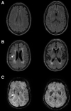
Magnetic resonance imaging (MRI) evidence of small vessel disease (SVD). A, Fluid-attenuated inversion recovery (FLAIR) MRI scans of an older patient with minimal white matter hyperintensities. B, FLAIR MRI scans from equivalent areas of another older patient with extensive white matter hyperintensities and a cavitated lacune (arrow). C, Susceptibility-weighted images from 2 different patients with SVD, showing multiple microbleeds (eg, marked with an arrow).
Figure 2.
Figure 2.
Small arterial vessels in human brain: examples of hypertensive small vessel disease. A, A normal, healthy penetrating artery with thin wall and multiple myocyte nuclei (eg, marked with arrow). B, Arteriolosclerosis, with approximately concentric wall thickening, with acellular hyaline material (asterisk). C, Lipohyalinosis, with asymmetrical wall thickening around the lumen (*) and mural lipid-containing macrophages (arrow). D, Fibrinoid necrosis in a larger penetrating vessel. The wall is asymmetrically thickened with eosinophilic fibrinoid material (*). E, Microatheroma in a penetrating vessel. Asymmetrical thickening and cholesterol clefts are seen as open slits (arrow) in the intimal layer. Internal lamina is visible (arrowhead) delimiting the intima. F, For comparison, large vessel atheroma in a leptomeningeal artery, with internal lamina clearly visible (arrowhead). Hematoxylin and eosin staining. Scale bars: A and B, 20 microns, C through E, 50 microns, F, 1000 microns. C and D were kindly supplied by Professor Colin Smith, University of Edinburgh.
Figure 3.
Figure 3.
Histopathology of arteriolosclerosis. Small penetrating arteries in subcortical white matter. A, Arteriolosclerosis, with hyaline, fibrotic wall thickening, and depleted, swollen myocytes. HE stain. B, The endothelial cell layer is preserved, immunolabeled with thrombomodulin (brown, arrow). C, Depletion of myocytes, immunolabeled with smooth muscle myosin (black, arrow). Light green counterstain. D and E, Collagenous fibrosis (green) in the vessel wall, Masson trichrome stain. D shows a severely fibrotic vessel (arrow), close to another that exhibits no fibrosis (arrowhead), adjacent to the cortical gray matter (marked with *). In E, the fibrotic vessel is enlarged. F, Collagen type IV α1/α2 immunolabeling shows duplication of the intimal basal lamina (brown, arrow), maintained adventitial layer (arrowhead), and absence of type IV collagen in the hyaline fibrotic media. Scale bars: 50 (A), 20 (B), 50 (C), 500 (D), 100 (E), and 50 μm (F).
Figure 4.
Figure 4.
Parenchymal lesions associated with small vessel disease. A, Ex vivo magnetic resonance imaging-directed tissue sampling of a frontal cortical block containing white matter hyperintensity (marked with *) from the Rush archive. B and C, The block from A, stained with hematoxylin-eosin (B) and Luxol fast blue (C), showing white matter pallor (*). D, In another case, astrocytes immunolabeled with GFAP (glial fibrillary acidic protein) (brown) around the border of a microinfarct (*). E, Axonal bulbs (arrow), immunopositive for hyperphosphorylated neurofilament-H (brown), centered around a small penetrating artery. F, Microhemorrhage (marked with arrow) labeled with Perl’s stain, around a small artery. Scale bars: B and C, 5 mm. D, 500 microns. E, 100 microns. F, 20 microns.

References

    1. Savva GM, Wharton SB, Ince PG, Forster G, Matthews FE, Brayne C; Medical Research Council Cognitive Function and Ageing Study. Age, neuropathology, and dementia. N Engl J Med. 2009;360:2302–2309. doi: 10.1056/NEJMoa0806142 - PubMed
    1. MRC-CFAS. Pathological correlates of late-onset dementia in a multicentre, community-based population in England and Wales. Neuropathology group of the Medical Research Council Cognitive Function and Ageing Study (MRC CFAS). Lancet. 2001;357:169–175. doi: 10.1016/s0140-6736(00)03589-3 - PubMed
    1. Oveisgharan S, Kim N, Agrawal S, Yu L, Leurgans S, Kapasi A, Arfanakis K, Bennett DA, Schneider JA, Buchman AS. Brain and spinal cord arteriolosclerosis and its associations with cerebrovascular disease risk factors in community-dwelling older adults. Acta Neuropathol. 2023;145:219–233. doi: 10.1007/s00401-022-02527-z - PMC - PubMed
    1. Gouw AA, Seewann A, van der Flier WM, Barkhof F, Rozemuller AM, Scheltens P, Geurts JJG. Heterogeneity of small vessel disease: a systematic review of MRI and histopathology correlations. J Neurol Neurosurg Psychiatry. 2011;82:126–135. doi: 10.1136/jnnp.2009.204685 - PubMed
    1. Fisher CM. The arterial lesions underlying lacunes. Acta Neuropathol. 1968;12:1–15. doi: 10.1007/BF00685305 - PubMed