. 2023 Dec;13(12):4765-4784.
doi: 10.1016/j.apsb.2023.08.015. Epub 2023 Aug 17.

Non-canonical STING-PERK pathway dependent epigenetic regulation of vascular endothelial dysfunction via integrating IRF3 and NF- κ B in inflammatory response

Affiliations

Non-canonical STING-PERK pathway dependent epigenetic regulation of vascular endothelial dysfunction via integrating IRF3 and NF- κ B in inflammatory response

Xuesong Li et al. Acta Pharm Sin B. 2023 Dec.

Abstract

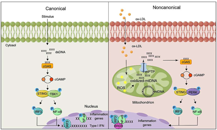
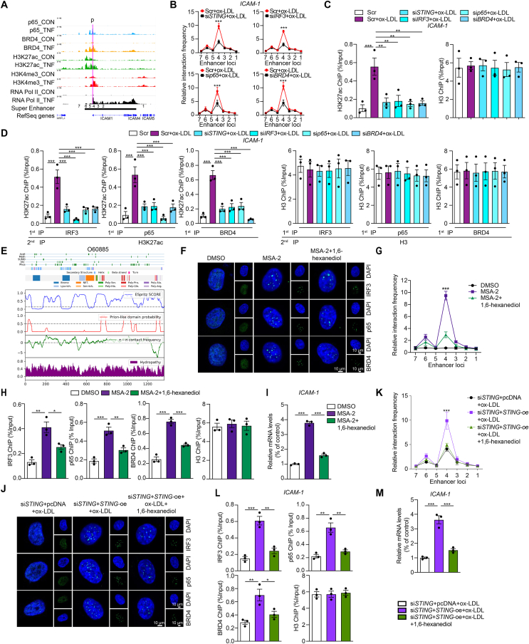
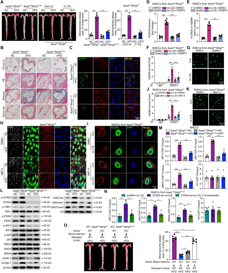
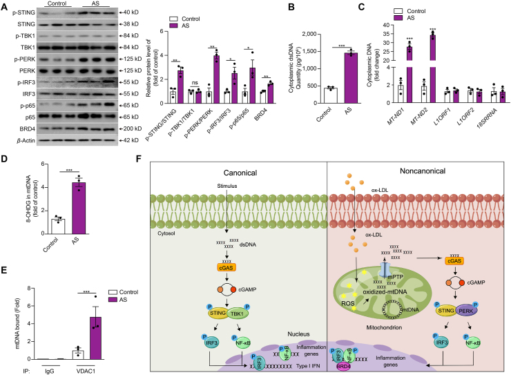
Inflammation-driven endothelial dysfunction is the major initiating factor in atherosclerosis, while the underlying mechanism remains elusive. Here, we report that the non-canonical stimulator of interferon genes (STING)-PKR-like ER kinase (PERK) pathway was significantly activated in both human and mice atherosclerotic arteries. Typically, STING activation leads to the activation of interferon regulatory factor 3 (IRF3) and nuclear factor-kappa B (NF-κB)/p65, thereby facilitating IFN signals and inflammation. In contrast, our study reveals the activated non-canonical STING-PERK pathway increases scaffold protein bromodomain protein 4 (BRD4) expression, which encourages the formation of super-enhancers on the proximal promoter regions of the proinflammatory cytokines, thereby enabling the transactivation of these cytokines by integrating activated IRF3 and NF-κB via a condensation process. Endothelium-specific STING and BRD4 deficiency significantly decreased the plaque area and inflammation. Mechanistically, this pathway is triggered by leaked mitochondrial DNA (mtDNA) via mitochondrial permeability transition pore (mPTP), formed by voltage-dependent anion channel 1 (VDAC1) oligomer interaction with oxidized mtDNA upon cholesterol oxidation stimulation. Especially, compared to macrophages, endothelial STING activation plays a more pronounced role in atherosclerosis. We propose a non-canonical STING-PERK pathway-dependent epigenetic paradigm in atherosclerosis that integrates IRF3, NF-κB and BRD4 in inflammatory responses, which provides emerging therapeutic modalities for vascular endothelial dysfunction.

Keywords: Atherosclerosis; BRD4; Endothelial dysfunction; Inflammation; Mitochondrial DNA; PERK; ROS; STING.


Conflict of interest statement

The authors declare no conflicts of interest.

Figures

Image 1
Graphical abstract
Figure 1
Figure 1
Atherosclerosis-related endothelial dysfunction mediated by oxidized LDL is dependent on the activation of the non-canonical STING–PERK pathway. (A, B) Heat map displaying the expression (microarray; log2 RMA signal) of inflammatory factors and IFN-response genes in distant macroscopically intact tissue as control (Con, light green) and atherosclerotic plaque (orange) in (A) and human coronary artery endothelial cells (HCAECs) treated with scramble control (Scr, green), Scr + oxidative low-density lipoprotein (ox-LDL, red), and stimulator of interferon genes siRNA (siSTING)+ox-LDL (yellow) in (B). (C, D) GO biological process enrichment in HCAECs treated with Scr + ox-LDL versus Scr (C) and in HCAECs treated with siSTING + ox-LDL versus Scr + ox-LDL (D) by DAVID analysis. (E) Quantitative real-time PCR (qRT-PCR) analysis of ISG15, ISG20, MX2, and IL-6 mRNA expression in ox-LDL-treated HCAECs combined with siSTING. (F) Western blot analysis of the non-canonical STING–PKR-like ER kinase (PERK) pathway in HCAECs treated with ox-LDL (100 μg/mL), lipopolysaccharide (LPS, 1 μg/mL), or tumor necrosis factor α (TNFα, 10 ng/mL). (G) Co-immunoprecipitation analysis of STING-PERK binding in HCAECs treated with ox-LDL, LPS and TNFα. (H) Western blot analysis of the phosphorylated interferon regulatory factor 3 (p-IRF3) in ox-LDL-treated HCAECs combined with siPERK. (I) Immunofluorescence staining of STING and IRF3 in ox-LDL-treated HCAECs versus control (Scale bar = 20 μm). (J) Western blot analysis of the non-canonical STING–PERK pathway in ox-LDL-treated HCAECs combined with siSTING, STING inhibitor C-176 (0.5 μmol/L) or STING activator MSA-2 (10 μmol/L). (K) Co-immunoprecipitation analysis of STING–PERK binding in ox-LDL-treated HCAECs combined with siSTING. (L, M) Permeability of HCAECs to FITC-Dexstran (L), THP-1 monocytes adhesion (M) in ox-LDL-treated HCAECs combined with siSTING or MSA-2 or C-176. (N) qRT-PCR analysis of adhesions molecules and chemokines mRNA expression in ox-LDL-treated HCAECs combined with siSTING or MSA-2 or C-176. Data are expressed as mean ± SEM, n = 3; ∗P < 0.05, ∗∗P < 0.01, ∗∗∗P < 0.001.
Figure 2
Figure 2
Mitochondrial dsDNA leaking mediated the non-canonical STING–PERK pathway activation in the oxidized LDL-induced endothelial injury. (A–D) The cytoplasmic double-stranded DNA (dsDNA) concentration (A), qPCR analysis of cytoplasmic mitochondrial DNA (mtDNA, MT-ND1 and MT-ND2), nuclear LINE1 elements (L1ORF1 and L1ORF2), ribosomal gene (RNA 18S) (B), cyclic GMP-AMP (cGAMP) concentration (C) and immunofluorescence staining of mitochondria by TOMM20 (mitochondria, red) and dsDNA (green) (D) in HCAECs treated with or without ox-LDL (Scale bar = 10 μm). (E–J) Western blot analysis of the non-canonical STING-PERK pathway (E), co-immunoprecipitation analysis of STING-PERK binding (F), cGAMP concentration (G), immunofluorescence staining of STING (H), THP-1 monocytes adhesion (I), and adhesions molecules and chemokines mRNA expression (J) in mtDNA (3 μg/mL)-transfected HCAECs combined with DNase I (1 μg/mL) or C-176. (K–O) Western blot analysis of the non-canonical STING–PERK pathway (K), co-immunoprecipitation analysis of STING-PERK binding (L), cyclic GMP–AMP synthase (cGAS) bound mtDNA by cytosolic DNA immunoprecipitation (M), cGAMP concentration (N) and adhesions molecules and chemokines mRNA expression (O) in ox-LDL-treated HCAECs combined with ethidium bromide (EtBr). Data are expressed as mean ± SEM, n = 3; ∗∗P < 0.01, ∗∗∗P < 0.001; ND, not detected; ns, no significance.
Figure 3
Figure 3
Oxidized LDL increases 8-OHDG of mtDNA via reactive oxygen species (ROS) and causes mtDNA release into the cytoplasm via voltage-dependent anion channel 1 (VDAC1)-dependent mitochondrial permeability transition pore (mPTP) opening. (A–F) The ROS level stained by DCFH-DA (A), mitochondrial membrane potential stained by TMRM (B), the opening of mPTP stained by MPTP assay kit (C), the expression level of cytoplasmic mtDNA by qPCR (D), heat map results of inflammatory genes (E), and mtDNA oxidation reflected by the level of 8-OHDG (F) in ox-LDL-treated HCAECs combined with mitochondrial ROS scavenger TEMPO at the dose of 0.01 mmol/L and 0.1 mmol/L. (G) Cytosolic DNA immunoprecipitation analysis of the relative amount of mtDNA binding to VDAC1 in ox-LDL-treated HCAECs combined with TEMPO. (H) Representative images of mPTP assay in ox-LDL-treated HCAECs combined with VDAC1 oligomerization inhibitor VBIT-4 (10 μmol/L). (I–M) Immunostaining of TOMM20 (mitochondria, red) and dsDNA (green) (I, J), qPCR analysis of mtDNA, nuclear LINE1 elements or a ribosomal gene (K), attachment of THP-1 monocytes (L), and intercellular adhesion molecule 1 (ICAM-1) mRNA expression (M) in ox-LDL-treated HCAECs combined with mPTP inhibitor cyclosporin and VBIT-4. Data are shown as mean ± SEM, n = 3; ∗∗∗P < 0.001.
Figure 4
Figure 4
Cooperation of IRF3, NF-κB and BRD4 in the transcription of inflammation genes in oxidized LDL-induced endothelial injury. (A) Tracks of chromatin immunoprecipitation sequencing (ChIP-seq) peaks for IRF3 (red), p65 (blue, upper; cyan, down), and BRD4 (yellow) at the ICAM1 gene loci in control (top) or stimulant-induced inflammation (bottom) samples. (B) Western blot analysis of total and phosphorylated p65 and IRF3 and BRD family in ox-LDL-treated HCAECs combined with siSTING or C-176. (C) Western blot analysis of total and phosphorylated p65 and IRF3 and BRD4 in ox-LDL-treated HCAECs combined with IRF3, p65, or BRD4 siRNA. (D) ChIP analysis of the enrichment of IRF3, p65, BRD2, BRD3, and BRD4 at −1 kb, promoter and +1 kb of inflammatory genes in ox-LDL-treated HACECs combined with STING, p65, IRF3 or BRD4 siRNA. (E, F) The ChIP-qPCR (E) and Re-ChIP (F) analysis of the binding of BRD4, IRF3, p65, and IRF3 on the +1 kb region of ICAM1 in ox-LDL-treated HCAECs combined with STING, p65, IRF3 or BRD4 siRNA. (G) qRT-PCR analysis of adhesions molecules and chemokines mRNA expression in ox-LDL-induced HCAECs combined with STING, p65, IRF3 or BRD4 siRNA. Data are shown as mean ± SEM, n = 3; ∗∗∗P < 0.001.
Figure 5
Figure 5
Oxidized LDL regulates the open chromatin state of inflammatory genes in ECs in a BRD4-dependent super-enhancer associated with condensation. (A) ChIP-seq peaks (p65, BRD4, H3K27ac, H3K4me3 and RNA Pol II levels) and putative super-enhancer location around ICAM-1. The putative super-enhancer region is marked as the horizontal line in black. The promoter is marked as the purple line. (B) Quantitative analysis of chromosome conformation capture assays-qPCR (3C-qPCR) analysis of long-distance interactions between ICAM-1 promoter and seven enhancer loci in ox-LDL-treated HACECs combined with STING, IRF3, p65 or BRD4 siRNA (x-axis means seven enhancer loci of ICAM-1). (C, D) The ChIP-qPCR (C) and Re-ChIP (D) analysis of the binding of H3K27ac and BRD4, IRF3, or p65, separately, on the +1 kb region of ICAM1 in ox-LDL-treated HCAECs combined with STING, p65, IRF3 or BRD4 siRNA. The Re-ChIP was performed with 1st round pull-down of antibodies, including IRF3, p65, and BRD4 as well as 2nd round pull-down of H3K27ac antibody. (E) The color-coded schematic representation of the aligned amino acid sequence and corresponding prion-like domain disorder propensity plots for BRD4 using the PLAAC (prion-like amino acid composition) pool. (F–I) Immunofluorescence staining of IRF3 (green), p65 (green), and BRD4 (green) in the nuclear (blue) (F), 3C-qPCR analysis of long-distance interactions between ICAM-1 promoter and seven enhancer loci (the positions of different loci were described as Fig. 5A, x-axis means seven enhancer loci of ICAM-1) (G), ChIP analysis of the enrichment of IRF3, p65, and BRD4 at the super-enhancer region of ICAM-1 (H), and ICAM-1 mRNA level (I) in ox-LDL-treated HCAECs combined with MSA-2 or condensate inhibitor 1,6-hexanediol. (J–M) Immunofluorescence staining of IRF3 (green), p65 (green) and BRD4 (green) in the nuclear (blue) (J), 3C-qPCR analysis of long-distance interactions between ICAM-1 promoter and seven enhancers (x-axis means seven enhancer loci of ICAM-1) (K), ChIP analysis of the enrichment of IRF3, p65 and BRD4 at the super-enhancer region of ICAM-1 (L), and ICAM-1 mRNA level (M) in siSTING- and ox-LDL-treated HCAECs combined with STING overexpression or 1,6-hexanediol. Data are shown as mean ± SEM, n = 3; ∗P < 0.05, ∗∗P < 0.01, ∗∗∗P < 0.001.
Figure 6
Figure 6
Endothelial deletion of STING reverses the atherosclerotic lesions in ApoeKOStingWT mice. (A) Representative images of en face aortas stained with oil red O from ApoeKOStingWT and ApoeKOStingEC-KO mice fed with normal chow diet (NC) or high-fat diet (HFD), as well as the ApoeKOStingWT mice fed with NC or HFD combined with STING inhibitor C-176. (B, C) Representative oil red O, hematoxylin and eosin (H&E), Masson and Sirius red staining of the aortic root (B), immunofluorescence staining of CD31 (red) and ICAM-1 (green) (C) in arteries from ApoeKOStingWT and ApoeKOStingEC-KO mice fed with NC or HFD. (D–G) The ROS level (D), mtDNA oxidation (E), VDAC1 bound mtDNA amount by cytosolic DNA immunoprecipitation (F), and the opening of mPTP (G) in mouse aortic endothelial cells (MAECs) derived from ApoeKOStingWT mouse thoracic aorta treated with ox-LDL and 0.1 mmol/L TEMPO. (H) Representative en face aortas immunofluorescence staining of endothelium (CD31), dsDNA and mitochondria (TOMM20) in the arteries from ApoeKOStingWT mice fed with NC or HFD combined with VBIT-4. (I–K) Immunofluorescence staining of dsDNA and mitochondria (TOMM20) (I), VDAC1 bound mtDNA amount by cytosolic DNA immunoprecipitation (J), the opening of mPTP (K) in MAECs derived from ApoeKOStingWT mouse thoracic aorta treated with ox-LDL and VBIT-4. (L, M) The protein levels of non-canonical STING pathway, BRD family, inflammation and super-enhancer specific makers (L), and ChIP analysis of the enrichment of IRF3, p65, and BRD4 at the super-enhancer region of Icam-1 (M) of MAECs derived from the thoracic aorta of ApoeKOStingWT and ApoeKOStingEC-KO mice fed with NC or HFD. (N) ChIP analysis of the enrichment of IRF3, p65, and BRD4 at the super-enhancer region of Icam-1 of ox-LDL-treated MAECs derived from the thoracic aorta of ApoeKOStingEC-KO mice, combined with STING overexpression and 1,6-hexanediol. (O) Representative images of en face aortas stained with oil red O from ApoeKOStingWT and ApoeKOStingEC-KO mice which were transplanted with bone marrow cells from ApoeKOStingWT and ApoeKOStingEC-KO mice. The recipient mice were fed with HFD. Data are shown as mean ± SEM, n = 6; ∗P < 0.05, ∗∗P < 0.01, ∗∗∗P < 0.001.
Figure 7
Figure 7
The activation of the non-canonical STING-PERK pathway in human atherosclerotic plaques. (A–E) The protein level of the non-canonical STING–PERK pathway (A), the cytoplasmic dsDNA concentration (B), the cytoplasmic levels of mtDNA, nuclear LINE1 elements or RNA 18S (C), the mtDNA oxidation (D), mtDNA binding to VDAC1 (E) in the aortic tissues from patients with and without atherosclerosis. (F) The proposed model of non-canonical STING–PERK pathway activation and development of atherosclerosis. ox-LDL damages mitochondria of ECs and increases the generation of ROS, which oxidizes the mtDNA. The oxidized mtDNA interacts with VDAC1 and induces oligomerization of VDAC1 to form mPTP, releasing mtDNA to the cytosol. DNA binds and activates DNA sensor cGAS through conformational changes, producing cGAMP from ATP and GTP to activate the adaptor STING. STING dimerizes and recruits PERK to phosphorylate and activate transcription factors IRF3 and NF-κB. Meanwhile, STING regulates the formation of the transcriptional complex of super-enhancer on the proximal promoter regions of proinflammatory cytokines, including p-IRF3, p-p65, and BRD4 via the condensation characteristic of BRD4 to promote the transactivation of proinflammatory cytokines. Data are shown as mean ± SEM, ∗P < 0.05, ∗∗P < 0.01, ∗∗∗P < 0.001; ns, no significance.

References

    1. Benjamin E.J., Virani S.S., Callaway C.W., Chamberlain A.M., Chang A.R., Cheng S., et al. Heart disease and stroke statistics-2018 update: a report from the American Heart Association. Circulation. 2018;137:e67–e492. - PubMed
    1. Xu S., Ilyas I., Little P.J., Li H., Kamato D., Zheng X., et al. Endothelial dysfunction in atherosclerotic cardiovascular diseases and beyond: from mechanism to pharmacotherapies. Pharmacol Rev. 2021;73:924–967. - PubMed
    1. Zheng S., Li Z., Song J., Wang P., Xu J., Hu W., et al. Endothelial METRNL determines circulating METRNL level and maintains endothelial function against atherosclerosis. Acta Pharm Sin B. 2023;13:1568–1587. - PMC - PubMed
    1. Zhang X., Bai X.C., Chen Z.J. Structures and mechanisms in the cGAS–STING innate immunity pathway. Immunity. 2020;53:43–53. - PubMed
    1. Hinkle J.T., Patel J., Panicker N., Karuppagounder S.S., Biswas D., Belingon B., et al. STING mediates neurodegeneration and neuroinflammation in nigrostriatal α-synucleinopathy. Proc Natl Acad Sci U S A. 2022;119 - PMC - PubMed