[Preprint]. 2023 Nov 21:2023.11.20.567970.
doi: 10.1101/2023.11.20.567970.

Staphylococcus aureus skin colonization is mediated by SasG lectin variation

Affiliations

Staphylococcus aureus skin colonization is mediated by SasG lectin variation

Krista B Mills et al. bioRxiv. .

Update in

Abstract

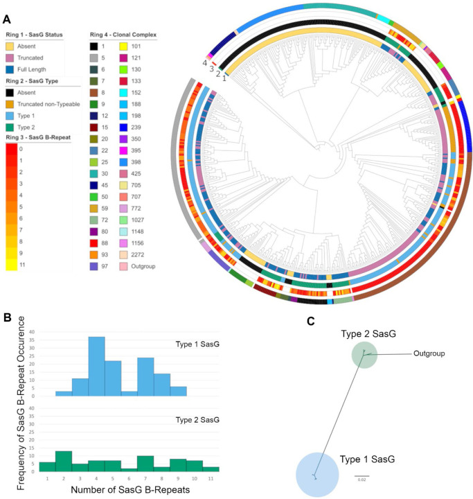
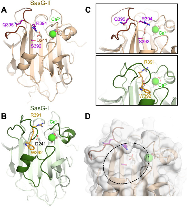
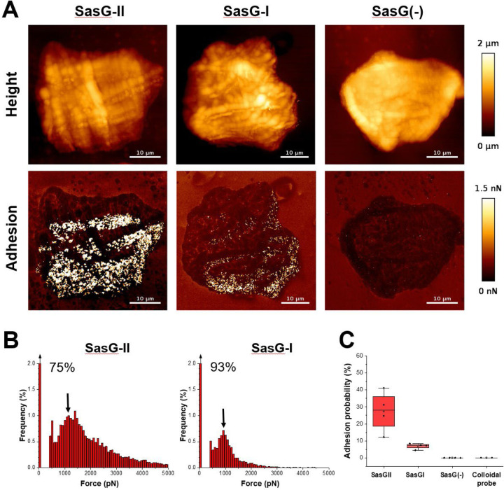
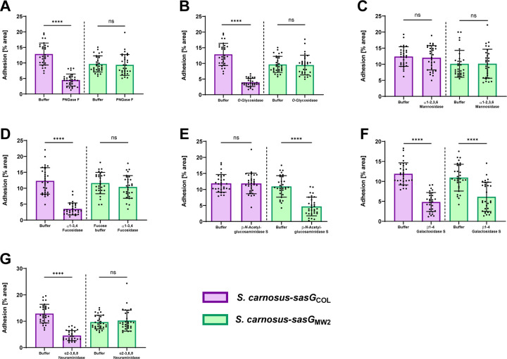
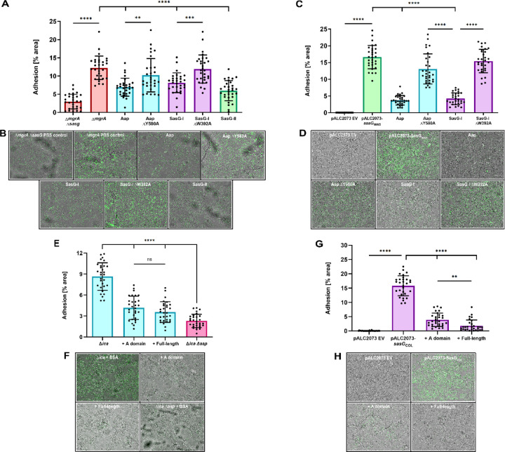
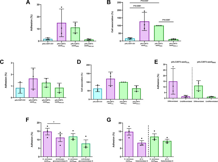
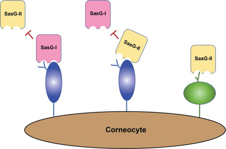
Staphylococcus aureus causes the majority of skin and soft tissue infections, but this pathogen only transiently colonizes healthy skin. However, this transient skin exposure enables S. aureus to transition to infection. Initial adhesion of S. aureus to skin corneocytes is mediated by surface protein G (SasG). Here, phylogenetic analyses reveal the presence of two major divergent SasG alleles in S. aureus, SasG-I and SasG-II. Structural analyses of SasG-II identified a unique non-aromatic arginine in the binding pocket of the lectin subdomain that mediates adhesion to corneocytes. Atomic force microscopy and corneocyte adhesion assays indicated SasG-II can bind to a broader variety of ligands than SasG-I. Glycosidase treatment resulted in different binding profiles between SasG-I and SasG-II on skin cells. Additionally, SasG-mediated adhesion was recapitulated using differentiated N/TERT keratinocytes. Our findings indicate that SasG-II has evolved to adhere to multiple ligands, conferring a distinct advantage to S. aureus during skin colonization.


Conflict of interest statement

Declaration of interests A.B.H. has served as a Scientific Advisory Board member for Hoth Therapeutics, Inc., holds equity in Hoth Therapeutics and Chelexa BioSciences, LLC, and was a co-inventor on seven patents broadly related to the subject matter of this work.

Figures

Figure 1.
Figure 1.. SasG Variation in the context of S. aureus phylogenetic diversity.
(A) Maximum likelihood phylogenetic tree of 574 S. aureus isolates based on their proteome. For each isolate, SasG status is mapped to ring 1, SasG type (defined in Panel B) is mapped to ring 2, SasG B-repeat number is mapped to ring 3, and the clonal complex of the isolate is mapped to ring 4. (B) Histograms of SasG B-repeats from both SasG allelic types. (C) Maximum likelihood phylogenetic tree of SasG from 191 aligned full-length sequences identifies two SasG allelic types.
Figure 2.
Figure 2.. The SasG-II lectin contains a unique non-aromatic residue in the glycan binding pocket.
(A) Crystal structure of the SasG-II lectin showing the structural Ca2+ ion, the conserved central D241 residue that adopts an atypical trans conformation, and the side chains of S392, R394, and Q395 near the end of β17. (B) Comparative view of SasG-II in the same orientation. Residues R391 and W392 are analogous to R394 and Q395 in SasG-II; note the distinct positioning of corresponding residues W392 (SasG-II) and Q395 (SasG-I). (C) Close-up view of the region near the end of β17, rotated by approximately 45° from panels A and B. Note the sharp bend of the main chain near R391 in SasG-I that is not observed in SasG-II. (D) Surface view of SasG-II showing the putative binding pocket lacking an aromatic residue at its base.
Figure 3.
Figure 3.. Multiparametric nanoimaging using single bacterial probes indicates SasG-II binds a broader variety of ligands than SasG-I.
(A) Height images (top) and adhesion images (bottom) of corneocytes recorded in PBS using a SasG-II, SasG-I, or EV (SasG[−]) cell probe. See also Figure S2–S4. (B) Histograms of adhesion forces registered on whole corneocytes (total of n = 9,590 curves for one representative SasG-II probe; n = 2,532 curves for one representative SasGI probe). The arrow at the top left of the histograms stands for the non-adhesive events. (C) Box plot comparing adhesion probabilities for SasG-II (n = 4 from 3 independent bacterial cultures), SasG-I (n = 4 from 2 independent bacterial cultures), SasG(−) (n = 4 from 2 independent bacterial cultures) or colloidal (n = 2) probes. For more data, see Figures S1, S2, and S3.
Figure 4.
Figure 4.. SasG-I and -II mediated adhesion to corneocytes shows differential responses upon treatment with glycosidases.
S. carnosus-sasGCOL (SasG-I) and S. carnosus-sasGMW2 (SasG-II) were tested for adhesion to corneocytes following pre-incubation with (A) PNGase F, (B) O-Glycosidase, (C) α1–2,3,6 Mannosidase, (D) α1–3,4 Fucosidase, (E) β-N-Acetylglucosaminidase S, (F) β1–4 Galactosidase S, and (G) α2–3,6,8 Neuraminidase. The percent area of adhesion in 10 images from three independent experiments (n = 30) was measured with Fiji ImageJ and analyzed in GraphPad Prism. Statistical significance was analyzed using the unpaired t-test or non-parametric Mann-Whitney test for data with non-normal distribution (****P<0.0001).
Figure 5.
Figure 5.. SasG-II-mediated adhesion is mediated by the lectin subdomain and may bind the same ligand as Aap and SasG-I.
(A) MRSA MW2 (SasG-II) was tested for adhesion to healthy human corneocytes after pre-incubation/blocking with 5 µM of purified lectins. MRSA MW2 ∆sasG was used as a negative control strain. **P=0.0019. (C) SasG-II-expressing S. carnosus-sasGMW2 was tested for adhesion to healthy human corneocytes after pre-incubation/blocking with 5 µM of purified lectins. S. carnosus-pALC2073 EV was used as a negative control strain. S. epidermidisica was tested for adhesion to corneocytes following pre-incubation/blocking with 100 µg/mL of purified full-length or A-domain SasG-II. S. epidermidisicaaap was used as a negative control strain. (G) SasG-I-expressing S. carnosus-sasGCOL was tested for adhesion to healthy human corneocytes after pre-incubation/blocking with 100 µg/mL of purified full-length or A-domain SasG-II. S. carnosus-pALC2073 EV was used as a negative control strain. **P=0.0091. (B, D, F, H) Representative bright-field (representing corneocytes) and green-channel (representing GFP-expressing bacteria) overlay microscopy images of experimental groups tested in Panels A, C, E, and G, respectively. (All panels) The percent area of adhesion in 10 images from three independent experiments (n = 30) was measured with Fiji ImageJ and analyzed in GraphPad Prism. Statistical significance was analyzed using ordinary one-way ANOVA (***P=0.0002; ****P<0.0001).
Figure 6.
Figure 6.. SasG-I and SasG-II-mediated adhesion to differentiated N/TERT keratinocytes following treatment with glycosidases suggests complex N-linked glycans and core 2 O-glycans may be important for SasG-I and SasG-II binding.
S. carnosus-pALC2073 EV, SasG-I-expressing S. carnosus-sasGCOL and SasG-II-expressing S. carnosus-sasGMW2, and SasG-II-expressing S. carnosus with the A-domain deleted (S. carnosus-sasGMW2∆A) at an MOI of 5 were tested for adhesion to either differentiated (A and B) or undifferentiated (C and D) N/TERT keratinocytes. (A) Adhesion to terminally differentiated cells as shown by overall percent adhesion. (B) Adhesion to terminally differentiated cells as shown by percent cell association to pALC2073-sasGMW2 input inoculum. Both SasG-expressing strains adhered more to differentiated cells than the EV and A domain mutant controls. (C) Adhesion to a monolayer of undifferentiated cells as shown by overall percent adhesion. (D) Adhesion to a monolayer of undifferentiated cells as shown by percent cell association to pALC2073-sasGMW2 input inoculum. There were no significant differences in adhesion between the EV and A domain mutant controls and the SasG-expressing strains. (A-D) The CFU/mL of three independent experiments (n = 3) were calculated and analyzed for statistical significance in GraphPad Prism using ordinary one-way ANOVA. (E) Data from panels A and C displaying differences in adhesion between differentiated and undifferentiated N/TERT keratinocytes for SasG-I-expressing S. carnosus-sasGCOL and SasG-II-expressing S. carnosus-sasGMW2. Both strains adhered well to differentiated N/TERT keratinocytes, and did not adhere well to undifferentiated N/TERT keratinocytes. No statistical analyses were performed for Panel E. SasG-I-expressing S. carnosus-sasGCOL and SasG-II-expressing S. carnosus-sasGMW2 were tested for overall percent adhesion to differentiated N/TERT keratinocytes following treatment with (F) β-N-Acetylglucosaminidase S and (G) β1–4 Galactosidase S. β1–4 Galactosidase S reduced adhesion of both strains, while β-N-Acetylglucosaminidase S resulted in a greater reduction in adhesion of S. carnosus-sasGMW2. The CFU/mL of three independent experiments (n = 3) were calculated and analyzed for statistical significance in GraphPad Prism using an unpaired t-test. *P=0.0500.
Figure 7.
Figure 7.. Model for SasG-I and SasG-II-mediated adhesion to healthy human skin corneocytes.
SasG-I blocks adhesion of SasG-II, and likewise SasG-II can block adhesion of SasG-I, indicating they can all bind the same corneocyte receptor in some capacity. However, removal of N-glycans and core 1 or 3 O-glycans does not block SasG-II binding as it does with SasG-I, suggesting that SasG-II may bind a core 2 O-glycan structure elsewhere on the corneocyte receptor.

Similar articles

References

    1. Parlet C.P., Brown M.M., and Horswill A.R. (2019). Commensal Staphylococci Influence Staphylococcus aureus Skin Colonization and Disease. Trends Microbiol 27, 497–507. 10.1016/j.tim.2019.01.008. - DOI - PMC - PubMed
    1. Brown A.F., Leech J.M., Rogers T.R., and McLoughlin R.M. (2014). Staphylococcus aureus Colonization: Modulation of Host Immune Response and Impact on Human Vaccine Design. Front Immunol 4, 507. 10.3389/fimmu.2013.00507. - DOI - PMC - PubMed
    1. Otto M. (2010). Staphylococcus colonization of the skin and antimicrobial peptides. Expert Rev Dermatol 5, 183–195. 10.1586/edm.10.6. - DOI - PMC - PubMed
    1. von Eiff C., Becker K., Machka K., Stammer H., and Peters G. (2001). Nasal carriage as a source of Staphylococcus aureus bacteremia. Study Group. N Engl J Med 344, 11–16. 10.1056/NEJM200101043440102. - DOI - PubMed
    1. Moran G.J., Krishnadasan A., Gorwitz R.J., Fosheim G.E., McDougal L.K., Carey R.B., Talan D.A., and Group E.M.I.N.S. (2006). Methicillin-resistant S. aureus infections among patients in the emergency department. N Engl J Med 355, 666–674. 10.1056/NEJMoa055356. - DOI - PubMed

Publication types