[Preprint]. 2023 Nov 22:2023.11.22.568342.
doi: 10.1101/2023.11.22.568342.

Engineering luminopsins with improved coupling efficiencies

Affiliations

Engineering luminopsins with improved coupling efficiencies

Ashley Slaviero et al. bioRxiv. .

Update in

  • Engineering luminopsins with improved coupling efficiencies.
    Slaviero AN, Gorantla N, Simkins J, Crespo EL, Ikefuama EC, Tree MO, Prakash M, Björefeldt A, Barnett LM, Lambert GG, Lipscombe D, Moore CI, Shaner NC, Hochgeschwender U. Slaviero AN, et al. Neurophotonics. 2024 Apr;11(2):024208. doi: 10.1117/1.NPh.11.2.024208. Epub 2024 Mar 29. Neurophotonics. 2024. PMID: 38559366 Free PMC article.

Abstract

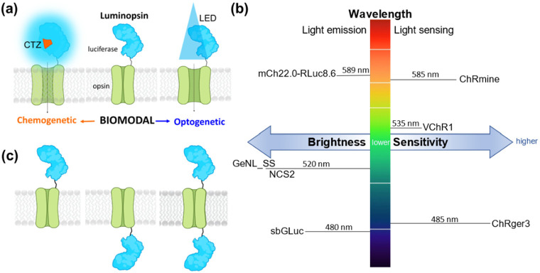
Significance: Luminopsins (LMOs) are bioluminescent-optogenetic tools with a luciferase fused to an opsin that allow bimodal control of neurons by providing both optogenetic and chemogenetic access. Determining which design features contribute to the efficacy of LMOs will be beneficial for further improving LMOs for use in research.

Aim: We investigated the relative impact of luciferase brightness, opsin sensitivity, pairing of emission and absorption wavelength, and arrangement of moieties on the function of LMOs.

Approach: We quantified efficacy of LMOs through whole cell patch clamp recordings in HEK293 cells by determining coupling efficiency, the percentage of maximum LED induced photocurrent achieved with bioluminescent activation of an opsin. We confirmed key results by multielectrode array (MEAs) recordings in primary neurons.

Results: Luciferase brightness and opsin sensitivity had the most impact on the efficacy of LMOs, and N-terminal fusions of luciferases to opsins performed better than C-terminal and multi-terminal fusions. Precise paring of luciferase emission and opsin absorption spectra appeared to be less critical.

Conclusions: Whole cell patch clamp recordings allowed us to quantify the impact of different characteristics of LMOs on their function. Our results suggest that coupling brighter bioluminescent sources to more sensitive opsins will improve LMO function. As bioluminescent activation of opsins is most likely based on Förster resonance energy transfer (FRET), the most effective strategy for improving LMOs further will be molecular evolution of luciferase-fluorescent protein-opsin fusions.

Keywords: Förster resonance energy transfer; bioluminescence; luciferase; opsin; optogenetics; whole cell patch clamp recording.


Conflict of interest statement

Disclosures The authors declare that they have no competing interests.

Figures

Fig. 1
Fig. 1
Luminopsin (LMO) features. (a) The luciferase-opsin fusion molecule (luminopsin) enables bimodal chemogenetic (CTZ) and optogenetic (LED) access to opsin activation. (b) Parameters for improving LMO performance are brightness of the light emitter, light sensitivity of the light sensor, and matching spectra for peak light emission and sensing. Moieties used in this study are indicated. (c) Possibilities for arrangement of moieties are N-terminal, C-terminal, or N- and C-terminal fusion of the luciferase to the opsin.
Fig. 2
Fig. 2
Quantifying LMO efficacy. (a) Membrane potential changes elicited chemo (CTZ)- or opto (LED, arc lamp)-genetically can be measured in HEK293 cells expressing an LMO. (b) In whole cell voltage patch clamp, inward current is determined in cells, here expressing LMO7, exposed to luciferin (hCTZ) and to light from an arc lamp (photocurrent). (c) Equation used to quantify LMO efficacy using currents measured in patch clamp. (d) Absence of effects of various luciferins and wavelengths of physical light source on the current of non-expressing HEK293 cells (controls, n=5).
Fig. 3
Fig. 3
Increasing brightness of the light emitter. (a) Schematics of LMO3, LMO7, and LMO10 with same opsin (VChR1) and different light emitters (sbGLuc, NCS2, GeNL_SS). (b) Fluorescent images (left), photocurrent patch clamp traces (middle), and luciferin-induced patch clamp traces (right) for LMO3, LMO7, and LMO10. (c) Coupling efficiencies of LMO3, LMO7, and LMO10 (n=5).
Fig. 4
Fig. 4
Increasing sensitivity of the light sensor. (a) Schematics of LMO7 and LMO8 with same luciferase-opsin fusion protein (NCS2) and different opsins (VChR1, ChRger3). (b) Fluorescent images (left), photocurrent patch clamp traces (middle), and luciferin-induced patch clamp traces (right) for LMO7 and LMO8. (c) Coupling efficiencies of LMO7 and LMO8 (n=5).
Fig. 5
Fig. 5
Increasing both brightness of the light emitter and sensitivity of the light sensor. (a) Schematics of LMO10 and LMO11 with brightest luciferase-opsin fusion protein (GeNL_SS) and opsins VChR1 and the super light sensitive ChRmine. (b) Fluorescent images (left), photocurrent patch clamp traces (middle), and luciferin-induced patch clamp traces (right) for LMO10 and LMO11. (d) Coupling efficiencies of LMO10 and LMO11 (n=5).
Fig. 6
Fig. 6
Different arrangements of moieties in LMOs. (a) Schematics of NCS2 tethered to VChR1 at the N-terminal (LMO7), the C-terminal (LMO7.2), and the N- and C-terminal (LMO7.3). (b) Fluorescent images (left), photocurrent patch clamp traces (middle), and luciferin-induced patch clamp traces (right) for LMO7, LMO7.2, and LMO7.3. (c) Coupling efficiencies of LMOs 7, 7.2, and 7.3 (n=5).
Fig. 7
Fig. 7
Matching emission and excitation spectra of moieties in LMOs. (a) Schematics of LMO11 (GeNL_SS-ChRmine) and LMO12 (ChRmine-mCh22.0-RLuc8.6). (b) Fluorescent images (left), photocurrent patch clamp traces (middle), and luciferin-induced patch clamp traces (right) for LMO11 and LMO12. (d) Coupling efficiencies of LMOs 11 and 12 (n=5).
Fig. 8
Fig. 8
Multi-electrode recordings from cortical neurons transduced with AAV-hSyn-LMO7 and -LMO11. Average spiking frequencies of MEA channels before any treatment (pre) and after application (post) of physical or biological light, or vehicle. Spiking frequency increased when neurons were exposed to an LED light and with application of 10 μM CTZ but did not change with application of vehicle.
Fig. 9
Fig. 9
Coupling efficiency versus bioluminescence produced by LMOs. Coupling efficiencies as determined throughout the above studies are plotted on the left, and bioluminescence measured from each LMO is plotted to the right. Bioluminescence produced by each LMO was measured in HEK293 cells in a plate reader with 100 μM concentration of luciferin (n=8 for each LMO). Readings were taken immediately after addition of substrate. Bioluminescence was normalized to expression of fluorescent reporter in each LMO using fluorescence ROI analysis in ImageJ.
Fig. 10
Fig. 10
Summary of patch clamp recordings in HEK293 cells. (a) Key features of LMOs tabulated in the order displayed in (b) and (c). (b) Representative example of luciferin-induced inward currents for each LMO tested. (c) Coupling efficiency graphs of LMOs tested. Red line in (b) and (c) indicates the level of performance of LMO3, the previously determined standard for a robustly functioning LMO.

References

    1. Fenno L., Yizhar O., and Deisseroth K., “The development and application of optogenetics,” Annu. Rev. Neurosci. 34 (2011) [doi:10.1146/annurev-neuro-061010-113817]. - DOI - PMC - PubMed
    1. Sternson S. M. and Roth B. L., “Chemogenetic tools to interrogate brain functions,” Annu. Rev. Neurosci. 37 (2014) [doi:10.1146/annurev-neuro-071013-014048]. - DOI - PubMed
    1. Berglund K. et al., “Light-Emitting Channelrhodopsins for Combined Optogenetic and Chemical-Genetic Control of Neurons,” PLoS One 8(3) (2013) [doi:10.1371/journal.pone.0059759]. - DOI - PMC - PubMed
    1. Tung J. K., Gutekunst C. A., and Gross R. E., “Inhibitory luminopsins: Genetically-encoded bioluminescent opsins for versatile, scalable, and hardware-independent optogenetic inhibition,” Sci. Rep. 5 (2015) [doi:10.1038/srep14366]. - DOI - PMC - PubMed
    1. Berglund K. et al., “Luminopsins integrate opto- and chemogenetics by using physical and biological light sources for opsin activation,” Proc. Natl. Acad. Sci. U.S.A. 113(3) (2016) [doi:10.1073/pnas.1510899113]. - DOI - PMC - PubMed

Publication types