[Preprint]. 2025 Jan 28:2023.11.20.567915.
doi: 10.1101/2023.11.20.567915.

Animal septins contain functional transmembrane domains

Affiliations

Animal septins contain functional transmembrane domains

Jenna A Perry et al. bioRxiv. .

Update in

Abstract

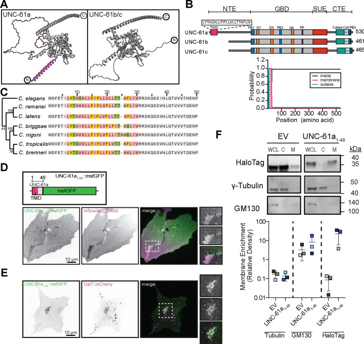
Septins, a conserved family of filament-forming proteins, contribute to eukaryotic cell division, polarity, and membrane trafficking. Septins scaffold other proteins to cellular membranes, but it is not fully understood how septins associate with membranes. We identified and characterized an isoform of Caenorhabditis elegans septin UNC-61 that was predicted to contain a transmembrane domain (TMD). The TMD isoform is expressed in a subset of tissues where the known septins were known to act, and TMD function was required for tissue integrity of the egg-laying apparatus. We found predicted TMD-containing septins across much of opisthokont phylogeny and demonstrated that the TMD-containing sequence of a primate TMD-septin is sufficient for localization to cellular membranes. Together, our findings reveal a novel mechanism of septin-membrane association with profound implications for septin dynamics and regulation.


Conflict of interest statement

Competing interests: The authors have declared no competing interests.

Figures

Fig. 1.
Fig. 1.. C. elegans UNC-61a contains a transmembrane domain.
(A) AlphaFold structures of UNC-61a and UNC-61b/c. Magenta: N-terminal alpha-helix of UNC-61a. (B) (top) Illustration of UNC-61a. Motif colors: pink: transmembrane domain (TMD), grey: GTP-binding domain, red: SUE, blue: PB1/2/3, orange: GTPase domains, purple: polyacidic region (PA), teal: coiled-coil domain, light green: amphipathic helix (AH). Brackets at top of schematic denote septin domains (NTE: N-terminal extension, GBD: GTP-binding domain, SUE: septin unique element, CTE: C-terminal extension) (bottom) TMHMM output probability of TMD for C. elegans UNC-61. Teal: extracellular, pink: TMD, black: cytosolic. (C) (right) Alignment of the first 50 amino acids of UNC-61 homologs in C. elegans and closely related nematodes. Non-TMD residues: grey, TMD sequence: colored based on amino acid properties (hydrophobic: rose, aromatic: orange, positively charged: blue, negatively charged: red, hydrophilic: green, cystine: yellow). (left) Neighbor-joining tree of evolutionary relationships. (D) (top) Illustration of UNC-61a 1–48::msfGFP construct, highlighting the position of msfGFP (green), TMD (magenta), and UNC-61a fragment (grey). (bottom) U2OS cells transiently co-expressing UNC-61a TMD1–48::msfGFP (green: and plasma membrane label mScarlet3::CAAX( magenta). Images shown are a single plane, inverted contrast, and scaled to display minimum and maximum pixel intensities. Dashed box (white) indicates region used for 2x inset. Scale bar = 10 μm. (E) U2OS cells transiently co-expressing UNC-61a TMD1–48::msfGFP (green) and GalT::mCherry (Trans-Golgi and Golgi transport intermediates; magenta). Images are maximum intensity projections, inverted contrast, and are scaled to display pixel intensity ranges. Dashed box (white) indicates region used for 2x inset. Scale bar = 10 μm. (F) (top) Western blots of cytosolic (C) and membrane (M) fractions of HeLa cells transiently expressing either HaloTag empty vector (EV) or UNC-61a TMD1–48::HaloTag (UNC-61a TMD1–48). Lanes 1–3: whole cell lysate (WCL) blotted singly with antibodies recognizing GM130 (membrane), γ-tubulin (cytosol) and HaloTag. Lanes 4–6 WCL,C, and M fractions are probed with a mixture of all three antibodies (GM130, γ-tubulin, and HaloTag). A shorter exposure (4.9 s) was used to image the HaloTag to prevent band saturation. (bottom) Membrane enrichment of HaloTag for the EV and UNC-61a TMD1–48::HaloTag was analyzed semi-quantitatively and plotted as the ratio of membrane enrichment to cytosolic enrichment. Plot: mean ± SD of three independent experiments.
Fig. 2.
Fig. 2.. UNC-61a TMD is required for normal vulval morphology and function.
(A) (top) Schematic of adult C. elegans hermaphrodite (pink: pharynx, blue: germline, orange: vulva.) (bottom) Images are inverted contrast of the fluorophore annotated. Arrow: vulval epithelium apical surfaces; arrowhead; linear septin decorated structures. Dashed lines outline the C. elegans terminal bulb and oogenic germline. The fluorescent signal beneath the germline corresponds to the autofluorescence of the C. elegans intestine. Scale bar = 10 μm (pharynx and vulva); 25 μm (germline). (B) (top) Schematic of representative images of protruded vulva (pvl) phenotypes and embryos in uterus. (middle) Representative transmitted light images of pvl phenotypes. Arrow denotes embryos that are more developed than they should be indicating an egg-laying defective (egl) phenotype. (bottom) Percent incidence of post-embryonic phenotypes (pvl: protruded vulva; egl: egg-laying defective; bag: bag-of-worms) of control (grey), unc-61Δ (pink), unc-61 Δa (yellow), and unc-61a ΔTMD (blue) animals (n=100 animals). (C) (left top) Schematic of control and unc-61a ΔTMD vulvae (green: epithelium, black: apical surface, grey: surrounding muscles.) (left bottom) Inverted contrast images of Lifeact::GFP (F-actin) in vulva region. Arrowhead: muscle cuticle connection in control, disconnection in unc-61a ΔTMD; arrows: apical surface of the vulval epithelium. (right) Percent incidence of muscle detachment and open vulva phenotypes. “n” above bar indicates the number of animals scored for each condition. Scale bar = 20 μm. (D) (left) Representative images of uterine and embryo permeability in control and unc-61a ΔTMD animals. Arrowheads denote vulval location, asterisks indicate germline location, and dashed lines outline uterus. Scale bar = 50 μm. (right) Quantification of uterine (dashed lines) mean fluorescence intensity in control (grey; n = 15 animals) and unc-61a ΔTMD animals (blue; n = 15 animals). Error bars are mean ± SD. ****, ≤ 0.0001, as determined by Student’s unpaired t-test.
Fig. 3.
Fig. 3.. Animal septins are predicted to contain transmembrane domains (TMD).
(A) Cladogram of all septin sequences deposited on UniProt (magenta: sequences containing TMDs). Rim colors: organismal phyla (dark blue: Ascomycota, brown: Basidiomycota, fuchsia: Mucoromycota, purple: Chytridiomycota, lime green: Viridiplantae, yellow: Chordata, teal: Arthropoda, light green: Nematoda, blue: Platyhelminthes, light pink: Mollusca, hot pink: Cnideria (B) msfGFP::CsSEPT10C (green) expressed in U2OS cells and colocalized with Translocase of Outer Mitochondrial Membrane 20 (TOMM20; magenta). (C) msfGFP::CsSEPT10C (green) co-expressed with HaloTag::Sec61β (ER; magenta) in U2OS cells. (D) (Left) Neighbor-joining tree of evolutionary relationships (Right) Schematic of CsSEPT10 and HsSEPT10 protein architecture. Colors as in Figure 1C. (E) Localization of msfGFP::HsSEPT10 and the chimera msfGFP::HsSEPT10ΔC-CsSEPT10C. (F) Colocalization of the chimera msfGFP::HsSEPT10ΔC-CsSEPT10C (green) and mApple::TOMM20 (magenta). All images are inverted contrast, unless merged, and insets are magnified 2x. Scale bar = 10 μm.

References

    1. Woods B. L. & Gladfelter A. S. The state of the septin cytoskeleton from assembly to function. Current Opinion in Cell Biology 68, 105–112 (2021). - PMC - PubMed
    1. Benoit B., Poüs C. & Baillet A. Septins as membrane influencers: direct play or in association with other cytoskeleton partners. Frontiers in Cell and Developmental Biology 11, (2023). - PMC - PubMed
    1. Tanaka-Takiguchi Y., Kinoshita M. & Takiguchi K. Septin-Mediated Uniform Bracing of Phospholipid Membranes. Current Biology 19, 140–145 (2009). - PubMed
    1. Casamayor A. & Snyder M. Molecular Dissection of a Yeast Septin: Distinct Domains Are Required for Septin Interaction, Localization, and Function. Mol Cell Biol 23, 2762–2777 (2003). - PMC - PubMed
    1. Akil A. et al. Septin 9 induces lipid droplets growth by a phosphatidylinositol-5-phosphate and microtubule-dependent mechanism hijacked by HCV. Nat Commun 7, 12203 (2016). - PMC - PubMed

Publication types