

Urinary single-cell sequence analysis of the urinary macrophage in different outcomes of membranous nephropathy
- PMID: 38046013
- PMCID: PMC10689170
- DOI: 10.1093/ckj/sfad132
Urinary single-cell sequence analysis of the urinary macrophage in different outcomes of membranous nephropathy
Abstract
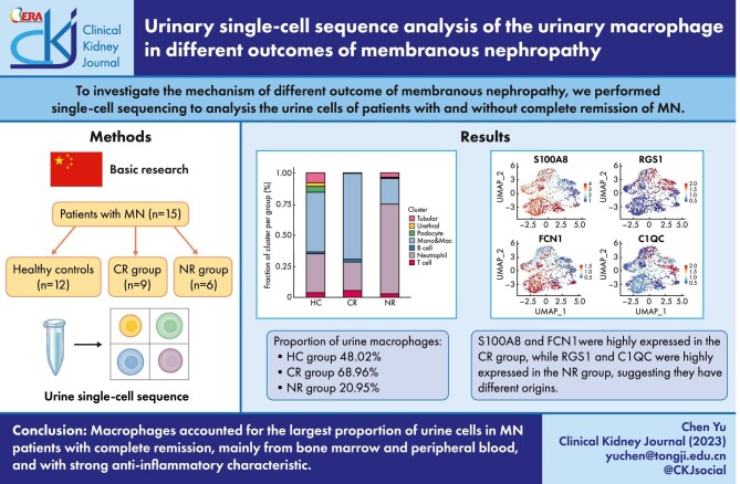
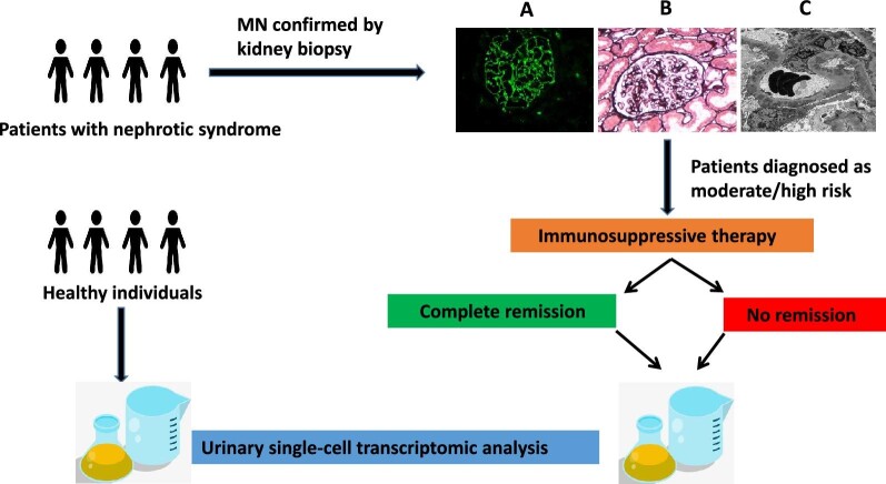
Background: Great progress has been made in the diagnosis and treatment of membranous nephropathy (MN). However, a significant number of patients do not respond to immunosuppressive therapy and eventually progress to end-stage kidney disease. To investigate the mechanism of different outcome of MN, we performed single-cell sequencing to analyze the urine cells of patients with and without complete remission of MN.
Methods: Urine single-cell RNA sequencing was performed on 12 healthy controls (HC) and 15 patients with MN. The patients were divided into a complete remission group (CR, n = 9) and a no remission group (NR, n = 6).
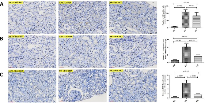
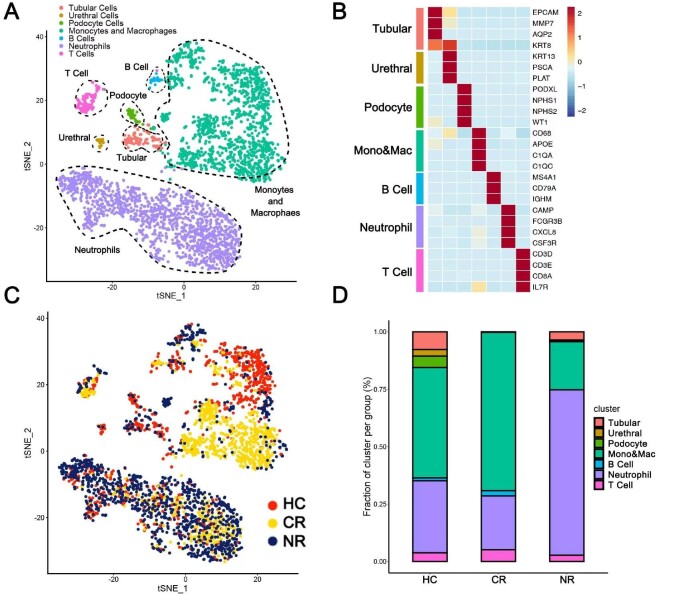
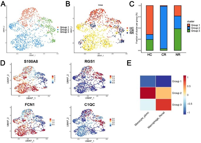
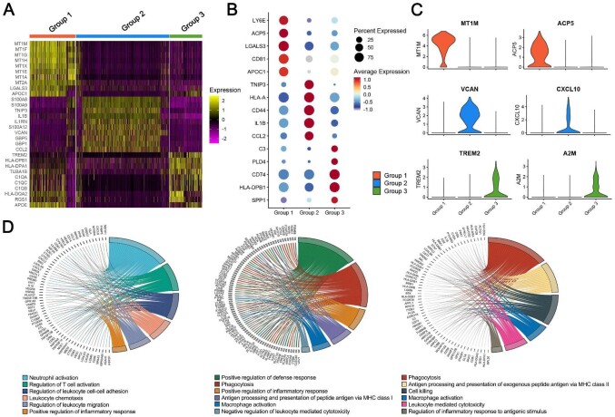
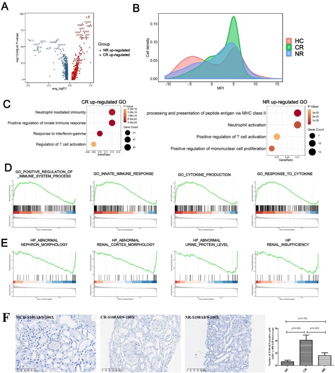
Results: (i) Macrophages were the largest group in urine cells, comprising 48.02%, 68.96% and 20.95% in the HC, CR and NR groups, respectively. (ii) Urinary macrophages expressing FIColin-1 and S100 calcium-binding protein A8 were mainly found in the HC and CR groups, indicating that they were derived from bone marrow and peripheral blood, while the urinary macrophages expressing the regulator of G-protein signaling 1 (RGS1) and HLA-DPA1, mainly found in the NR group, were derived from renal resident macrophages. (iii) In healthy adults, urine macrophages expressed the metallothionein family, indicating that they can regulate anti-inflammatory and proinflammatory functions bidirectionally. In the CR group, the urine macrophages showed strong proinflammatory properties. In the NR group, the urinary macrophages mainly associated with the level of proteinuria and the impaired renal function.
Conclusions: Our study firstly delineated the differences in urinary cell maps between healthy individuals and MN patients with CR or NR outcomes. Not only the origin but also the function of urine macrophages were different in the HC, CR and NR groups.
Keywords: macrophages; membranous nephropathy; proteinuria; urine; urine single-cell sequencing.
© The Author(s) 2023. Published by Oxford University Press on behalf of the ERA.
Conflict of interest statement
This paper has not been presented elsewhere expect in abstract format at ASN Kidney Week 2022. The authors declare that there are no other potential conflicts of interest with respect to the research, authorship or publication of this manuscript.
Figures
Similar articles
-
Urinary soluble HLA class I antigen in patients with minimal change disease: a predictor of steroid response.Nephron. 1998;79(1):44-9. doi: 10.1159/000044990. Nephron. 1998. PMID: 9609461
-
New insights into immune mechanisms underlying response to Rituximab in patients with membranous nephropathy: A prospective study and a review of the literature.Autoimmun Rev. 2016 Jun;15(6):529-38. doi: 10.1016/j.autrev.2016.02.014. Epub 2016 Feb 11. Autoimmun Rev. 2016. PMID: 26876383 Review.
-
Urine complement analysis implies complement activation is involved in membranous nephropathy.Front Med (Lausanne). 2025 Feb 13;12:1515928. doi: 10.3389/fmed.2025.1515928. eCollection 2025. Front Med (Lausanne). 2025. PMID: 40018357 Free PMC article.
-
Renal outcomes of idiopathic and atypical membranous nephropathy in adult Chinese patients: a single center retrospective cohort study.BMC Nephrol. 2021 Apr 22;22(1):148. doi: 10.1186/s12882-021-02348-4. BMC Nephrol. 2021. PMID: 33888083 Free PMC article.
-
IgG4-related membranous nephropathy with high blood and low urine IgG4/IgG ratio: a case report and review of the literature.Clin Rheumatol. 2014 Jan;33(1):145-8. doi: 10.1007/s10067-013-2406-0. Epub 2013 Oct 9. Clin Rheumatol. 2014. PMID: 24105363 Review.
Cited by
-
Integrative profiling of untreated primary membranous nephropathy at the single-cell transcriptome level.Clin Kidney J. 2024 Jun 14;17(7):sfae168. doi: 10.1093/ckj/sfae168. eCollection 2024 Jul. Clin Kidney J. 2024. PMID: 39027416 Free PMC article.
-
Blood and urine biomarkers of disease progression in IgA nephropathy.Biomark Res. 2024 Jul 29;12(1):72. doi: 10.1186/s40364-024-00619-4. Biomark Res. 2024. PMID: 39075557 Free PMC article. Review.
References
-
- Kidney Disease: Improving Global Outcomes (KDIGO) Glomerular Diseases Work Group . KDIGO 2021 Clinical Practice Guideline for the Management of Glomerular Diseases. Kidney Int 2021;100:S1–276. - PubMed
LinkOut - more resources
Full Text Sources
Research Materials
