Top/Middle-Down Characterization of α-Synuclein Glycoforms
- PMID: 38047498
- PMCID: PMC10836061
- DOI: 10.1021/acs.analchem.3c02405
Top/Middle-Down Characterization of α-Synuclein Glycoforms
Abstract
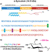
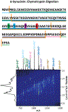
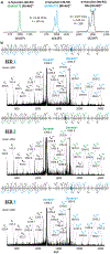
α-Synuclein is an intrinsically disordered protein that plays a critical role in the pathogenesis of neurodegenerative disorders, such as Parkinson's disease. Proteomics studies of human brain samples have associated the modification of the O-linked N-acetyl-glucosamine (O-GlcNAc) to several synucleinopathies; in particular, the position of the O-GlcNAc can regulate protein aggregation and subsequent cell toxicity. There is a need for site specific O-GlcNAc α-synuclein screening tools to direct better therapeutic strategies. In the present work, for the first time, the potential of fast, high-resolution trapped ion mobility spectrometry (TIMS) preseparation in tandem with mass spectrometry assisted by an electromagnetostatic (EMS) cell, capable of electron capture dissociation (ECD), and ultraviolet photodissociation (213 nm UVPD) is illustrated for the characterization of α-synuclein positional glycoforms: T72, T75, T81, and S87 modified with a single O-GlcNAc. Top-down 213 nm UVPD and ECD MS/MS experiments of the intact proteoforms showed specific product ions for each α-synuclein glycoforms associated with the O-GlcNAc position with a sequence coverage of ∼68 and ∼82%, respectively. TIMS-MS profiles of α-synuclein and the four glycoforms exhibited large structural heterogeneity and signature patterns across the 8+-15+ charge state distribution; however, while the α-synuclein positional glycoforms showed signature mobility profiles, they were only partially separated in the mobility domain. Moreover, a middle-down approach based on the Val40-Phe94 (55 residues) chymotrypsin proteolytic product using tandem TIMS-q-ECD-TOF MS/MS permitted the separation of the parent positional isomeric glycoforms. The ECD fragmentation of the ion mobility and m/z separated isomeric Val40-Phe94 proteolytic peptides with single O-GlcNAc in the T72, T75, T81, and S87 positions provided the O-GlcNAc confirmation and positional assignment with a sequence coverage of ∼80%. This method enables the high-throughput screening of positional glycoforms and further enhances the structural mass spectrometry toolbox with fast, high-resolution mobility separations and 213 nm UVPD and ECD fragmentation capabilities.
Conflict of interest statement
The authors declare no competing financial interest.
Figures


References
-
- Stephens AD; Zacharopoulou M; Kaminski Schierle GS Trends Biochem. Sci 2019, 44 (5), 453–466. - PubMed
Publication types
MeSH terms
Substances
Grants and funding
LinkOut - more resources
Full Text Sources
Medical
Molecular Biology Databases

