Review
. 2023 Dec 4;4(1):46.
doi: 10.1186/s43556-023-00156-w.

Structure, function and drug discovery of GPCR signaling

Affiliations
Review

Structure, function and drug discovery of GPCR signaling

Lin Cheng et al. Mol Biomed. .

Abstract

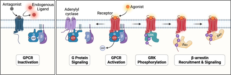
G protein-coupled receptors (GPCRs) are versatile and vital proteins involved in a wide array of physiological processes and responses, such as sensory perception (e.g., vision, taste, and smell), immune response, hormone regulation, and neurotransmission. Their diverse and essential roles in the body make them a significant focus for pharmaceutical research and drug development. Currently, approximately 35% of marketed drugs directly target GPCRs, underscoring their prominence as therapeutic targets. Recent advances in structural biology have substantially deepened our understanding of GPCR activation mechanisms and interactions with G-protein and arrestin signaling pathways. This review offers an in-depth exploration of both traditional and recent methods in GPCR structure analysis. It presents structure-based insights into ligand recognition and receptor activation mechanisms and delves deeper into the mechanisms of canonical and noncanonical signaling pathways downstream of GPCRs. Furthermore, it highlights recent advancements in GPCR-related drug discovery and development. Particular emphasis is placed on GPCR selective drugs, allosteric and biased signaling, polyphamarcology, and antibody drugs. Our goal is to provide researchers with a thorough and updated understanding of GPCR structure determination, signaling pathway investigation, and drug development. This foundation aims to propel forward-thinking therapeutic approaches that target GPCRs, drawing upon the latest insights into GPCR ligand selectivity, activation, and biased signaling mechanisms.


Conflict of interest statement

The authors declare no conflicts of interests.

Figures

Fig. 1
Fig. 1
Scheme of ligand-mediated GPCR inactivation or activation. Agonists bind to GPCRs and trigger downstream G-protein or β-arrestin signaling. Antagonists occupy the agonism-associated pocket and prevent endogenous ligand binding
Fig. 2
Fig. 2
Techniques for GPCR structure determination. a Crystal packing for GPCRs in the presence of fusion proteins. GnRH1R-PGS (left, PDB: 7BR3), GnRH1R: purple, PGS: yellow. ghrelin-BRIL (right, PDB: 7F83), ghrelin: red, BRIL: green. b Schematic diagrams of fusion proteins. The dashed line shows the distances between the N-terminus and C-terminus of the fusion proteins. c Strategies for cryo-EM structure determination of GPCRs. The complex of D1R with Gs and Nb35 (PDB: 7CKZ). mSMO with PGS fusion protein (PDB: 8CXO). hFZD5 with BRIL fusion protein binding with anti-BRIL Fab and anti-Fab Nanobody (PDB: 6WW2). β2AR links ICL3 to engaging BRIL mBRIL and the C-terminus to the K3 helix with an ALFA tag. The complex involves an anti-BRIL Fab, along with a bivalent ‘glue’ molecule containing anti-Fab (NbFab) and anti-ALFA (NbALFA) (PDB: 8J7E and 8JJO). SSTR2 is bound to nanobody6 (PDB: 7UL5)
Fig. 3
Fig. 3
The structural features of Class A GPCRs. a Hallmark for Class A GPCR activation. The cytoplasmic region of TM6 moves outward during receptor activation. b The “push-pull” activation model of the glycoprotein hormone receptor subfamily. c The ECL2 region acts as a “built-in” agonist for GPR52 (PDB: 6LI2). d The N-terminus, ECL2 and ECL3 contribute to the activation of OR51E2 (PDB: 8F76)
Fig. 4
Fig. 4
Activation mechanisms of Class B GPCRs. a Scheme of the “two domain binding model” as the common activation mechanism of Class B1 receptors. PCO371 binds to the intracellular pocket (e.g., PCO371-PTH1R-Gs complex, PDB: 8GW8) of Class B1 receptors and reveals a noncanonical activation mode. c Stalk undergoes a transition from a β-sheet to a partial α helix when mediating autologous activation of aGPCRs. Binding of the α helical stalk in the ligand pocket of ADGRG1 is from the stalk-ADGRG1-miniG13 complex (PDB: 7SF8). d Cortisol binds to the orthosteric pocket of ADGRG3 (GPR97) and triggers receptor activation as the endogenous ligand. The binding of cortisol in the ligand pocket of ADGRG3 is from the cotisol-ADGRG3-miniGo complex (PDB: 7D77)
Fig. 5
Fig. 5
General activation mechanisms of Class C GPCRs. Class C GPCRs are color coded as follows. The mGlu2, mGlu3, CaSR, GB1 and GB2 subunits are blue, light purple, green, yellow and orange, respectively. The endogenous agonists L-Glu, Ca2+, and GABA are indicated by colored balls; the G protein heterotrimers Gαi, Gβ, and Gγ subunits are indicated by purple, red, and fuchsia, respectively. The general conformational alterations of the class C GPCR dimer are shown above, the intracellular view of the TMD is displayed below, and the conformation of key amino acids is presented on the right. a Structures of the mGlu2-3 heterodimer in the inactivated state (PDB: 8JCV), intermediate activated state (PDB: 8JD2), and fully activated state (PDB: 8JD3). b Structures of the mGlu2 homodimer in the inactivated state (PDB: 7EPA), intermediate activated state (PDB: 7EPB), and fully activated state (PDB: 7E9G). c CaSR homodimer in the inactivated state (PDB: 7M3J) and the structure of the activated state (PDB: 7M3G). d Structures of the GABAB heterodimer in the inactivated state (PDB: 6VJM), the intermediate activated state (PDB: 6UO9), and the fully activated state (PDB: 7EB2)
Fig. 6
Fig. 6
Conformational alterations during Class F receptor activation. Class F receptors are composed of a cystine-rich domain (CRD), linker domain (LD) and transmembrane domain (TMD), and the inactive state is shown on the left. When activated by the ligand, TM6 in SMO and FZDs undergoes an outward shift, and a hydrogen bond between the conserved residues R6.32 and W7.55 in the inactive receptor is disrupted. The difference between SMO and FZDs is that the TM6 in SMO exhibits a parallel outward shift, while FZDs achieve a similar displacement of its cytoplasmic segment through a helical kink. This difference may be caused by the conserved residue P6.43 in the FZDs (as opposed to F6.43 in SMO). PDB ID of these structures: active mSMO (6O3C), inactive hSMO (5I7D); active FZD7 (7EVW), inactive FZD5 (6WW2)
Fig. 7
Fig. 7
Structural features of TAS2R46. a Orthosteric binding pocket of TAS2R46 (PDB: 7XP6). The indole ring of W883.32 is horizontally parallel to the benzene ring of strychnine. b The conformational changes of the “toggle switch” Y2416.51 between apo-TAS2R46 (PDB: 7XP4) and strychnine-TAS2R46; its side chain changed from pointing outward to pointing toward the core of the transmembrane helix. c The HPFIL motif in strychnine-TAS2R46
Fig. 8
Fig. 8
The elements in receptors determine the G-protein subtype selectivity. a Classification and sequence alignment of the C-terminus of the Gα subunit family. b, c, d Structural comparison of Gs (left)-D1R (PDB: 7CKW) and Gi (right)-D2R (PDB: 7JVR). b TM5 of D1R is longer than D2R in the cytoplasmic region. c. The distinct motifs in TM5 contribute to G-protein subtype selectivity between D1R and D2R. d Detailed interaction between the residue at the 34.51 position of ICL2 and Gα-proteins in Gs (left)- and Gi (right)-bound dopamine receptors. e Schematic diagrams displaying the propagating pathways that contribute to G-protein subtype-biased signal transduction for GPR120
Fig. 9
Fig. 9
Activation mechanisms of GPCR-arrestin signaling. a Two common GPCR-arrestin binding conformations: “Core” mode (e.g., β1AR-V2RCTerpp-βarr1, PDB: 6TKO) and “Tail” mode (e.g., GCGR-V2RCTerpp-βarr1, PDB: 8JRU). b Arrestin activation mediated by polar interaction between the key phosphorylation of receptor C-terminus and the lysine in the lariat loop. (β1AR-V2RCTerpp-βarr1, PDB:6TKO; M2R-V2RCTerpp-βarr1, PDB:6U1N; GCGR-V2RCTerpp-βarr1, PDB:8JRU; Rhodopsin-arrestin1, PDB: 4ZWJ; 5HT2BR-CTer−truncatedpp-βarr1, PDB: 7SRS; V2RCTerpp-βarr2, PDB: 8I10)
Fig. 10
Fig. 10
Structural basis of µOR G-protein biased agonism. The interactions between the balanced agonist fentanyl (PDB: 8EF5) or the G-protein-biased ligand C5 guano (PDB: 7U2L) and the involved residues in the µOR orthosteric pocket. The guanidine of C5 guano interacts with the key D2.50 in the Na+ binding pocket. Salt-bridge interactions and cation-π interactions are represented by black and red dashed lines, respectively. Schematic diagrams of cAMP inhibition and β-arrestin-2 recruitment on µOR with balanced or G-protein biased ligands are displayed compared with the reference ligand
Fig. 11
Fig. 11
Effects of allosteric modulators on receptor signaling. PAMs (a) and NAMs (b) modulate the effect of orthosteric agonists, while NALs (c) have no influence on receptor signaling mediated by orthosteric agonists. BAMs (d) potentiate signaling pathway a while inhibiting signaling pathway b of the receptor. The dose-dependence curves (below) display the effect of orthosteric agonists in the presence of allosteric modulations. The color saturation indicates the concentrations of allosteric modulators. OA: orthosteric agonist. e The binding sites of BAMs in receptors. SBI-553 (blue) binds to the pocket located at an intracellular region composed of TM6-7 and H8 of NTSR1 (PDB: 8FN0), while compound 9n sits in the upper portion between TM5-6 and ECL2 of HCAR2 (PDB: 8JII)

References

    1. Hauser AS, Kooistra AJ, Munk C, Heydenreich FM, Veprintsev DB, Bouvier M, et al. GPCR activation mechanisms across classes and macro/microscales. Nat Struct Mol Biol. 2021;28(11):879–888. doi: 10.1038/s41594-021-00674-7. - DOI - PMC - PubMed
    1. Garcia-Nafria J, Tate CG. Structure determination of GPCRs: cryo-EM compared with X-ray crystallography. Biochem Soc Trans. 2021;49(5):2345–2355. doi: 10.1042/BST20210431. - DOI - PMC - PubMed
    1. Wingler LM, Lefkowitz RJ. Conformational basis of G protein-coupled receptor signaling versatility. Trends Cell Biol. 2020;30(9):736–747. doi: 10.1016/j.tcb.2020.06.002. - DOI - PMC - PubMed
    1. Zhou Q, Yang D, Wu M, Guo Y, Guo W, Zhong L, et al. Common activation mechanism of class A GPCRs. Elife. 2019 doi: 10.7554/eLife.50279. - DOI - PMC - PubMed
    1. Yang D, Zhou Q, Labroska V, Qin S, Darbalaei S, Wu Y, et al. G protein-coupled receptors: structure- and function-based drug discovery. Signal Transduct Target Ther. 2021;6(1):7. doi: 10.1038/s41392-020-00435-w. - DOI - PMC - PubMed

LinkOut - more resources