Multicenter Study
. 2024 Jan 1;81(1):59-68.
doi: 10.1001/jamaneurol.2023.4398.

Neuronally Derived Extracellular Vesicle α-Synuclein as a Serum Biomarker for Individuals at Risk of Developing Parkinson Disease

Affiliations
Multicenter Study

Neuronally Derived Extracellular Vesicle α-Synuclein as a Serum Biomarker for Individuals at Risk of Developing Parkinson Disease

Shijun Yan et al. JAMA Neurol. .

Abstract

Importance: Nonmotor symptoms of Parkinson disease (PD) often predate the movement disorder by decades. Currently, there is no blood biomarker to define this prodromal phase.

Objective: To investigate whether α-synuclein in neuronally derived serum-extracellular vesicles identifies individuals at risk of developing PD and related dementia.

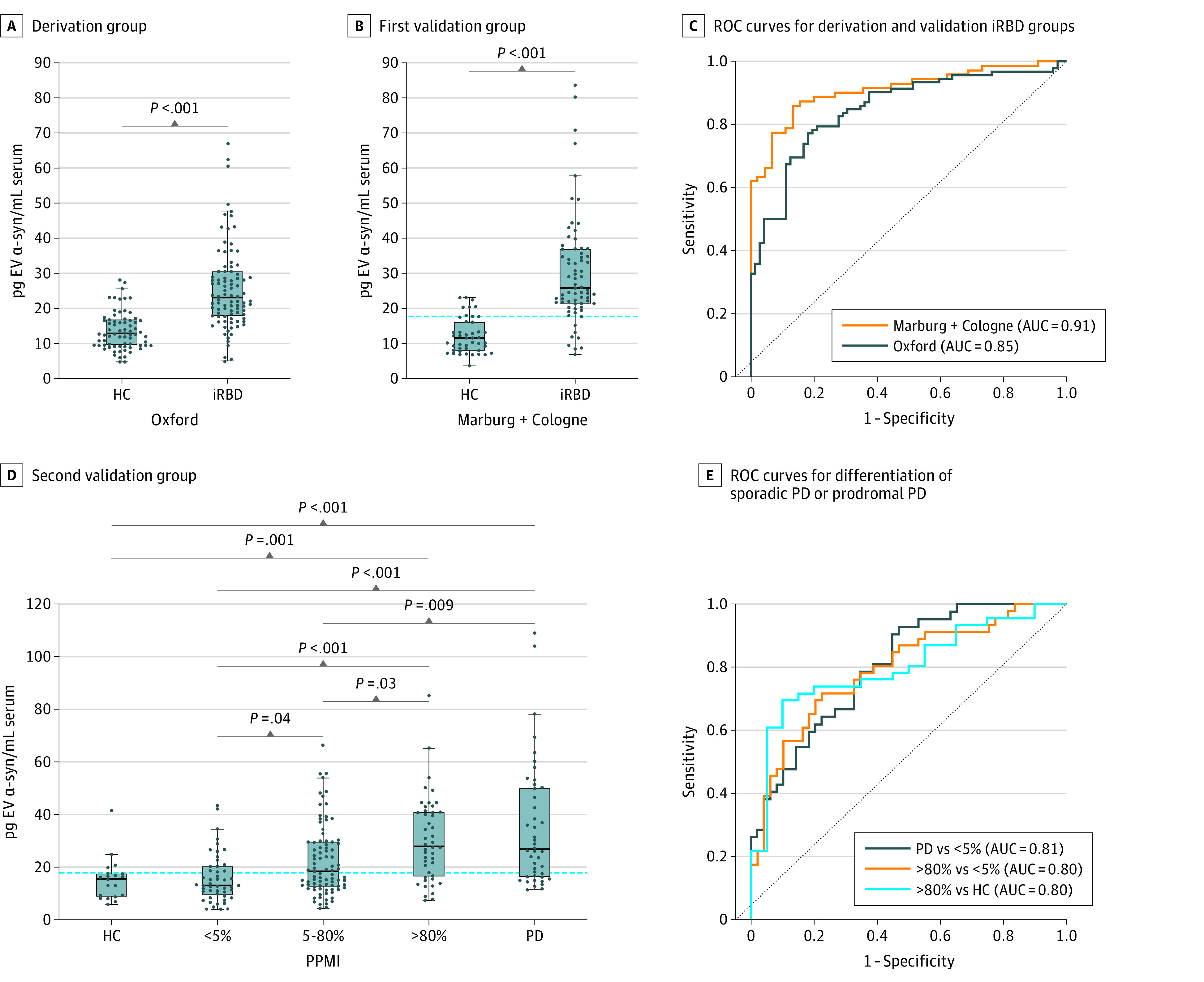
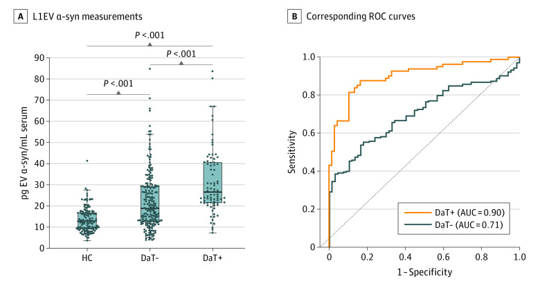
Design, setting, and participants: This retrospective, cross-sectional multicenter study of serum samples included the Oxford Discovery, Marburg, Cologne, and Parkinson's Progression Markers Initiative cohorts. Participants were recruited from July 2013 through August 2023 and samples were analyzed from April 2022 through September 2023. The derivation group (n = 170) included participants with isolated rapid eye movement sleep behavior disorder (iRBD) and controls. Two validation groups were used: the first (n = 122) included participants with iRBD and controls and the second (n = 263) included nonmanifest GBA1N409S gene carriers, participants with iRBD or hyposmia, and available dopamine transporter single-photon emission computed tomography, healthy controls, and patients with sporadic PD. Overall the study included 199 participants with iRBD, 20 hyposmic participants with available dopamine transporter single-photon emission computed tomography, 146 nonmanifest GBA1N409S gene carriers, 21 GBA1N409S gene carrier patients with PD, 50 patients with sporadic PD, and 140 healthy controls. In the derivation group and validation group 1, participants with polysomnographically confirmed iRBD were included. In the validation group 2, at-risk participants with available Movement Disorder Society prodromal markers and serum samples were included. Among 580 potential participants, 4 were excluded due to alternative diagnoses.

Exposures: Clinical assessments, imaging, and serum collection.

Main outcome and measures: L1CAM-positive extracellular vesicles (L1EV) were immunocaptured from serum. α-Synuclein and syntenin-1 were measured by electrochemiluminescence. Area under the receiver operating characteristic (ROC) curve (AUC) with 95% CIs evaluated biomarker performance. Probable prodromal PD was determined using the updated Movement Disorder Society research criteria. Multiple linear regression models assessed the association between L1EV α-synuclein and prodromal markers.

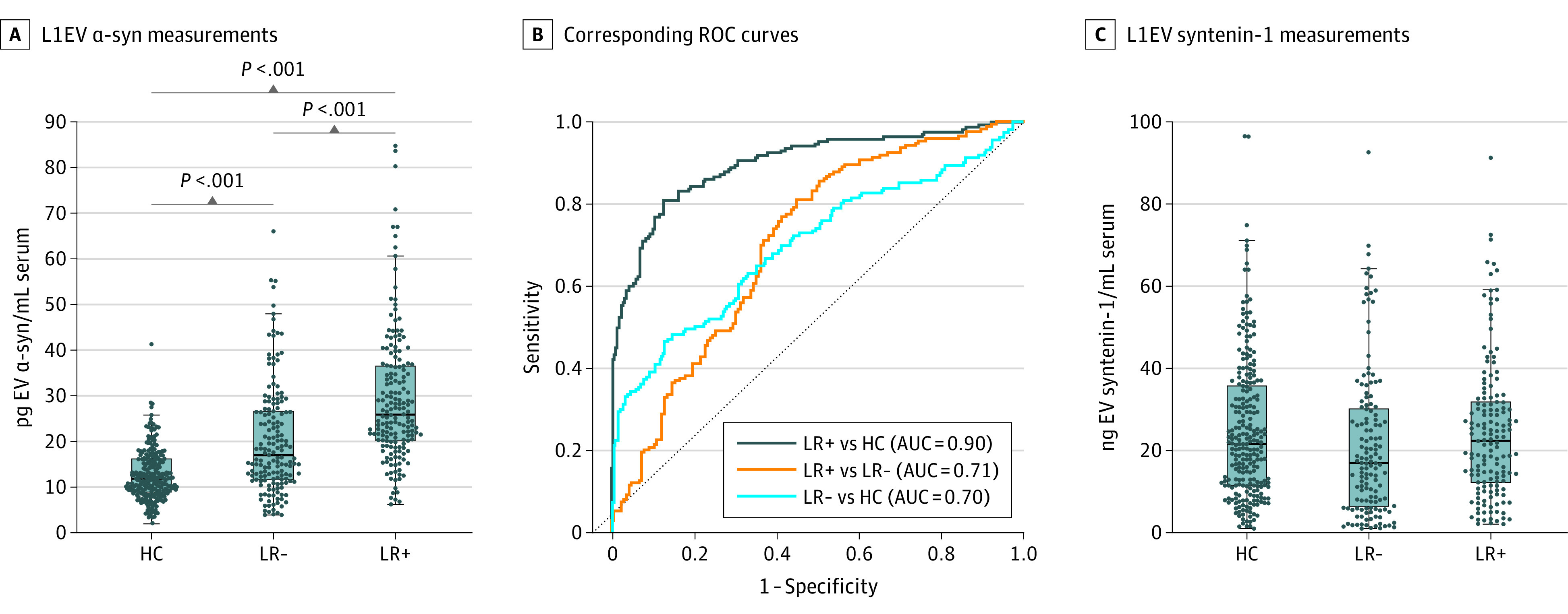
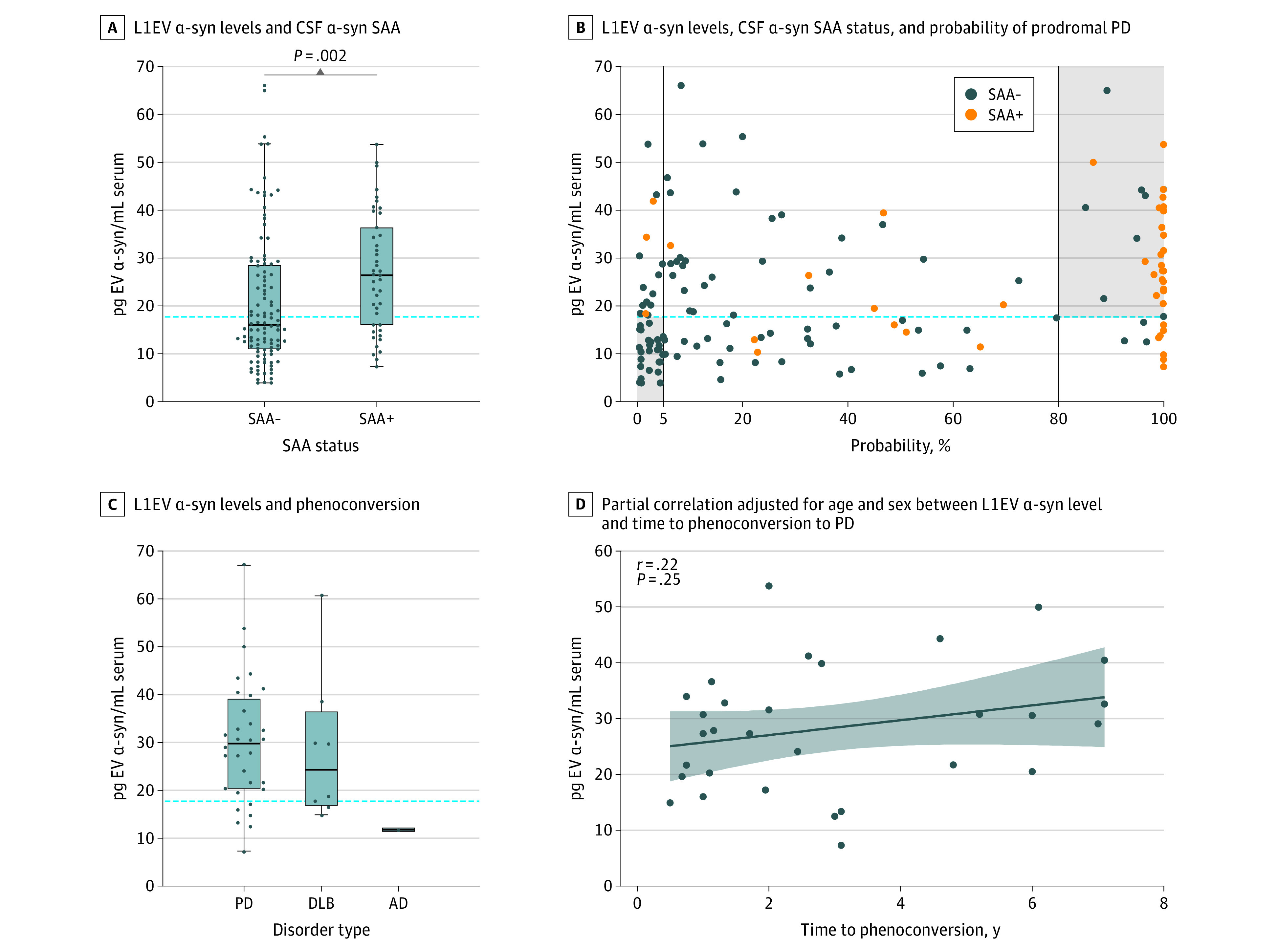
Results: Among 576 participants included, the mean (SD) age was 64.30 (8.27) years, 394 were male (68.4%), and 182 were female (31.6%). A derived threshold of serum L1EV α-synuclein distinguished participants with iRBD from controls (AUC = 0.91; 95% CI, 0.86-0.96) and those with more than 80% probability of having prodromal PD from participants with less than 5% probability (AUC = 0.80; 95% CI, 0.71-0.89). Subgroup analyses revealed that specific combinations of prodromal markers were associated with increased L1EV α-synuclein levels. Across all cohorts, L1EV α-synuclein differentiated participants with more than 80% probability of having prodromal PD from current and historic healthy control populations (AUC = 0.90; 95% CI, 0.87-0.93), irrespective of initial diagnosis. L1EV α-synuclein was increased in at-risk participants with a positive cerebrospinal fluid seed amplification assay and was above the identified threshold in 80% of cases (n = 40) that phenoconverted to PD or related dementia.

Conclusions and relevance: L1EV α-synuclein in combination with prodromal markers should be considered in the stratification of those at high risk of developing PD and related Lewy body diseases.


Conflict of interest statement

Conflict of Interest Disclosures: Dr Janzen reported grants from ParkinsonFonds Deutschland outside the submitted work. Dr Sommerauer reported grants from Else Kröner Fresenius Stiftung and the Köln Fortune Program during the conduct of the study. Dr Marek reported consultant fees from Michael J Fox Foundation, Roche, Invicro, Calico, Neuron23, Inhibikase, Takeda, Prothena, Koneksa, Biohaven, and XingImaging outside the submitted work. Dr Hu reported grants from the National Institute for Health and Care Research Oxford Biomedical Research Centre Grant and Parkinson's UK Cohort Studies Council Grant during the conduct of the study. Dr Oertel reported grants from the Michael J Fox Foundation, ParkinsonFonds Deutschland, and Stichting ParkinsonFonds during the conduct of the study, and grants from Charitable Hertie-Foundation and fees from educational lectures on Parkinson disease from Stada Pharma outside the submitted work. Dr Tofaris reported grants from the Michael J Fox Foundation, Weston Family Foundation, the National Institute for Health, and Care Research Oxford Biomedical Centre, UK Research and Innovation Engineering and Physical Sciences Research Council, and UK Research and Innovation Medical Research Council during the conduct of the study; grants from UK Research and Innovation Medical Research Council outside the submitted work; in addition, Dr Tofaris had a patent for Methodology for L1EV immunocapture pending and a patent for L1CAM extracellular vesicle α-synuclein in Parkinson disease pending. No other disclosures were reported.

Figures

Figure 1.
Figure 1.. L1CAM Extracellular Vesicle (L1EV) α-Synuclein Levels in the Derivation and Validation Groups
Boxplots showing the distribution of L1EV α-synuclein in (A) the derivation group (Oxford Discovery rapid eye movement sleep behavior disorder [iRBD] cohort) and (B) the first validation group (Marburg+Cologne iRBD cohorts) when compared with controls. The derived threshold of 17.75 pg/mL was applied to the first validation group, showing the consistency of the biomarker across iRBD cohorts. (C) Corresponding receiver operating characteristic (ROC) curves for the derivation and validation iRBD groups. (D) The derived threshold of 17.75 pg/mL also distinguished participants with more than 80% probability of having prodromal Parkinson disease (PD) who exhibited increased L1EV α-synuclein levels compared with those with minimal or intermediate risk in the second validation group (Parkinson’s Progression Markers Initiative [PPMI] cohort). Healthy controls and participants with sporadic PD were included as reference groups. (E) Corresponding ROC curves for the differentiation of participants with sporadic PD or more than 80% probability of having prodromal PD from healthy controls or those with less than 5% probability of having prodromal PD. The midline of the box plots indicates the median and the box indicates the 25th and 75th percentiles. Outliers and whiskers were plotted using the Tukey method. Statistical significance was determined by Mann-Whitney or Kruskal-Wallis test. AUC indicates area under the ROC curve; α-syn, α-synuclein; HC, healthy controls.
Figure 2.
Figure 2.. L1CAM Extracellular Vesicle (L1EV) α-Synuclein Measurements in At-Risk Participants With Available Dopamine Transporter Single-Photon Emission Computed Tomography
(A) Boxplot of L1EV α-synuclein measurements and (B) corresponding ROC curves for healthy controls (HC) and at-risk participants with a negative/normal (DaT-) or positive/abnormal (DaT+) dopamine transporter single-photon emission computed tomography. The midline of the box plots indicates the median and the box indicates the 25th and 75th percentiles. Outliers and whiskers were plotted using the Tukey method. Statistical significance was determined by Kruskal-Wallis test. AUC indicates area under the receiver operating characteristic curve; α-syn, α-synuclein; ROC, receiver operating characteristic.
Figure 3.
Figure 3.. L1CAM Extracellular Vesicle (L1EV) α-Synuclein Levels Are Increased in At-Risk Participants With Positive Likelihood Ratio (LR) of Developing Parkinson Disease (PD)
(A) Boxplot of L1EV α-synuclein (α-syn) measurements and (B) corresponding receiver operating characteristic (ROC) curves for those with positive likelihood ratio (LR+) or negative likelihood ratio (LR-) of developing PD based on the updated Movement Disorder Society prodromal research criteria or current and historic healthy controls (HC). (C) Boxplot showing measurements for L1EV syntenin-1, which is a generic extracellular vesicles-associated marker. The midline of the box plots indicates the median and the box indicates the 25th and 75th percentiles. Outliers and whiskers were plotted using the Tukey method. Statistical significance was determined by Kruskal-Wallis test.
Figure 4.
Figure 4.. L1CAM Extracellular Vesicle (L1EV) α-Synuclein Levels and Cerebrospinal Fluid (CSF) Seed Amplification Assay (SAA) or Phenoconversion
(A) Serum L1EV α-synuclein (α-syn) levels were higher in participants with positive CSF α-synuclein SAA. The midline of the box plots indicates the median and the box indicates the 25th and 75th percentiles. The horizontal dotted line represents the derived threshold (17.75 pg/mL) of L1EV α-synuclein. Outliers and whiskers were plotted using the Tukey method. Statistical significance was determined by Mann-Whitney test. (B) Association between serum L1EV α-synuclein levels, CSF α-synuclein SAA status, and probability of having prodromal Parkinson disease (PD). The horizontal dotted line represents the derived threshold (17.75 pg/mL) of L1EV α-synuclein. The vertical lines represent the age and sex adjusted probability of having prodromal PD at less than 5% and more than 80%. (C) Boxplot of serum L1EV α-synuclein levels in at-risk participants who phenoconverted to PD (n = 32), dementia with Lewy bodies (DLB) (n = 8), and Alzheimer disease (AD) (n = 1). The horizontal dotted line represents the derived threshold (17.75 pg/mL) of L1EV α-synuclein. The midline of the box plots indicates the median and the box indicates the 25th and 75th percentiles. Outliers and whiskers are plotted using the Tukey method. (D) Partial correlation adjusted for age and sex between serum L1EV α-synuclein levels and time to phenoconversion to PD. Least squares regression line with 95% CI was plotted and Pearson coefficient was reported.

Comment in

References

    1. Heinzel S, Berg D, Gasser T, Chen H, Yao C, Postuma RB; MDS Task Force on the Definition of Parkinson’s Disease . Update of the MDS research criteria for prodromal Parkinson’s disease. Mov Disord. 2019;34(10):1464-1470. doi: 10.1002/mds.27802 - DOI - PubMed
    1. Postuma RB, Iranzo A, Hu M, et al. Risk and predictors of dementia and parkinsonism in idiopathic REM sleep behaviour disorder: a multicentre study. Brain. 2019;142(3):744-759. doi: 10.1093/brain/awz030 - DOI - PMC - PubMed
    1. Sidransky E, Nalls MA, Aasly JO, et al. Multicenter analysis of glucocerebrosidase mutations in Parkinson’s disease. N Engl J Med. 2009;361(17):1651-1661. doi: 10.1056/NEJMoa0901281 - DOI - PMC - PubMed
    1. Jiang C, Fu Y, Liu G, Shu B, Davis J, Tofaris GK. Multiplexed profiling of extracellular vesicles for biomarker development. Nanomicro Lett. 2021;14(1):3. doi: 10.1007/s40820-021-00753-w - DOI - PMC - PubMed
    1. Yan S, Jiang C, Davis JJ, Tofaris GK. Methodological considerations in neuronal extracellular vesicle isolation for α-synuclein biomarkers. Brain. 2023;awad169. doi: 10.1093/brain/awad169 - DOI - PMC - PubMed

Publication types