. 2024 Feb 8;143(6):507-521.
doi: 10.1182/blood.2023021054.

A dual-receptor T-cell platform with Ab-TCR and costimulatory receptor achieves specificity and potency against AML

Affiliations

A dual-receptor T-cell platform with Ab-TCR and costimulatory receptor achieves specificity and potency against AML

Tao Dao et al. Blood. .

Abstract

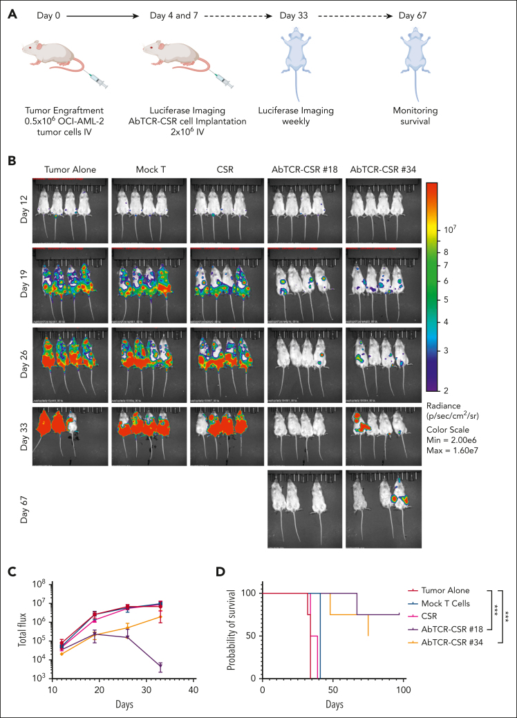
Chimeric antigen receptor T-cell (CAR T) therapy has produced remarkable clinical responses in B-cell neoplasms. However, many challenges limit this class of agents for the treatment of other cancer types, in particular the lack of tumor-selective antigens for solid tumors and other hematological malignancies, such as acute myeloid leukemia (AML), which may be addressed without significant risk of severe toxicities while providing sufficient abundance for efficient tumor suppression. One approach to overcome this hurdle is dual targeting by an antibody-T-cell receptor (AbTCR) and a chimeric costimulatory signaling receptor (CSR) to 2 different antigens, in which both antigens are found together on the cancer cells but not together on normal cells. To explore this proof of concept in AML, we engineered a new T-cell format targeting Wilms tumor 1 protein (WT1) and CD33; both are highly expressed on most AML cells. Using an AbTCR comprising a newly developed TCR-mimic monoclonal antibody against the WT1 RMFPNAPYL (RMF) epitope/HLA-A2 complex, ESK2, and a secondary CSR comprising a single-chain variable fragment directed to CD33 linked to a truncated CD28 costimulatory fragment, this unique platform confers specific T-cell cytotoxicity to the AML cells while sparing healthy hematopoietic cells, including CD33+ myelomonocytic normal cells. These data suggest that this new platform, named AbTCR-CSR, through the combination of a AbTCR CAR and CSR could be an effective strategy to reduce toxicity and improve specificity and clinical outcomes in adoptive T-cell therapy in AML.


Conflict of interest statement

Conflict-of-interest disclosure: D.A.S. serves on a board of, or has equity in, or income from, Lantheus, Sellas, Iovance, Pfizer, Actinium Pharmaceuticals, OncoPep, Repertoire, Sapience, Atengen, and Eureka Therapeutics. A.K. is a consultant for Novartis, Rgenta, and Blueprint Medicines. T.D. is a consultant for Eureka Therapeutics. A.D. has patents covering CD33 binders for CAR T cells and bispecifics and is on a board, or has equity, or income from NomoCan Pharmaceuticals and PromiCell Therapeutics Inc. The remaining authors declare no competing financial interests.

The current affiliation of A.Y.C. is Regeneron, Tarrytown, NY.

Figures

None
Graphical abstract
Figure 1.
Figure 1.
Binding of the ESK2 clone no. 18 and no. 34 and specificity of the epitope. (A) Binding of clones to T2 cells pulsed with or without indicated peptides. WT1 RMF or mutant peptides at a concentration of 50 μg/mL were pulsed onto T2 cells overnight. Cells were washed and stained with BiTEs of ESK2 clone no. 18 and no. 34 or ESK1 at 1 μg/mL. T2 cells alone or pulsed with irrelevant HLA-A2–binding peptide HPV-E7 (39) were used as controls. (B) In parallel, HLA-A2 expression was determined by staining the cells with the anti–HLA-A2 mAb BB7 clone. (C) WT1 RMF sequences were substituted with alanine at positions 1, 3, 4, 5, 7, and 8, or with glycine at position 6 indicated as WT1-A1 to WT1-A8 or WT1-G6, respectively (supplemental Table 1) and the binding of (C) ESK2-clone no. 18 and no. 34 and (D) HLA-A2 was determined by flow cytometric analysis. The data are representative results from 6 similar experiments. HPV, human papilloma virus.
Figure 2.
Figure 2.
Specific recognition and killing of tumor cells by clone no. 34 BiTE. Recognition and cytolytic activity of the naturally presented WT1 RMF/A2 complex on the tumor cell surface by the ESK2 clone no. 18 and no. 34 BiTEs were probed. (A) MAC-1 T-cell lymphoma, (B) JMN mesothelioma, or (C) SW-620 colon cancer cell lines were incubated with PBMCs at an effector-to-target (E:T) ratio of 20:1, in the presence or absence of BiTEs and control BiTEs at the indicated concentrations overnight, and the cytotoxicity was measured by BLI. (D) BV173 CLL, (E) SET-2 AML, (F) or HL-60 AML cell lines were incubated with PBMCs at an E:T ratio of 20:1, in the presence or absence of clone no. 34 BiTE or control at the concentrations of 1 μg/mL, 0.3 μg/mL, or 0.1 μg/mL for 5 hours and the cytotoxicity was measured using a 51Cr-release assay. The mean shown is the average of triplicate microwells ± standard deviation. The data are representative of 10 experiments. The effector cells were used from several different donors; whereas differences among the experimental groups were similar, the baselines were variable among the individuals; therefore, only representative data are shown.
Figure 3.
Figure 3.
Cytotoxicity of AbTCR or AbTCR-CSR against AML cells. (A) A cartoon depicting the design of the AbTCR and the costimulatory receptor CSR only, which, together, form the AbTCR-CSR. AbTCR is the primary signal, CSR is the secondary signal. The combination is known as ARTEMIS 2.0. ESK2 AbTCR or ESK2 AbTCR-CSR T cells (B-C) were incubated with the indicated leukemia target cells at an E:T ratio of 1:1, overnight. The cytotoxicity was measured by luciferase-based assay. Each data point was the average of triplicate cultures ± standard deviation, and representative of 3 similar experiments with different donors. To test whether the cytotoxicity of AbTCR-CSR T cells required the primary signal, (D) AML-14, (E) HL-60, or (F) SKOV3/A2 cell lines were labeled with CFSE, washed, and incubated with mock T cells, CSR cells (ESK2 is replaced with an irrelevant Fab AbTCR as the recognition receptor), ESK2 AbTCR-CSR T cells no. 18, or ESK2 AbTCR-CSR T cells no. 34 at an E:T ratio of 1:1, overnight. The cells were harvested, washed, and stained with mAb to CD33 and subjected to flow cytometry. The analysis was performed by gating on larger tumor cells based on forward and side scatters, and the percentage of CD33+ cells was shown in the CFSE+ target population.
Figure 4.
Figure 4.
No cytotoxicity of AbTCR-CSR was observed against normal PBMCs. Mock T cells, CSR (with irrelevant Fab AbTCR), ESK2 AbTCR-CSR clone no. 18, or AbTCR-CSR clone no. 34 were incubated with (A) CFSE-labeled AML-14 (as a positive control), (B) PBMCs from HLA-A2 positive donor, or (C) negative donor at an E:T ratio of 1:1, overnight. The cells were harvested, washed, and stained with mAb to CD33 and zombie dye (to stain dead cells) and analyzed by flow cytometry. (D) The flow plots show a representative profile of CD33 vs CFSE double-positive cells and the percentage of CD33+ cell death was summarized. (E) In the same experiments, CFSE+ target PBMCs were also stained with CD3 and CD19 and the percentage of each lineage cells are shown in bar graphs. The data represent 1 of 4 separate experiments with different donors.
Figure 5.
Figure 5.
No cytotoxicity of AbTCR-CSR against neutrophils from normal donors. (A) Neutrophils from HLA-A∗02:01–positive donor or (B) HLA-A∗02:01–negative donor were isolated using a human whole-blood neutrophil isolation kit (Miltenyi) and incubated with mock T cells, CSR only, or AbTCR-CSR clone no. 18 or no. 34 at an E: T ratio of 1:1, overnight. The cells were stained with CD15 (for neutrophils) or CD3 (AbTCR-CSR T cells). The percentage of CD15+CFSE+ cells after coculture are shown in panel C and the positive control cell line AML-14 is shown in panel D. Because AML-14 cells do not express CD15, CD33 was used for its marker. The data represents 1 set of results from 4 independent donors.
Figure 6.
Figure 6.
AbTCR-CSR therapeutic trial in an AML animal model. (A) Human AML cell line AML-14 cells (5 million) were injected IV into NSG mice and leukemia engraftment was confirmed on day 13 by bioluminescent imaging. (B) AbTCR-CSR clone no. 18, AbTCR-CSR clone no. 34, or control mock T cells (6 million cell per mouse) were injected IV, and tumor burden was monitored by BLI on days 18 and 21. (C) Mean tumor burden was calculated by summing the luminescence signal of each mouse and presenting the average signal for each group (n = 5 per group). (D) Survival of mice from experimental groups; a comparison of the differences of clone no. 18 or no. 34 vs the 2 control groups was performed using the Mantel-Cox test; ∗∗∗∗P < .0001.
Figure 7.
Figure 7.
AbTCR-CSR therapeutic trial in an OCI-AML-2 human AML animal model. OCI-AML-2 cells (0.5 million) were injected IV into NSG mice. Groups were blindly assigned to treatment groups. (A) Mock T cells, CSR-only cells, or ESK2 AbTCR-CSR cells (1 million) were injected IV on day 4 and day 12 after tumor cell injection. (B) Tumor burden was assessed by BLI on the indicated days. (C) Mean tumor burden was calculated by summing the luminescent signal of each mouse, and the average signal for each group (n = 4 per group) was plotted. (D) Survival of mice from experimental groups. A comparison of the differences of clone no. 18 or no. 34 vs the 2 control groups was calculated using the Mantel-Cox test; ∗∗∗P < .001. Max, maximum; min, minimum.

Comment in

References

    1. Brentjens RJ, Davila ML, Riviere I, et al. CD19-targeted T cells rapidly induce molecular remissions in adults with chemotherapy-refractory acute lymphoblastic leukemia. Sci Transl Med. 2013;5(177) - PMC - PubMed
    1. Sadelain M, Brentjens R, Rivière I. The basic principles of chimeric antigen receptor design. Cancer Discov. 2013;3(4):388–398. - PMC - PubMed
    1. Brudno JN, Kochenderfer JN. Recent advances in CAR T-cell toxicity: mechanisms, manifestations and management. Blood Rev. 2019;34:45–55. - PMC - PubMed
    1. Cummins KD, Gill S. Will CAR T cell therapy have a role in AML? Promises and pitfalls. Semin Hematol. 2019;56(2):155–163. - PubMed
    1. Marofi F, Rahman HS, Al-Obaidi ZMJ, et al. Novel CAR T therapy is a ray of hope in the treatment of seriously ill AML patients. Stem Cell Res Ther. 2021;12(1):465. - PMC - PubMed

Publication types

Substances