. 2024 Apr;11(2):1249-1257.
doi: 10.1002/ehf2.14614. Epub 2023 Dec 4.

Previous cardiovascular injury is a prerequisite for immune checkpoint inhibitor-associated lethal myocarditis in mice

Affiliations

Previous cardiovascular injury is a prerequisite for immune checkpoint inhibitor-associated lethal myocarditis in mice

Nestor Rubio-Infante et al. ESC Heart Fail. 2024 Apr.

Abstract

Aims: Immune checkpoint inhibitors (ICIs) are antineoplastic drugs designed to activate the immune system's response against cancer cells. Evidence suggests that they may lead to immune-related adverse events, particularly when combined (e.g., anti-CTLA-4 plus anti-PD-1), sometimes resulting in severe conditions such as myocarditis. We aimed to investigate whether a previously sustained cardiac injury, such as pathological remodelling due to hypertension, is a prerequisite for ICI therapy-induced myocarditis.

Methods: We evaluated the cardiotoxicity of ICIs in a hypertension (HTN) mouse model (C57BL/6). Weekly doses were administered up to day 21 after the first administration. Our analysis encompassed the following parameters: (i) survival and cardiac pathological remodelling, (ii) cardiac function assessed using pressure-volume (PV)-loops, with brain natriuretic peptide (BNP) serving as a marker of haemodynamic dysfunction and (iii) cardiac inflammation (cytokine levels, infiltration, and cardiac antigen autoantibodies).

Results: After the first administration of ICI combined therapy, the treated HTN group showed a 30% increased mortality (P = 0.0002) and earlier signs of hypertrophy and pathological remodelling compared with the untreated HTN group. BNP (P = 0.01) and TNF-α (<0.0001) increased 2.5- and 1.7-fold, respectively, in the treated group, while IL-6 (P = 0.8336) remained unchanged. Myocarditis only developed in the HTN group treated with ICIs on day 21 (score >3), characterised by T cell infiltration and increased cardiac antigen antibodies (86% showed a titre of 1:160). The control group treated with ICI was unaffected in any evaluated feature.

Conclusions: Our findings indicate that pre-existing sustained cardiac damage is a necessary condition for ICI-induced myocarditis.

Keywords: Heart damage; Heart failure; Immune checkpoint inhibitors; Inflammation; Myocarditis.


Conflict of interest statement

None declared.

Figures
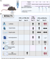
Figure 1
Figure 1
(A) Schematic representation of the HTN + ICIs dual therapy mouse model. Mice were administered with L‐NAME for 5 weeks, represented by the black lines, and AngII 1 week after for 4 weeks, indicated by the purple lines. Blue circles represent the day of the weekly ICI dual treatment (anti‐CTL‐4 and anti‐PD‐1). Mice were analysed on days 7 and/or 21 of ICI treatment (black circles). (B) Comparative results from the CTRL group versus CTRL + ICIs group (day 7 and day 21), CTRL group versus HTN group (day 21) and HTN versus HTN + ICIs groups (day 7 and day 21). (1) In the absence of inflammatory stimuli, although some circulating cardiac autoantibodies (AAbs) might be present and increased by ICIs, cardiac tissue remains without signs of inflammation or damage. (2) Under sustained cardiac injury induced by L‐NAME and angiotensin II (ANG II), cardiac dysfunction and autoantigens might be released without evidence of myocarditis. (3) Increased mortality due to dual ICI therapy in the HTN group associated with evidence of myocarditis and increased IgG cardiac autoantigens. Created with BioRender.com.
Figure 2
Figure 2
ICIs increase mice mortality and cardiac remodelling from the first treatment dose. (A) Kaplan Meir curves of mice survival (n = 8–17). (B) Body weight gain (n = 6–9). (C) Heart weight/tibia length [HW/TL; (n = 6–13)]. (D) Myocyte area normalized to the control group (n = 4–9). The dotted line are guides to the eye for each experimental group. Data are presented as mean ± SEM. The logrank test was used for survival curves comparison and ANOVA test with post hoc Holm–Sidak multiple comparisons test. *P ≤ 0.05, *versus CTRL, #versus CTRL + ICIs and &versus HTN.
Figure 3
Figure 3
One dose of ICIs treatment triggers cardiac dysfunction associated with hypertrophy. (A) Representative images of PV‐Loop recordings with preload impediment by transiently occluding the inferior vena cava after 7 days of ICIs administration are shown. The first loop represents a steady‐state loop. The following hemodynamic parameters are displayed for each group: (B) stroke volume (μL), (C) cardiac output (mL/min), (D) ejection fraction (%), and (E) EDPVR slope. (F) BNP mRNA relative expression by qPCR from the cardiac apical section obtained from each group and normalized with GAPDH (n = 5–8). The dotted line are guides to the eye for each experimental group. Data are presented as mean ± SEM; ANOVA test with post hoc Holm–Sidak multiple comparisons test. *P ≤ 0.05, **P ≤ 0.01, ***P ≤ 0.001.
Figure 4
Figure 4
Inflammation is exacerbated from the first treatment dose with ICIs, triggering myocarditis with evidence of T‐cell infiltration and increased autoantibodies. mRNA relative expression by qPCR from the cardiac apical section obtained from each group and normalised with GAPDH gene expression (n = 5–8) were assessed for (A) TNF‐α and (B) IL‐6. (C) Representative images of histological H&E staining analysis of immune cell infiltration by myocarditis score determination, 20×. (D) Immunohistochemistry images of T cells (CD3 positive cells), 40×. (E) Autoantibodies from each group identified by indirect ELISA (1:80 dilution factor) are shown (n = 3–7). (F) Antibody titre on day 21 of different mice per group. The dotted line are guides to the eye for each experimental group. Data are presented as mean ± SEM; ANOVA test with Tukey's (gene expression) or post hoc Holm–Sidak (autoantibodies) multiple comparisons test. *P ≤ 0.05, versus CTRL, #versus CTRL + ICIs and &versus HTN.

References

    1. Rubio‐Infante N, Ramírez‐Flores YA, Castillo EC, Lozano O, García‐Rivas G, Torre‐Amione G. Cardiotoxicity associated with immune checkpoint inhibitor therapy: A meta‐analysis. Eur J Heart Fail 2021;23:1739‐1747. doi:10.1002/ejhf.2289 - DOI - PubMed
    1. Rubio‐Infante N, Ramírez‐Flores YA, Castillo EC, Lozano O, García‐Rivas G, Torre‐Amione G. A systematic review of the mechanisms involved in immune checkpoint inhibitors cardiotoxicity and challenges to improve clinical safety. Front Cell Dev Biol 2022;10:851032. doi:10.3389/fcell.2022.851032 - DOI - PMC - PubMed
    1. Buchbinder EI, Desai A. CTLA‐4 and PD‐1 pathways: Similarities, differences, and implications of their inhibition. Am J Clin Oncol 2016;39:98‐106. doi:10.1097/COC.0000000000000239 - DOI - PMC - PubMed
    1. Kushnareva E, Kushnarev V, Artemyeva A, Mitrofanova L, Moiseeva O. Myocardial PD‐L1 expression in patients with ischemic and non‐ischemic heart failure. Front Cardiovasc Med 2021;8:759972. doi:10.3389/fcvm.2021.759972 - DOI - PMC - PubMed
    1. Tay WT, Fang Y‐H, Beh ST, Liu Y‐W, Hsu L‐W, Yen C‐J, et al. Programmed cell Death‐1: Programmed cell death‐ligand 1 interaction protects human cardiomyocytes against T‐cell mediated inflammation and apoptosis response in vitro. Int J Mol Sci 2020;21:2399. doi:10.3390/ijms21072399 - DOI - PMC - PubMed

Substances