. 2023 Dec;25(12):1821-1832.
doi: 10.1038/s41556-023-01274-x. Epub 2023 Dec 4.

Prostate lineage-specific metabolism governs luminal differentiation and response to antiandrogen treatment

Affiliations

Prostate lineage-specific metabolism governs luminal differentiation and response to antiandrogen treatment

Jenna M Giafaglione et al. Nat Cell Biol. 2023 Dec.

Abstract

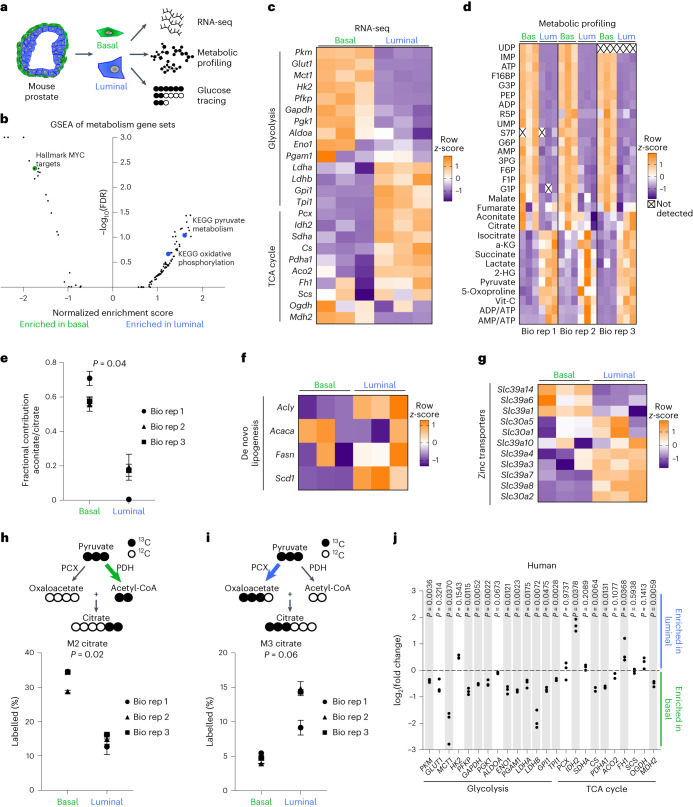
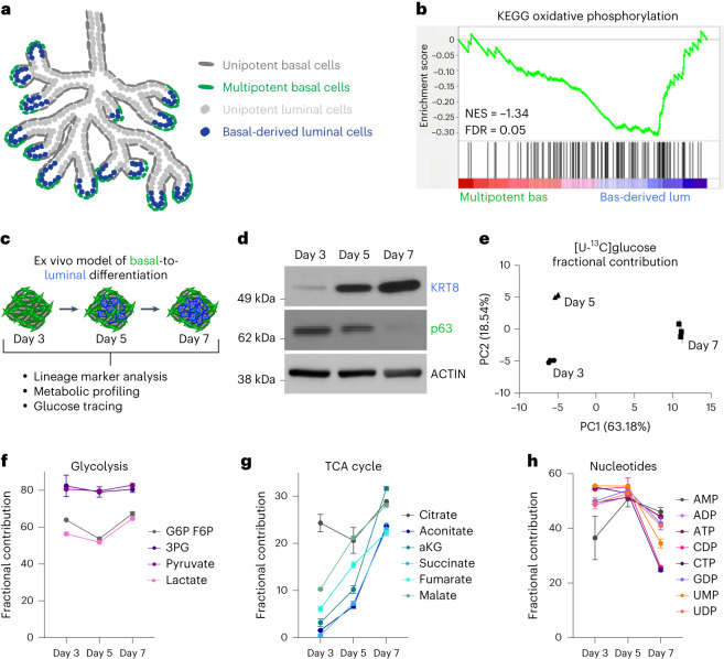
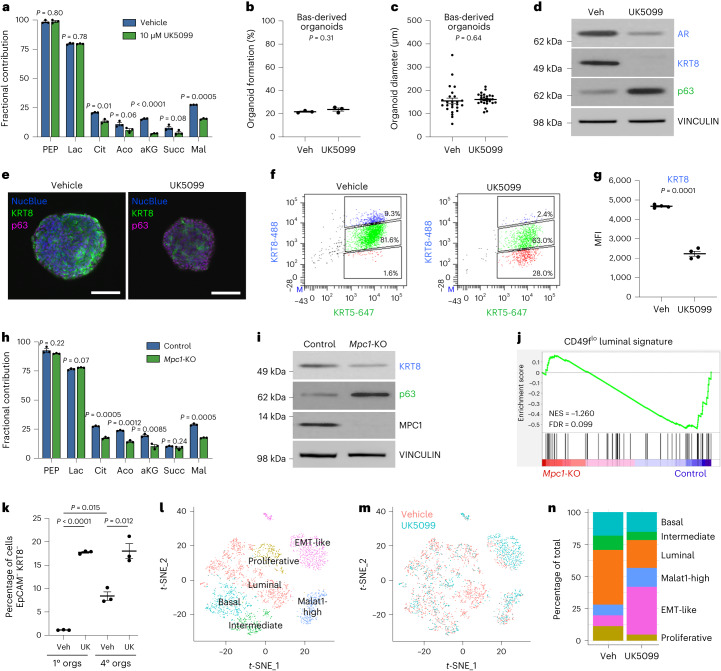
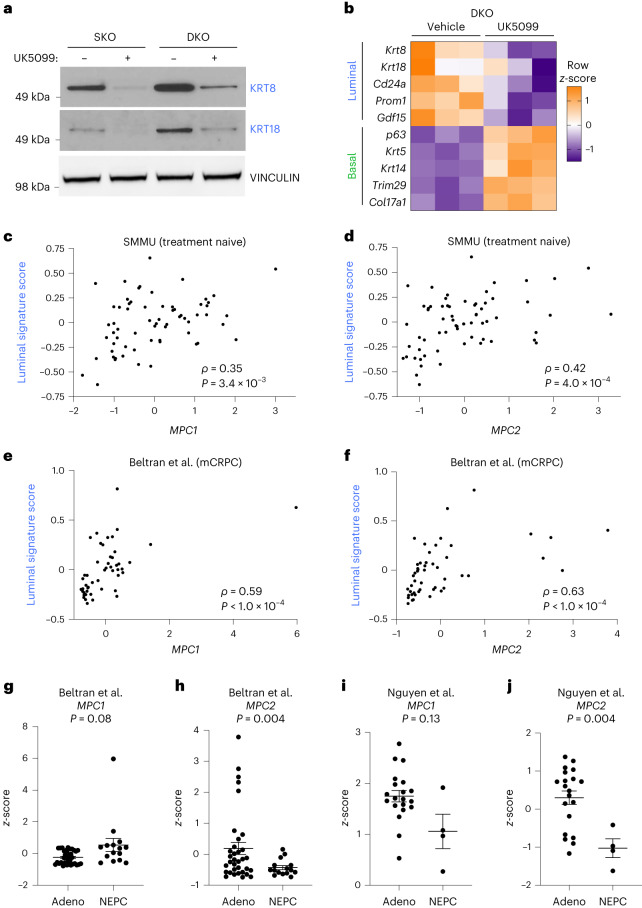
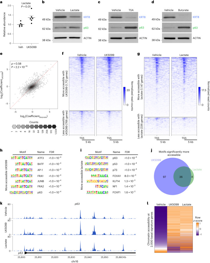
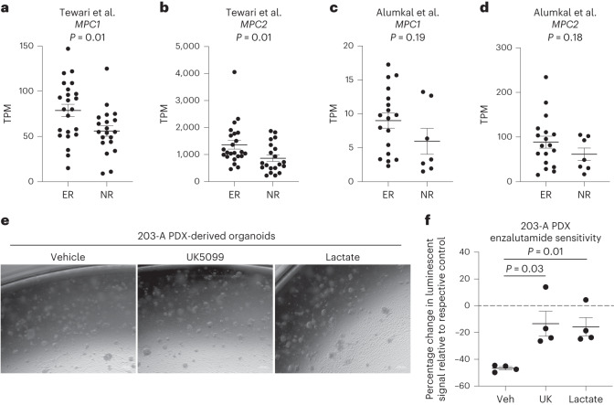
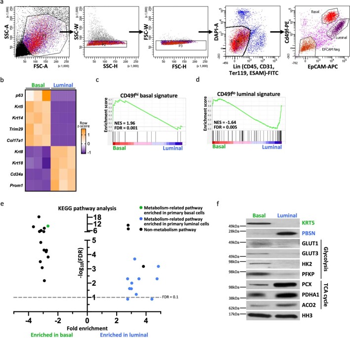
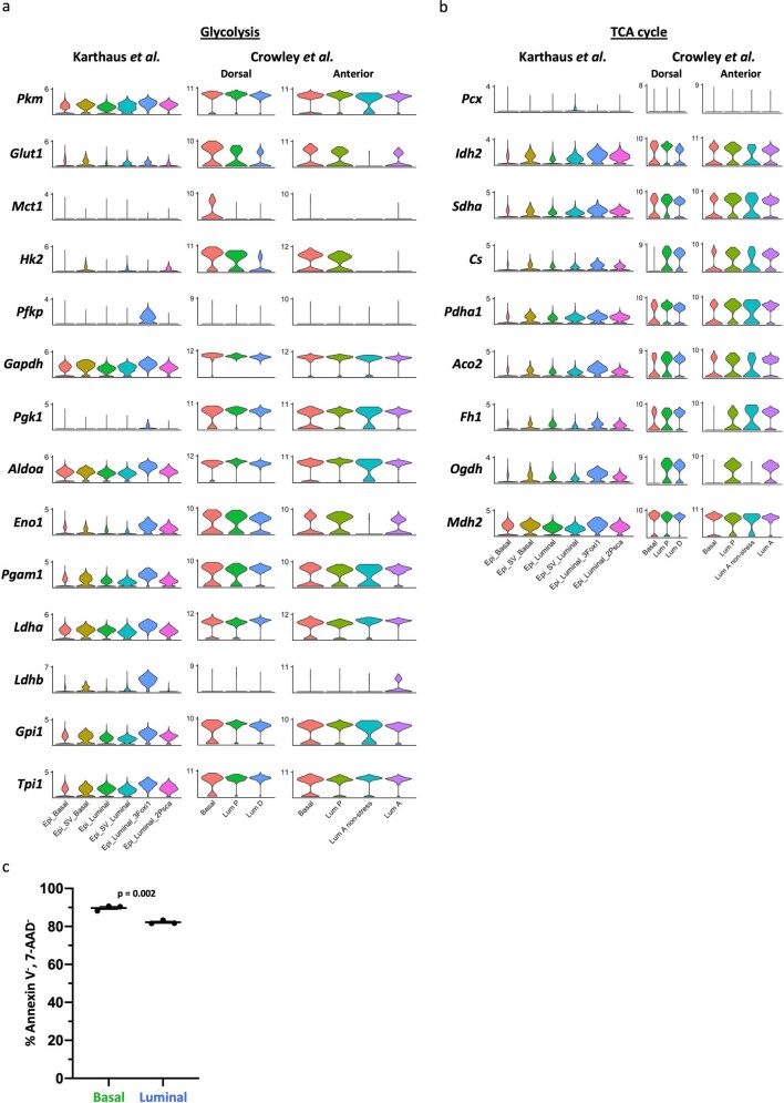
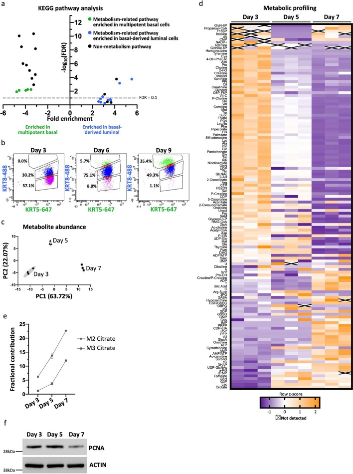
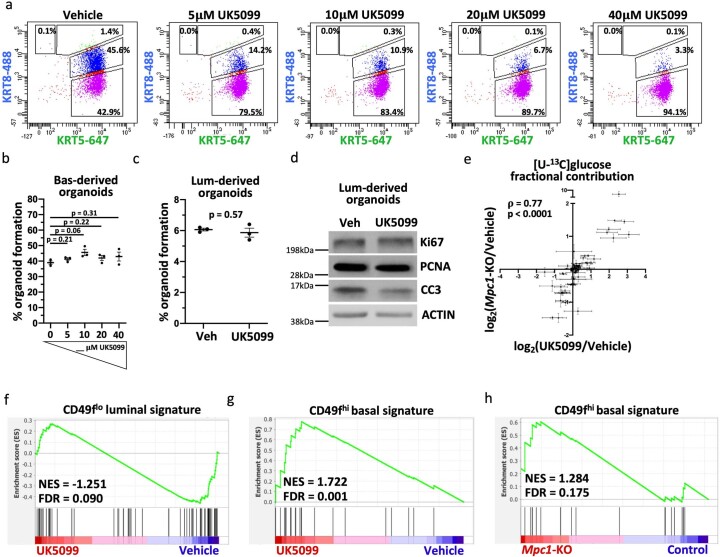
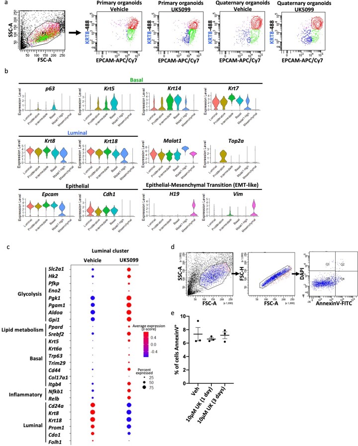
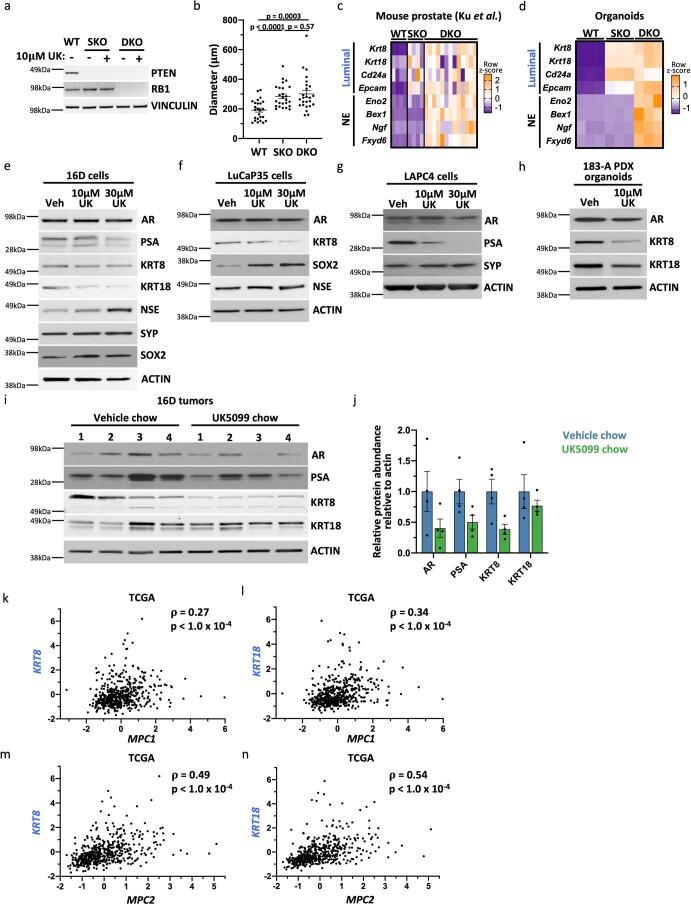
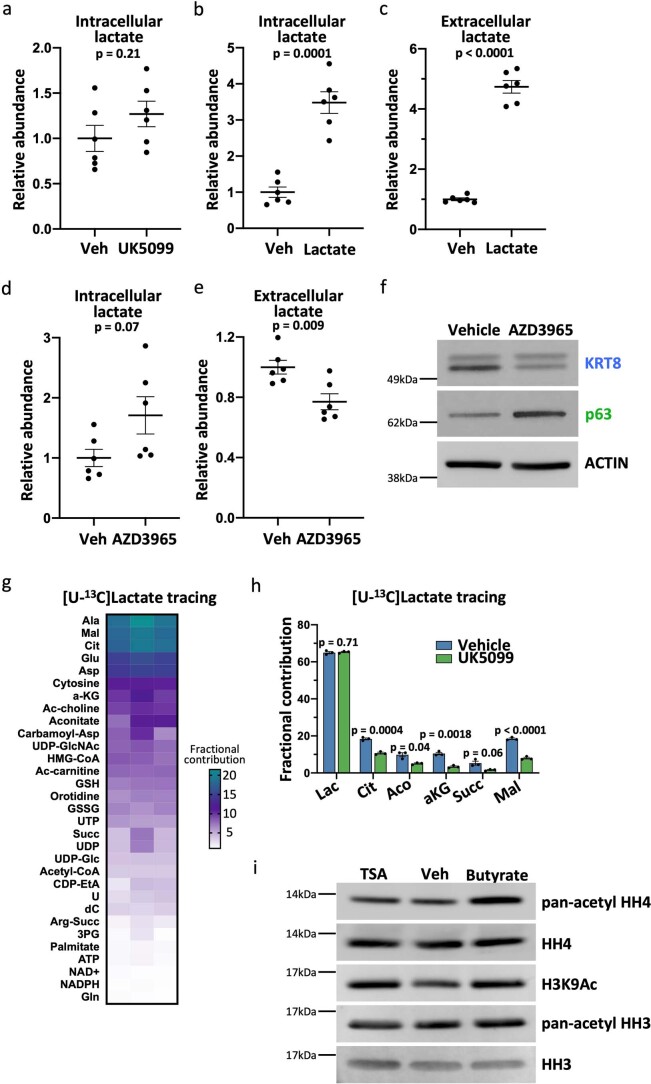
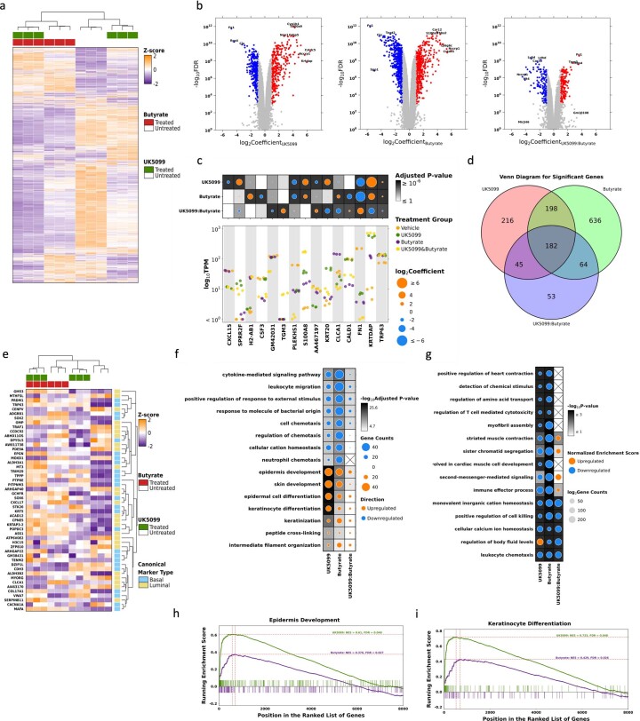
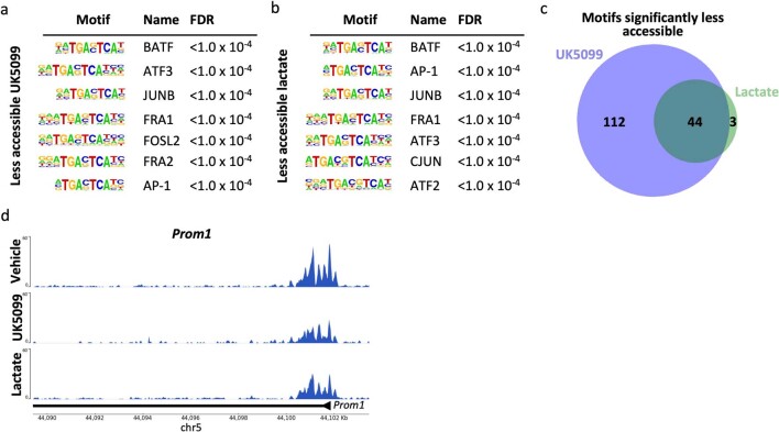
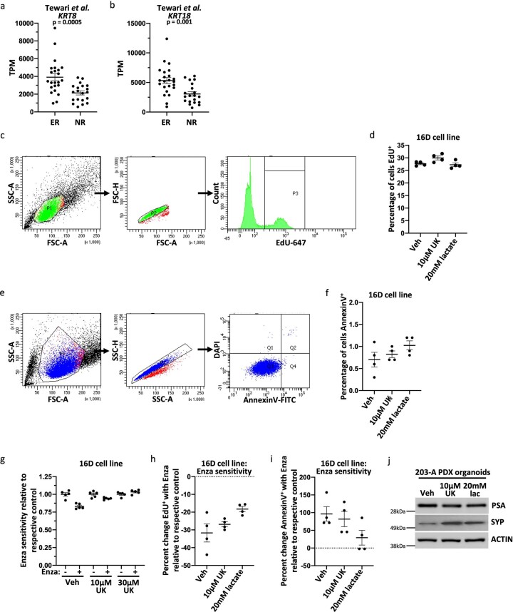
Lineage transitions are a central feature of prostate development, tumourigenesis and treatment resistance. While epigenetic changes are well known to drive prostate lineage transitions, it remains unclear how upstream metabolic signalling contributes to the regulation of prostate epithelial identity. To fill this gap, we developed an approach to perform metabolomics on primary prostate epithelial cells. Using this approach, we discovered that the basal and luminal cells of the prostate exhibit distinct metabolomes and nutrient utilization patterns. Furthermore, basal-to-luminal differentiation is accompanied by increased pyruvate oxidation. We establish the mitochondrial pyruvate carrier and subsequent lactate accumulation as regulators of prostate luminal identity. Inhibition of the mitochondrial pyruvate carrier or supplementation with exogenous lactate results in large-scale chromatin remodelling, influencing both lineage-specific transcription factors and response to antiandrogen treatment. These results establish reciprocal regulation of metabolism and prostate epithelial lineage identity.


Conflict of interest statement

P.C.B. sits on the Scientific Advisory Boards of Sage Bionetworks, BioSymetrics Inc. and Intersect Diagnostics Inc. E.C. obtains funding from Genentech, Sanofi, AbbVie, Astra Zeneca, Foghorn Pharmaceuticals, Kronos Bio, MacroGenics, Janssen Research, Bayer Pharmaceuticals, Forma Pharmaceuticals, Gilead and Zenith Epigenetics, and is a consultant of DotQuant. P.S.N. has received consulting fees from Janssen, Merck, Bristol Myers Squib and Venable Fitzpatrick, and received research funding from Janssen for work unrelated to the present study. All other authors declare no competing interests.

Figures

Fig. 1
Fig. 1. Primary basal and luminal prostate cells have distinct metabolic features.
a, Schematic of RNA-seq, metabolic profiling and glucose tracing performed on primary basal and luminal cells isolated from mouse prostate. b, GSEA of all KEGG, Hallmark and Reactome metabolism gene sets in basal and luminal cells. c, Heatmap of glycolytic and TCA cycle enzymes from RNA-seq of three biological replicates of basal and luminal cells. d, Heatmap of metabolite abundance in primary basal and luminal mouse prostate cells with three technical replicates for each of the three biological replicates. e, Aconitate-to-citrate fractional contribution ratio in primary basal and luminal mouse prostate cells fed [U-13C]glucose tracer for 16 h. f,g, Heatmaps of genes involved in de novo lipogenesis (f) and zinc transport (g) from RNA-seq of primary basal and luminal mouse prostate cells. h,i, Percentage M2 citrate (h) and percentage M3 citrate (i) from [U-13C]glucose in basal and luminal cells (n = 3 technical replicates for each of the 3 biological replicates). j, Fold change in glycolytic and TCA cycle enzymes from RNA-seq of basal and luminal cells from three human prostates. Shaded grey rectangles indicate genes that have statistically significant (P < 0.05) differential abundance. For all panels, data are shown as mean ± s.e.m. P values were calculated using a paired two-tailed t-test. Bas, basal; Bio rep, biological replicate; Lum, luminal. Source data
Fig. 2
Fig. 2. Basal-to-luminal differentiation is accompanied by increased pyruvate oxidation.
a, Schematic of in vivo model of basal-to-luminal differentiation in P10–P12 murine prostate. b, GSEA showing enrichment of KEGG oxidative phosphorylation in basal-derived luminal cells relative to multipotent basal cells in vivo. c, Schematic of lineage marker analysis, metabolic profiling and glucose tracing performed on primary basal-derived mouse organoids 3, 5 and 7 d after plating into organoid culture. d, Western blot analysis of the luminal marker KRT8 and the basal marker p63 in basal-derived organoids. e, PCA of fractional contribution from [U-13C]glucose metabolic tracing data of basal-derived organoids with three technical replicates per timepoint. Organoids were cultured with [U-13C]glucose 48 h before collecting metabolites at each timepoint. fh, Fractional contribution from [U-13C]glucose to glycolytic (f), TCA cycle (g) and nucleotide intermediates (h) in basal-derived organoids with three technical replicates per timepoint. For all panels, data are shown as mean ± s.e.m. NES, normalized enrichment score; PC1, principal component 1; PC2, principal component 2. Source data
Fig. 3
Fig. 3. Inhibition or knockout of the MPC prevents basal-to-luminal differentiation.
a, [U-13C]glucose tracer analysis of vehicle- and 10 μM UK5099-treated basal-derived organoids 7 d after plating (n = 3 independent biological replicates). Data are shown as mean ± s.e.m. b,c, Percentage organoid formation (n = 3 independent biological replicates) (b) and organoid diameter (n = 25 independent biological samples) (c) of vehicle- and 10 μM UK5099-treated basal-derived organoids 7 d after plating. d, Western blot analysis of luminal markers androgen receptor (AR) and KRT8 and basal marker p63 in vehicle- and 10 μM UK5099-treated basal-derived organoids 7 d after plating. e, Immunofluorescence of luminal marker KRT8 and basal marker p63 in representative vehicle- and 10 μM UK5099-treated basal-derived organoids 7 d after plating. Scale bars, 100 μm. f, Intracellular flow cytometry of KRT8 and basal marker cytokeratin 5 (KRT5) in vehicle- and 10 μM UK5099-treated basal-derived organoids 7 d after plating. g, Quantification of mean fluorescence intensity (MFI) of KRT8 from panel f (n = 4 independent biological replicates). h, [U-13C]glucose tracer analysis of control and Mpc1-KO basal-derived organoids (n = 3 independent biological replicates). Data are shown as mean ± s.e.m. i, Western blot analysis of basal and luminal markers in control and Mpc1-KO basal-derived organoids. j, GSEA showing negative enrichment of CD49flow luminal signature in Mpc1-KO relative to control basal-derived organoids. k, Flow cytometry quantification of percentage of EpCAMKRT8 cells in vehicle- and 10 μM UK5099-treated primary and quaternary basal-derived organoids (n = 3 independent biological replicates). l, t-Distributed stochastic neighbour embedding (t-SNE) plot of scRNA-seq data on quaternary prostate organoids illustrating distinct cell populations. m, t-SNE plot of vehicle- and 10 μM UK5099-treated cells from scRNA-seq data. n, Quantification of percentage of vehicle- and 10 μM UK5099-treated cells in each cluster from scRNA-seq data. For all panels, error bars represent s.e.m. P values were calculated using an unpaired two-tailed t-test with Welch’s correction. Aco, aconitate; Cit, citrate; EMT, epithelial–mesenchymal transition; Lac, lactate; Mal, malate; orgs, organoids; Succ, succinate; UK, UK5099; Veh, vehicle. Source data
Fig. 4
Fig. 4. MPC is a regulator of luminal lineage identity in prostate cancer.
a, Western blot analysis of luminal markers KRT8 and KRT18 in SKO and DKO mouse prostate organoids treated with vehicle or 10 μM UK5099 for 5 d. b, Heatmap of canonical basal and luminal markers from RNA-seq of vehicle- and 10 μM UK5099-treated DKO organoids. c,d, Correlation analysis of luminal signature score and MPC1 (c) or MPC2 (d) z-scores in treatment-naive prostate cancer samples from the SMMU dataset. e,f, Correlation analysis of luminal signature score and MPC1 (e) or MPC2 (f) z-scores in metastatic castration-resistant prostate cancer samples from the Beltran et al. dataset. g,h, RNA abundance of MPC1 (g) or MPC2 (h) in adenocarcinoma (adeno) or neuroendocrine prostate cancer (NEPC) samples from the Beltran et al. dataset. i,j, RNA abundance of MPC1 (i) or MPC2 (j) in adeno or NEPC samples from the Nguyen et al. PDX dataset. Correlation analysis was performed using Spearman’s correlation with a two-tailed P value. For all panels, error bars represent s.e.m. P values in gj were calculated using an unpaired two-tailed t-test with Welch’s correction. mCRPC, metastatic castration-resistant prostate cancer. Source data
Fig. 5
Fig. 5. Intracellular lactate accumulation results in large-scale chromatin remodelling of key lineage-specific transcription factors.
a, Extracellular lactate abundance in primary basal-derived mouse organoids treated with vehicle or 10 μM UK5099 for 7 d (n = 6 independent biological replicates). Error bars represent s.e.m. P value was calculated using an unpaired two-tailed t-test with Welch’s correction. bd, Western blot analysis of the luminal marker KRT8 and the basal marker p63 in basal-derived organoids treated with vehicle or 20 mM sodium lactate (b), 10 nM Trichostatin A (TSA) (c) or 1 mM sodium butyrate (d) for 7 d. e, Spearman’s correlation between log2Coefficients of UK5099 and Butyrate effects for each gene (r = 0.58, P < 2.2 × 10−16). Each hexagonal bin represents a region of the plot with the colour denoting the number of genes that fall within that region. The red dotted line represents x = y. f, Heatmap of 1,712 hyper-accessible genes and 766 hypo-accessible genes (fold change ≥ 1.5 or fold change ≤ 0.5) in basal-derived mouse organoids treated with vehicle or 10 μM UK5099 for 7 d. g, Heatmap of 1,147 hyper-accessible genes and 336 hypo-accessible genes (fold change ≥ 1.5 or fold change ≤ 0.5) in basal-derived mouse organoids treated with vehicle or 20 mM sodium lactate for 7 d. h,i, Seven most significantly enriched transcription factor binding motifs in more accessible regions in organoids treated with 10 μM UK5099 (h) or 20 mM sodium lactate (i). The FDR was controlled using the Benjamini–Hochberg method. j, Venn diagram depicting overlap in significantly enriched transcription factor binding motifs in more accessible regions in UK5099-treated and lactate-supplemented organoids. k, Browser track depicting ATAC-seq peaks in p63 gene in vehicle-treated, UK5099-treated and lactate-supplemented organoids. l, Heatmap of chromatin accessibility of 2,000 basal signature genes in vehicle-treated, UK5099-treated and lactate-supplemented organoids. TSS, transcription start site. Source data
Fig. 6
Fig. 6. Modulation of lactate metabolism alters antiandrogen response in prostate cancer.
a,b, RNA abundance of MPC1 (a) or MPC2 (b) in nonresponders (NR) or exceptional responders (ER) from the Tewari et al. dataset, which contains RNA-seq of pre-treatment localized prostate cancer biopsies from 43 patients enrolled in neoadjuvant trials of androgen pathway inhibition. c,d, mRNA abundance of MPC1 (c) or MPC2 (d) in NR or ER from the Alumkal et al. dataset, which contains RNA-seq of metastatic castration-resistant prostate cancer biopsies from 25 patients enrolled in neoadjuvant trials of androgen pathway inhibition. e, Representative phase contrast images of MDA PCa 203-A PDX-derived organoids treated with vehicle, 10 μM UK5099 or 20 mM sodium lactate for 7 d. f, Percentage change in luminescence signal with 10 μM enzalutamide treatment from CellTiter-Glo assay in castration-resistant MDA PCa 203-A PDX-derived organoids treated with vehicle, 10 μM UK5099 or 20 mM sodium lactate for 7 d before beginning 10 μM enzalutamide treatment (n = 4 independent biological replicates). For all panels, error bars represent s.e.m. P values were calculated using an unpaired two-tailed t-test with Welch’s correction. Source data
Extended Data Fig. 1
Extended Data Fig. 1. Validation of basal and luminal mouse prostate cell isolation and evaluation of metabolic pathways.
(a) Gating scheme for isolating primary basal and luminal cells from mouse prostate. (b) Heatmap of select canonical basal and luminal markers from RNA sequencing of primary basal and luminal mouse prostate cells with three biological replicates. (c) Gene set enrichment analysis (GSEA) showing positive enrichment of CD49fhigh signature in basal cells relative to luminal cells. (d) GSEA showing positive enrichment of CD49flow signature in luminal cells relative to basal cells. (e) 30 pathways most enriched in differentially abundant genes (log2(fold change) ≥ 1, FDR < 0.2) in basal and luminal cells identified by KEGG pathway analysis. Metabolism-related pathways highlighted in green (basal-enriched) and blue (luminal-enriched). (f) Western blot analysis of select glycolytic and TCA cycle enzymes in basal and luminal cells. Source data
Extended Data Fig. 2
Extended Data Fig. 2. Basal and luminal prostate epithelial cells have distinct metabolic features.
(a-b) Analysis of glycolytic (a) and TCA cycle (b) enzymes in Karthaus et al. and Crowley et al. mouse single cell RNA sequencing data. (c) Percentage of Annexin V, 7-AAD primary basal and luminal cells after overnight culture (n = 3 independent biological replicates). Error bars represent SEM. p-values were calculated using an unpaired two-tailed t-test with Welch’s correction. Source data
Extended Data Fig. 3
Extended Data Fig. 3. Basal to luminal differentiation is associated with metabolic reprogramming.
(a) 30 pathways most enriched in differentially abundant genes (log2(fold change) ≥ 1) in multipotent basal cells and basal-derived luminal cells identified by KEGG pathway analysis. Metabolism-related pathways highlighted in green (enriched in multipotent basal-enriched) and blue (enriched in basal-derived luminal-enriched). (b) Intracellular flow cytometry analysis of the basal marker cytokeratin 5 (KRT5) and the luminal marker cytokeratin 8 (KRT8) in primary basal-derived mouse organoids three, six and nine days after plating into organoid culture. (c) Principal component analysis of metabolic profiling data for basal-derived organoids with three technical replicates per timepoint. (d) Heatmap of metabolite abundance in primary basal-derived mouse organoids (n = 3 independent biological replicates per timepoint). (e) Fractional contribution from [U-13C]glucose to M2 and M3 citrate in basal-derived organoids (n = 3 independent biological replicates per timepoint). Error bars represent SEM. (f) Western blot analysis of proliferation marker PCNA in basal-derived organoids. Source data
Extended Data Fig. 4
Extended Data Fig. 4. Inhibition of the mitochondrial pyruvate carrier antagonizes luminal lineage identity.
(a) Intracellular flow cytometry of KRT8 and KRT5 in basal-derived organoids treated with 0-40 μM UK5099 for seven days. (b) Percent organoid formation of basal-derived organoids treated with 0-40 μM UK5099 (n = 3 independent biological replicates). (c) Percent organoid formation of vehicle- and 10 μM UK5099-treated luminal-derived organoids (n = 3 independent biological replicates). (d) Western blot analysis of proliferation markers (Ki67 and PCNA) and apoptosis marker (CC3, cleaved caspase-3) in vehicle- and 10 μM UK5099-treated luminal-derived organoids seven days after plating. (e) Correlation analysis of [U-13C]glucose fractional contribution comparing Mpc1-KO and 10 μM UK5099-treated basal-derived organoids (n = 3 independent biological replicates). (f) GSEA showing negative enrichment of CD49flow luminal signature in 10 μM UK5099-treated relative to vehicle-treated basal-derived organoids. (g) GSEA showing enrichment of CD49fhigh basal signature in vehicle-treated relative to 10 μM UK5099-treated basal-derived organoids. (h) GSEA showing enrichment of CD49fhigh basal signature in control relative to Mpc1-KO basal-derived organoids. For all panels, error bars represent SEM. p-values were calculated using an unpaired two-tailed t-test with Welch’s correction. Correlation analysis was performed using Spearman’s correlation. Source data
Extended Data Fig. 5
Extended Data Fig. 5. Single cell RNA-sequencing illustrates that inhibition of the mitochondrial pyruvate carrier reduces luminal marker expression and increases expression of basal markers, glycolytic enzymes, and inflammatory signaling genes in the luminal subpopulation in mouse prostate organoids.
(a) Representative flow cytometry plots illustrating EPCAM+ KRT8+ (red), EPCAM+ KRT8(green), and EPCAM KRT8 (blue) populations in primary and quaternary organoids. (b) Expression level (log2(read count)) of canonical basal, luminal, epithelial and epithelial-mesenchymal transition (EMT-like) markers in distinct cell populations from scRNA-seq data to validate cluster identification. (c) Dot plot of glycolytic enzymes, lipid metabolism genes, canonical basal markers, inflammatory genes and canonical luminal markers with vehicle or UK5099 treatment within the phenotypic luminal cluster. (d) Flow cytometry gating scheme for apoptosis analysis in panel (e). (e) Quantification of percent of AnnexinV+ cells in quaternary organoids treated with vehicle, 10 μM UK5099 for one day, or 10 μM UK5099 for three days (n = 3 independent biological replicates). Error bars in represent SEM. Source data
Extended Data Fig. 6
Extended Data Fig. 6. Inhibition of the mitochondrial pyruvate carrier antagonizes luminal lineage identity in models of prostate cancer.
(a) Western blot validation of knockout of Pten in single knockout (SKO) and double knockout (DKO) mouse prostate organoids and validation of knockout of Rb1 in DKO organoids. (b) Organoid diameter (micrometers) of wildtype (WT), SKO and DKO organoids (n = 25 independent biological samples). Error bars represent SEM. (c) Heatmap of canonical luminal and neuroendocrine markers in WT, SKO and DKO mouse prostates from RNA sequencing data published in Ku et al. (d) Heatmap of canonical luminal and neuroendocrine markers in WT, SKO and DKO mouse organoids from RNA sequencing data. (e-i) Western blot analysis of lineage markers AR, PSA, KRT8, KRT18, NSE, SYP, and SOX2 in human 16D cell line (e), LuCaP35 cell line (f), LAPC4 cell line (g), 183-A PDX organoids (h), and 16D subcutaneous tumours (i). (j) Quantification of Western blot in panel I (n = 4 independent tumors). Data are shown as mean ± SEM. (k-l) Correlation analysis of z-score expression of the luminal marker KRT8 (k) or KRT18 (l) with MPC1 in 499 primary prostate carcinomas from The Cancer Genome Atlas (TCGA). (m-n) Correlation analysis of z-score expression of the luminal marker KRT8 (m) or KRT18 (n) with MPC2 in 499 primary prostate carcinomas from TCGA. Correlation analysis was performed using Spearman’s correlation with a two-tailed p-value. p-values in (b) were calculated using an unpaired two-tailed t-test with Welch’s correction. Source data
Extended Data Fig. 7
Extended Data Fig. 7. Intracellular lactate accumulation antagonizes luminal lineage identity in mouse prostate organoids.
(a-b) Intracellular lactate abundance in primary basal-derived mouse organoids treated with vehicle or 10 μM UK5099 (a) or 20 mM sodium lactate (b) for seven days (n = 6 independent biological replicates). (c) Extracellular lactate abundance in organoids treated with vehicle or 20 mM sodium lactate (n = 6 independent biological replicates). (d-e) Intracellular (d) and extracellular (e) lactate abundance in organoids treated with vehicle or 10 μM AZD3965 (n = 6 independent biological replicates). Error bars in panels (a-e) represent SEM. (f) Western blot analysis of the luminal marker KRT8 and the basal marker p63 in basal-derived organoids treated with vehicle or 10 μM AZD3965 for seven days. (g) Heatmap of fractional contribution of [U-13C]lactate tracing data from organoids treated with 20 mM sodium lactate for six days followed by treatment with 20 mM [U-13C]lactate for 24 hours. (h) Fractional contribution from [U-13C]lactate to TCA cycle intermediates in organoids treated with 20 mM sodium lactate for six days followed by treatment with 20 mM [U-13C]lactate and vehicle or 10 μM UK5099 for 24 hours (n = 3 independent biological replicates). Data are shown as mean ± SEM. p-values were calculated using an unpaired two-tailed t-test with Welch’s correction. (i) Western blot analysis of pan-acetyl histone H4 (pan-acetyl HH4), total histone H4 (HH4), H3K9Ac, pan-acetyl histone H3 (pan-acetyl HH3) and total histone H3 (HH3) in histone extracts from basal-derived organoids treated with vehicle, 1 mM sodium butyrate or 10 nM TSA for seven days. Source data
Extended Data Fig. 8
Extended Data Fig. 8. Histone deacetylase inhibition and mitochondrial pyruvate carrier inhibition have similar effects on gene expression in mouse prostate organoids.
(a) The distribution of gene abundance (n = 13687) among 12 samples. The primary heatmap color represents the Z-score normalized TPM value of each gene. Missing values were omitted. Hierarchical clustering has been applied to both samples (columns) and genes (rows). The top covariate heatmap indicates the treatment status of each sample. (b) Genes with significant differential RNA abundance were identified based on coefficient and adjusted p-value values (|log2(Coefficient)| > 1, adjusted p-value < 0.01). Genes with downregulated RNA abundance are denoted by blue dots, while genes with upregulated RNA abundance are represented by red dots. Grey dots represent genes without a significant difference. Labeled genes have either the least adjusted p-values or the greatest |log2(Coefficient)| values. (c) Significant genes (adjusted p-value < 0.01) were selected to show univariate effects under different treatments. The primary strip plot shows raw TPM distribution among selected genes with dot colors indicating treatment types. The dotmap above presents the effect sizes and significance of each gene under different treatments. The dot size represents log2Coefficient values, while background color is indicative of the adjusted p-value. (d) The Venn diagram illustrates the overlap between effects, as reflected by the number of significant genes (n = 1,394; |log2(Coefficient)| > 1, adjusted p-value < 0.01). The model was adjusted using empirical Bayes moderation for standard error, and the false discovery rate (FDR) was controlled using the Benjamini-Hochberg method. (e) The distribution of top basal and luminal gene abundance (n = 50) among 12 samples. The color in the primary heatmap signifies the Z-score normalized TPM value for each gene. Missing values were omitted. Hierarchical clustering has been applied to both samples (columns) and genes (rows). The top covariate heatmap indicates the treatment status for each sample, while the right covariate heatmap identifies the canonical marker type for each gene. (f) Top 15 enriched gene ontology terms in UK5099, Butyrate, and UK5099:Butyrate effects. The direction of regulation is calculated by the normalized enrichment score and is denoted by different colors: orange indicates upregulation, while blue represents downregulation. The dot size corresponds to the number of genes enriched in each gene set, while the background shading indicates the -log10adjusted p-value. (g) Top 15 enriched GSEA gene sets in UK5099, Butyrate, and UK5099:Butyrate effects. The direction of regulation is calculated by the normalized enrichment score (NES) and denoted by different colors: orange (upregulation), and blue (downregulation). The dot size corresponds to the number of genes enriched in each gene set, while the background shading indicates the -log10p-value. (h) Epidermis development gene set enrichment results. Genes were ranked from high to low based on log2Coefficient of UK5099 or Butyrate effect in the general linear model. (i) Keratinocyte differentiation gene set enrichment results. Genes were ranked from high to low based on log2Coefficient of UK5099 or Butyrate effect in the general linear model. Source data
Extended Data Fig. 9
Extended Data Fig. 9. Analysis of less accessible transcription factor binding motifs in mouse prostate organoids treated with UK5099 or lactate supplementation.
(a-b) Seven most significantly enriched transcription factor binding motifs in less accessible regions in organoids treated with 10 μM UK5099 (a) or 20 mM sodium lactate (b). The false discovery rate (FDR) was controlled using the Benjamini-Hochberg method. (c) Venn diagram depicting overlap in significantly enriched transcription factor binding motifs in less accessible regions in UK5099-treated and lactate-supplemented organoids. (d) Browser track depicting ATAC-seq peaks in Prom1 gene in vehicle-treated, UK5099-treated, and lactate-supplemented organoids.
Extended Data Fig. 10
Extended Data Fig. 10. Modulation of lactate metabolism alters antiandrogen response and lineage identity in human models of prostate cancer.
(a-b) RNA abundance of KRT8 (a) or KRT18 (b) in non-responders (NR) or exceptional responders (ER) from the Tewari et al. dataset, which contains RNA sequencing of pre-treatment localized prostate cancer biopsies from 43 patients enrolled in neoadjuvant trials of androgen pathway inhibition. p-values were calculated using an unpaired two-tailed t-test with Welch’s correction. (c) Flow cytometry gating scheme for data in panels (d,h). (d) Percentage of cells EdU+ in 16D cells treated with vehicle, 10 μM UK5099, or 20 mM lactate for seven days (n = 4 independent biological replicates). (e) Flow cytometry gating scheme for data in panels (f,i). (f) Percentage of cells AnnexinV+ in 16D cells treated with vehicle, 10 μM UK5099, or 20 mM lactate for seven days (n = 4 independent biological replicates). (g) Luminescence signal relative to respective control from CellTiter-Glo assay in 16D cell line treated with vehicle, 10 μM UK5099, or 30 μM UK5099 for seven days before beginning 48 hours of 10 μM Enzalutamide treatment (n = 5 independent biological replicates). (h-i) Percent change EdU+ (h) and AnnexinV+ (i) with 10 μM Enzalutamide treatment relative to respective control in 16D cell line (n = 4 independent biological replicates). (j) Western blot analysis of PSA and Synaptophysin (SYP) in 203-A PDX-derived organoids. For all panels, error bars represent SEM. Source data

References

    1. Abate-Shen C, Shen MM. Molecular genetics of prostate cancer. Genes Dev. 2000;14:2410–2434. - PubMed
    1. Choi N, et al. Adult murine prostate basal and luminal cells are self-sustained lineages that can both serve as targets for prostate cancer initiation. Cancer Cell. 2012;21:253–265. - PMC - PubMed
    1. Ousset, M. et al. Multipotent and unipotent progenitors contribute to prostate postnatal development. Nat. Cell Biol. 10.1038/ncb2600 (2012). - PubMed
    1. Wang, J. et al. Symmetrical and asymmetrical division analysis provides evidence for a hierarchy of prostate epithelial cell lineages. Nat. Commun.10.1038/ncomms5758 (2014). - PubMed
    1. Toivanen R, Mohan A, Shen MM. Basal progenitors contribute to repair of the prostate epithelium following induced luminal anoikis. Stem Cell Rep. 2016;6:660–667. - PMC - PubMed

Publication types

MeSH terms