

Automatic Quantification of Normal Brain Gyrification Patterns and Changes in Fetuses with Polymicrogyria and Lissencephaly Based on MRI
- PMID: 38050002
- PMCID: PMC10714858
- DOI: 10.3174/ajnr.A8046
Automatic Quantification of Normal Brain Gyrification Patterns and Changes in Fetuses with Polymicrogyria and Lissencephaly Based on MRI
Abstract
Background and purpose: The current imaging assessment of fetal brain gyrification is performed qualitatively and subjectively using sonography and MR imaging. A few previous studies have suggested methods for quantification of fetal gyrification based on 3D reconstructed MR imaging, which requires unique data and is time-consuming. In this study, we aimed to develop an automatic pipeline for gyrification assessment based on routinely acquired fetal 2D MR imaging data, to quantify normal changes with gestation, and to measure differences in fetuses with lissencephaly and polymicrogyria compared with controls.
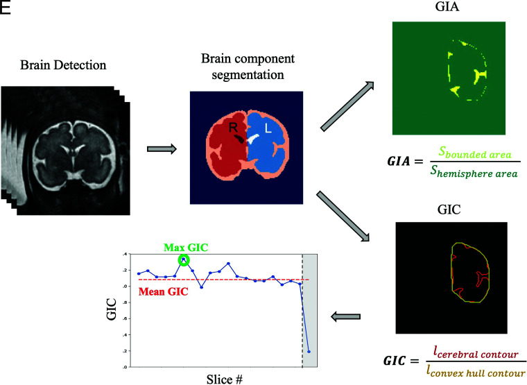
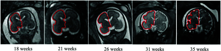
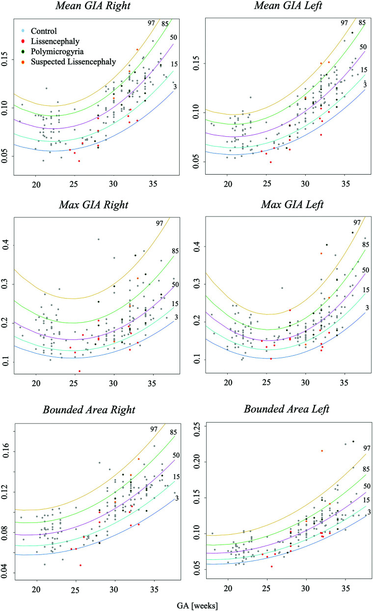
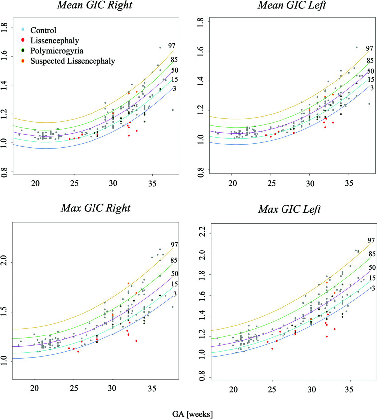
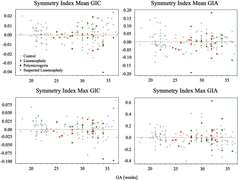
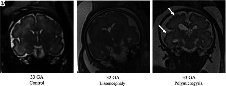
Materials and methods: We included coronal T2-weighted MR imaging data of 162 fetuses retrospectively collected from 2 clinical sites: 134 controls, 12 with lissencephaly, 13 with polymicrogyria, and 3 with suspected lissencephaly based on sonography, yet with normal MR imaging diagnoses. Following brain segmentation, 5 gyrification parameters were calculated separately for each hemisphere on the basis of the area and ratio between the contours of the cerebrum and its convex hull. Seven machine learning classifiers were evaluated to differentiate control fetuses and fetuses with lissencephaly or polymicrogyria.
Results: In control fetuses, all parameters changed significantly with gestational age (P < .05). Compared with controls, fetuses with lissencephaly showed significant reductions in all gyrification parameters (P ≤ .02). Similarly, significant reductions were detected for fetuses with polymicrogyria in several parameters (P ≤ .001). The 3 suspected fetuses showed normal gyrification values, supporting the MR imaging diagnosis. An XGBoost-linear algorithm achieved the best results for classification between fetuses with lissencephaly and control fetuses (n = 32), with an area under the curve of 0.90 and a recall of 0.83. Similarly, a random forest classifier showed the best performance for classification of fetuses with polymicrogyria and control fetuses (n = 33), with an area under the curve of 0.84 and a recall of 0.62.
Conclusions: This study presents a pipeline for automatic quantification of fetal brain gyrification and provides normal developmental curves from a large cohort. Our method significantly differentiated fetuses with lissencephaly and polymicrogyria, demonstrating lower gyrification values. The method can aid radiologic assessment, highlight fetuses at risk, and may improve early identification of fetuses with cortical malformations.
© 2023 by American Journal of Neuroradiology.
Figures
References
-
- Dubois J, Dehaene-Lambertz G. Fetal and postnatal development of the cortex: MRI and genetics. Brain Mapping: An Encyclopedic Reference 2015;2:11–19 10.1016/B978-0-12-397025-1.00194-9 - DOI
Publication types
MeSH terms
LinkOut - more resources
Full Text Sources
Medical