. 2024 Jan;38(1):e23358.
doi: 10.1096/fj.202301934R.

Hypoxia-inducible microRNA-155 negatively regulates epithelial barrier in eosinophilic esophagitis by suppressing tight junction claudin-7

Affiliations

Hypoxia-inducible microRNA-155 negatively regulates epithelial barrier in eosinophilic esophagitis by suppressing tight junction claudin-7

Gary E Markey et al. FASEB J. 2024 Jan.

Abstract

MicroRNA (miRNA)-mediated mRNA regulation directs many homeostatic and pathological processes, but how miRNAs coordinate aberrant esophageal inflammation during eosinophilic esophagitis (EoE) is poorly understood. Here, we report a deregulatory axis where microRNA-155 (miR-155) regulates epithelial barrier dysfunction by selectively constraining tight junction CLDN7 (claudin-7). MiR-155 is elevated in the esophageal epithelium of biopsies from patients with active EoE and in cell culture models. MiR-155 localization using in situ hybridization (ISH) in patient biopsies and intra-epithelial compartmentalization of miR-155 show expression predominantly within the basal epithelia. Epithelial miR-155 activity was evident through diminished target gene expression in 3D organotypic cultures, particularly in relatively undifferentiated basal cell states. Mechanistically, generation of a novel cell line with enhanced epithelial miR-155 stable overexpression induced a functionally deficient epithelial barrier in 3D air-liquid interface epithelial cultures measured by transepithelial electrical resistance (TEER). Histological assessment of 3D esophageal organoid cultures overexpressing miR-155 showed notable dilated intra-epithelial spaces. Unbiased RNA-sequencing analysis and immunofluorescence determined a defect in epithelial barrier tight junctions and revealed a selective reduction in the expression of critical esophageal tight junction molecule, claudin-7. Together, our data reveal a previously unappreciated role for miR-155 in mediating epithelial barrier dysfunction in esophageal inflammation.

Keywords: Barrier; claudin-7; eosinophilic esophagitis; epithelium; hypoxia; miR-155; microRNA 155; tight junction.


Conflict of interest statement

Conflict of Interest Statement

Authors have declared that no conflict of interest exists.

Figures
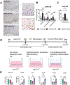
Figure 1:
Figure 1:. Epithelial miR-155 is elevated in active EoE and is highest in immature esophageal epithelial cells at 3D-ALI.
(A) Representative images of miR-155 in situ hybridisation on biopsies from uninflamed controls, active EoE, and inactive EoE. (B) Compartmental analysis on the individual esophageal layers shows that miR-155 was highest within the basal and suprabasal regions in active EoE and further reduced through therapeutic intervention in inactive EoE. (C) Total epithelial miR-155 was also evaluated by merging the data of the three constituent esophageal layers and found that EoE patients display significantly higher miR-155 than uninflamed controls and inactive EoE individuals. (D) EPC2-hTERTs were grown in organotypic 3D-ALI culture and harvested on day 2, 7 and 11. The expression of miR-155 was elevated during immature epithelial developmental timepoints on (E) days 2 and 7. Predicted miR-155 targets CLDN1, TJP1, OCLN, and PTEN were investigated by qRT-PCR and found to display inverse kinetics to the expression of miR-155 at 3D-ALI. Statistical significance of ISH data was assessed using a Kruskal-Wallis test with a Dunn’s multiple comparisons test. 3D-ALI data was assessed for significance using a One-Way ANOVA with a Tukey’s multiple comparisons test. *P ≤ 0.05, **P ≤ 0.01, ***P ≤ 0.001. Data are presented as means ± SEM and represent 3 experimental repeats.
Figure 2:
Figure 2:. Extended experimental hypoxia induces miR-155 and differentially regulates its targets.
(A) Complimentary binding of the HIF1A 3’ UTR to that of the miR-155 seed region. (B) HIF1A and miR-155 expression was assessed by qRT-PCR from esophageal epithelial cells exposed to a time course (0, 4, 24, 48, 72 hours) of experimental hypoxia compared with time matched normoxic controls. Inverse linear regression analysis was carried out to show the significantly inverse relationship between the expression of miR-155 and HIF1A in experimental hypoxia. (C) HIF-1α expression was assessed by Western blotting of nuclear isolates from esophageal epithelial cells exposed to a time course (0, 4, 24, 48, 72 hours) of experimental hypoxia compared to time matched normoxic controls. (D and E) qRT-PCR analysis of HIF2A and HIF1B (D), and miRNA processing genes DROSHA, DICER, and AGO2 (E). For Western blots, a representative for a single time course are presented. Statistical significance was assessed using a Students’ t test comparing time point–matched normoxic controls with hypoxic samples. *P < 0.05, **P < 0.01, ***P < 0.001 (n = 3). Data are presented as means ± SEM and represent 3 experimental repeats.
Figure 3:
Figure 3:. Stable miR-155–5p overexpression reduces HIF-1α signaling and induces a deficient epithelial barrier.
(A) Submerged monolayer cultures of miR-155OE cells shows a significant elevation in miR-155. qRT-PCR of miR-155 targets showed and attenuation of HIF1A in miR-155OE cells, and a significant decrease in OCLN. We did not observe significant differential expression in the remaining targets CLDN1, TJP1, PTEN, or WEE1 in submerged culture. (B) Using stratified squamous 3-dimensional air liquid interface (3D-ALI) cultures, miR-155 overexpression diminished barrier as measured by transepithelial electrical resistance (TEER) (n=4). (C) H&E stained sections from miR-155OE cells cultured at 3D-ALI indicate perturbed stratification. Black scale bar represents 60 μm. These data suggest that in the setting of miR-155OE, epithelial defects exist. (D) qRT-PCR of barrier-forming molecules showed a significant decrease in the tight junction CLDN7. (E) Whole cell protein was analysed by Western blot whereby claudin-7 was shown to be attenuated in miR-155OE cells in 3D-ALI. Submerged culture data was analysed for significance using a Students’ t test comparing time point–matched controls. *P < 0.05, **P < 0.01, ***P < 0.001. Data are presented as means ± SEM and represent a minimum of 3 experimental repeats.
Figure 4:
Figure 4:. MiR-155 overexpression dysregulates esophageal epithelial function.
RNA sequencing of miR-155OE and control cells cultured at 3D-ALI shows impaired epithelial function. (A) Significance was attributed to differentially expressed genes with an adjusted p-value ≤0.1. and yielded 627 downregulated genes and 1026 upregulated genes. A selection of genes were selected which have functions within cytoskeleton and epithelial organisation or have been validated in the literature to contribute to EoE pathophysiology. (B) GO analysis for pathway enrichment for upregulated and downregulated pathways in miR-155OE cells. (C) Venn diagram showing the number of genes differentially expressed as identified by publicly available RNA-seq of patients with inflammatory EoE as compared to controls (cut-off of fold change > 2 or < 0.5, adjusted p-value < 0.05) and in miR-155OE cells in 3D-ALI cultures for 11 days. Genes overlapping between these two data sets were identified (167 genes) and a selection with functions relevant to EoE pathophysiology were graphed into a heatmap. GO analyses of the miR-155OE-EoE overlap gene set depicted localization of cellular components and enrichment of diseases based on the 167 gene overlap based on adjusted p-value (-log10) showed a selection of diseases with perturbed epithelial function including Clouston syndrome, Netherton syndrome, Palmoplantar keratosis, and Eosinophilic esophagitis. GO analysis of the 167 intersected genes’ enriched pathways involved ‘intermediate filament’, ‘keratin filament’, and ‘intermediate filament cytoskeleton’ which shared 5 genes. These pathways reveal a dysregulation in cytoskeletal arrangement of miR-155OE cells. (D) Immunofluorescent stained miR-155OE organoids show no difference in basal KRT14, however miR-155OE organoids display less intense and mislocalized KRT4. (E) qRT-PCR of miR-155OE showing differential expression of an array of basal and suprabasal keratins.
Figure 5:
Figure 5:. MiR-155–5p overexpression attenuates CLDN7 expression and localisation in esophageal epithelial organoids.
(A) Schematic showing normal esophageal organoid culture development. (B) qRT-PCR of the key tight junction molecules of the esophagus showed a significant decrease in OCLN and CLDN7. Western blots of whole cell protein isolated from miR-155OE organoids displayed (C) attenuated claudin −7 expression. (D) H&E staining shows dilated intracellular-like spaces evident within the basal region of miR-155OE organoids. Immunofluorescent staining shows perturbed claudin-7 localisation in miR-155OE organoids, note the attenuated peri membranous staining of claudin-7 within the miR-155OE organoids compared to controls (E) Representative H&E staining of Claudin-7KD and shRNA control organoid showing dilated spaces and in Claudin-7KD compared to controls. (F) Claudin-7 promoter showing putative binding sites for transcription actors HNF4α and PU.1 of which are predicted miR-155 targets. Data was analysed for significance using a Students’ t test comparing controls. *P < 0.05, **P < 0.01, ***P < 0.001. Data are presented as means ± SEM and represent 3 experimental repeats.

References

    1. Dellon ES, Liacouras CA, Molina-Infante J, et al. Updated International Consensus Diagnostic Criteria for Eosinophilic Esophagitis: Proceedings of the AGREE Conference. Gastroenterology. Oct 2018;155(4):1022–1033.e10. doi:10.1053/j.gastro.2018.07.009 - DOI - PMC - PubMed
    1. Collins MH. Histopathology of eosinophilic esophagitis. Dig Dis. 2014;32(1–2):68–73. doi:10.1159/000357012 - DOI - PubMed
    1. Clevenger MH, Karami AL, Carlson DA, et al. Suprabasal cells retain progenitor cell identity programs in eosinophilic esophagitis-driven basal cell hyperplasia. JCI Insight. Sep 5 2023;doi:10.1172/jci.insight.171765 - DOI - PMC - PubMed
    1. Blanchard C, Stucke EM, Burwinkel K, et al. Coordinate Interaction between IL-13 and Epithelial Differentiation Cluster Genes in Eosinophilic Esophagitis. The Journal of Immunology. 2010;184(7):4033–4041. doi:10.4049/jimmunol.0903069 - DOI - PMC - PubMed
    1. Kottyan LC, Davis BP, Sherrill JD, et al. Genome-wide association analysis of eosinophilic esophagitis provides insight into the tissue specificity of this allergic disease. Nature Genetics. 2014;46(8):895–900. doi:10.1038/ng.3033 - DOI - PMC - PubMed

Publication types

MeSH terms