

doi: 10.1107/S2053230X23009536.
Epub 2023 Dec 5.
The crystal structure of the human smacovirus 1 Rep domain
Affiliations
- PMID: 38051309
- PMCID: PMC10833120
- DOI: 10.1107/S2053230X23009536
Item in Clipboard
The crystal structure of the human smacovirus 1 Rep domain
Acta Crystallogr F Struct Biol Commun.
.
Abstract
Replication initiator proteins (Reps) from the HUH endonuclease family process specific single-stranded DNA sequences to initiate rolling-circle replication in viruses. Here, the first crystal structure of the apo state of a Rep domain from the smacovirus family is reported. The structure of the human smacovirus 1 Rep domain was obtained at 1.33 Å resolution and represents an expansion of the HUH endonuclease superfamily, allowing greater diversity in bioconjugation-tag applications.
Keywords: HUH endonucleases; HUH-tags; Rep domains; bioconjugation; crystal structure; smacoviruses; ssDNA.
open access.
Figures
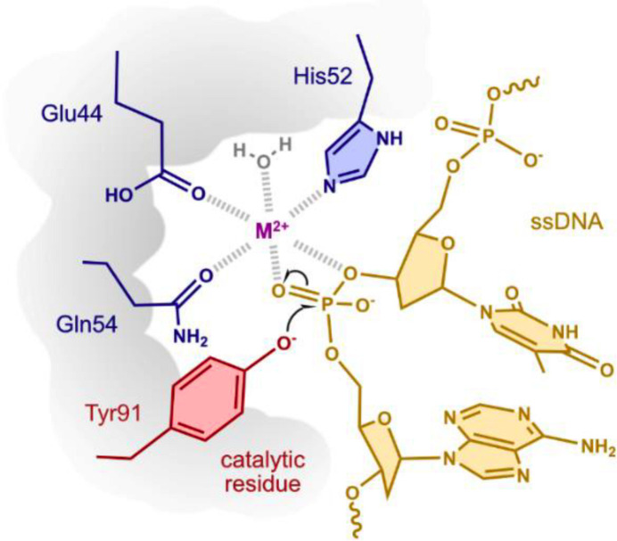
The catalytic activity of HUH endonucleases relies on the HUH/Q and tyrosine motifs to coordinate the nucleophilic attack on the DNA phosphate backbone (adapted from Tompkins et al., 2021 ▸).

The coordination of the ssDNA by the Rep during the pre- and post-cleavage complexes. A mutation from tyrosine to phenylalanine does not allow the cleavage reaction to proceed but retains the ssDNA-binding ability of the Rep (Larkin et al., 2005 ▸).

Ribbon model of HSV1 Rep showing the secondary-structure organization (left) consisting of β1, α1, β2, β3, α2, β4 and α3, and the orientation of the catalytic HUQ and tyrosine (Y81F in our model) motif (right). In our noncoordinated structure, the noncoordinating amino acid ‘U’ is oriented away from the binding site and is therefore not shown here.

Structural alignment of HSV1 Rep (gray) with WDV Rep (orange, PDB entry 6q1m ) on the left and PCV2 Rep (green, PDB entry 5xor ) on the right. Superimpositions illustrate the structural conservation among the Reps (top) and the orientation of the catalytic HUH/Q and tyrosine residues (bottom). The r.m.s.d. value on superimposition of HSV1 and WDV is 2.4 Å and that for HSV1 and PCV2 is 3.3 Å.

Structural and sequence comparison of HSV1 Rep with WDV Rep and PCV2 Rep using PROMALS3D (Pei et al., 2008 ▸). β-Strands are shown in blue and α-helices in red. Consensus amino-acid symbols: conserved amino acids are shown as bold uppercase letters; h, hydrophobic; s, small; p, polar; c, charged; –, negatively charged.
References
-
- Boer, R., Russi, S., Guasch, A., Lucas, M., Blanco, A. G., Pérez-Luque, R., Coll, M. & de la Cruz, F. (2006). J. Mol. Biol. 358, 857–869. - PubMed
MeSH terms
Substances
Grants and funding
LinkOut - more resources
Full Text Sources
