Review
. 2024 Feb;26(2):37-52.
doi: 10.1007/s11926-023-01125-6. Epub 2023 Dec 5.

Role of IFN-α in Rheumatoid Arthritis

Affiliations
Review

Role of IFN-α in Rheumatoid Arthritis

Chung M A Lin et al. Curr Rheumatol Rep. 2024 Feb.

Abstract

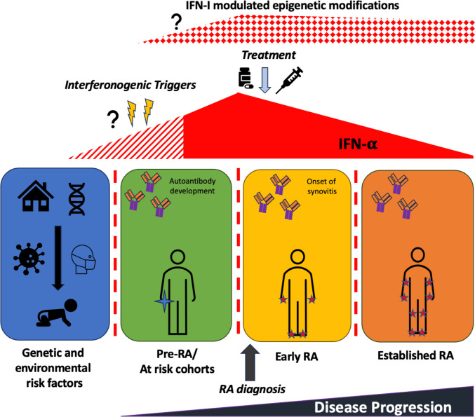
Purpose of review: Type 1 interferons (IFN-I) are of increasing interest across a wide range of autoimmune rheumatic diseases. Historically, research into their role in rheumatoid arthritis (RA) has been relatively neglected, but recent work continues to highlight a potential contribution to RA pathophysiology.

Recent findings: We emphasise the importance of disease stage when examining IFN-I in RA and provide an overview on how IFN-I may have a direct role on a variety of relevant cellular functions. We explore how clinical trajectory may be influenced by increased IFN-I signalling, and also, the limitations of scores composed of interferon response genes. Relevant environmental triggers and inheritable RA genetic risk relating to IFN-I signalling are explored with emphasis on intriguing data potentially linking IFN-I exposure, epigenetic changes, and disease relevant processes. Whilst these data cumulatively illustrate a likely role for IFN-I in RA, they also highlight the knowledge gaps, particularly in populations at risk for RA, and suggest directions for future research to both better understand IFN-I biology and inform targeted therapeutic strategies.

Keywords: Biomarkers; Early rheumatoid arthritis; Interferon gene signature; Rheumatoid arthritis; Type 1 interferons.


Conflict of interest statement

JDI discloses research grants from Pfizer, Janssen, and GSK; conference support from Eli Lilly and Gilead; speaker/consulting fees from AbbVie, BMS, Gilead, Roche, and UCB. FAHC discloses speaker fees from AstraZeneca. The remaining authors have no competing interests.

Figures

Fig. 1
Fig. 1
Schematic of interferon (IFN) triggers and downstream signalling pathways. A The production of IFN-I can occur following recognition of pathogen-associated molecular patterns (PAMPs), often associated with foreign bacteria or viruses, such as cytosolic DNA and double stranded RNA. These are detected by pattern recognition receptors (PRRs) which comprise of a large repertoire of germline-encoded receptors. These PRRs can be divided into subclasses including cell surface toll-like receptors (TLRs), cytosolic nod-like receptors (NLRs), retinoic acid inducible gene I receptors (RLRs), AIM2 like receptors (ALRs), and cGAS-STING pathway. Recognition of damage-associated molecular patterns (DAMPs) or PAMPS by PRRs results in transcription factor activation, such as TRAF (tumour necrosis factor receptor-associated factor), NF-kB nuclear factor kappa B, activating protein-1 (AP-1), and interferon regulatory factors (IRFs), STING (stimulator of interferon genes), and TBK1 (tank binding kinase 1), all involved in the transcription of IFN-I genes. B IFNs are categorised based on their receptor signalling, into IFN-I, IFN-II, and IFN-III. IFN-I signal via a heterodimeric receptor composed of two distinct multi-chain structures, IFN-α receptor 1 and 2 (IFNAR-1 and IFNAR-2) subunits. IFNAR associates with Janus Kinases (JAKs), with the former constitutively associated with JAK1 and the latter associated with tyrosine kinase 2 (TYK2). In response to ligand binding, these JAKs undergo activation and phosphorylate two latent transcription factors, signal transducers, and activators of transcription 1 and 2 (STAT1 and STAT2), resulting in their activation and subsequent heterodimer formation. This binds with IRF9 (IFN regulatory factor 9) or p48 to form a multi-component transcription complex called interferon-stimulated gene factor 3 (ISGF3). This complex translocates to the nucleus and binds to specific sites called IFN-stimulated response elements (ISREs), leading to the transcriptional induction of several IRGs ultimately responsible for IFN-I’s antiviral and immunomodulatory properties. The phosphorylated STAT proteins can alternatively form STAT1-STAT1 homodimers which bind gamma-activated sequences (GASs) to induce pro-inflammatory genes. As IFN-II can also signal via this alternative route (via their own heterodimeric receptor, composed of IFNGR1 and IFNGR2 subunits and associated with JAK1 and JAK 2 signalling), there can be a crossover between IFN-I and IFN-II signalling. Finally, IFN-III signals via its own heterodimeric receptor composed of IL-10R2 and IFNLR1 subunits, associated with the activation of TYK2 and JAK1, respectively. This can result in the formation and activation of STAT1-STAT2 heterodimers which associate with IRF9 to form ISGF3 complexes, with subsequent signalling as per IFN-I. AP-1, activating protein-1; DNA, deoxyribonucleic acid; ER, endoplasmic reticulum; NF-kB, nuclear factor kappa-light-chain-enhancer of activated B cells; NLR, nod-like receptor; P, phosphate; RLR, rig-I-like receptor; RNA, ribonucleic acid; TRAF, tumour necrosis factor receptor-associated factor.
Fig. 2
Fig. 2
Figure highlighting factors that may influence interferon response gene (IRG) expression, as well as additional aspects that can influence the subsequent calculation of the interferon gene signature (IGS). Primarily, class of IFN will dictate IRG expression and thus the resulting IGS calculated, however, additional contributory factors, for example genetic background or IFNAR expression, are highlighted. DAMP, damage associated molecular patterns; IFNAR, IFN alpha receptor; IFNGR, IFN gamma receptor; IFNLR, IFN lambda receptor; IGS, interferon gene signature; IRG, interferon response gene; PAMP, pattern associated molecular pattern; STAT, signal transducer and activators of transcription
Fig. 3
Fig. 3
Schematic depicting interaction of cellular subsets in the presence of IFN-I. IFN-α influences the activity of surrounding innate and adaptive immune cells. It remains unknown what initially triggers the cascade of IFN production; however, it has been suggested that the generation of DNA/RNA via cell death pathways including apoptosis, necrosis, and NETosis (with subsequent ROS generation) plays a role. Exposure to these self-antigens increases the risk of developing autoantibodies, which form immune complexes that have potential to interact with IFN-producing cells to enhance further IFN-I production. Monocytes develop an inflammatory phenotype and activated cDCs promote activation of CD4+ and CD8+ T cell subsets. These T cells themselves upon exposure to IFN-I can further enhance B cell activation and mediation of cell death, respectively. cDCs, conventional dendritic cells; IFN-𝛂, interferon-𝛂; NET, neutrophil extracellular traps; ROS, reactive oxygen species
Fig. 4
Fig. 4
Figure depicting some potential triggers of IFN-α production. Here, potential triggers are split into three subtypes: (1) cellular comprising of neutrophils, (2) environmental including infections increasing IFN-α production via cellular death and debris and a reduction in physical activity reportedly linked to increased IFN-α levels, and (3) proposed non-cellular host triggers including endogenous retroelement activity and the development of autoantibodies or immune complexes resulting in increased IFN-α production
Fig. 5
Fig. 5
Schematic depicting associations between RA disease progression and IFN-𝛂 levels over time. There is increasing evidence that IFN-I is increased at RA disease onset and in at-risk cohorts. Proposed triggers include environmental influences including infection on the background of genetic risk; however, when these events may occur in relation to disease onset or initial immune dysfunction, with regards to autoantibody generation, is unclear. There is emerging evidence that this IFN-α exposure in early RA populations may cause potentially pathogenic epigenetic changes in key cellular subsets which could persist into established disease. IFN-𝛂, interferon-𝛂; RA, rheumatoid arthritis

References

    1. Armstrong MAL. A Type-I interferon responses: from friend to foe in the battle against chronic viral infection. Front Immunol. 2016;7:609. - PMC - PubMed
    1. Platanias LC. Mechanisms of type-I- and type-II-interferon-mediated signalling. Nat Rev Immunol. 2005;5(5):375–386. doi: 10.1038/nri1604. - DOI - PubMed
    1. Mesev EV, LeDesma RA, Ploss A. Decoding type I and III interferon signalling during viral infection. Nat Microbiol. 2019;4(6):914–924. doi: 10.1038/s41564-019-0421-x. - DOI - PMC - PubMed
    1. •• Cooles FA, Isaacs JD. The interferon gene signature as a clinically relevant biomarker in autoimmune rheumatic disease. Lancet Rheumatol. 2021;4(1) 10.1016/S2665-9913(21)00254-X. A recent review exploring how a raised IGS can function as a clinically relevant biomarker in rheumatic diseases. - PubMed
    1. •• Rodriguez-Carrio J, Burska A, Conaghan PG, Dik WA, Biesen R, Eloranta ML, et al. EULAR points to consider for the measurement, reporting and application of IFN-I pathway activation assays in clinical research and practice. Ann Rheum Dis. 2023;82(6):754–62. 10.1136/ard-2022-223628. A thorough review of using IFN-I assays in clinical research and their clinical utility. - PubMed

Publication types