. 2023 Dec 20;15(50):58260-58273.
doi: 10.1021/acsami.3c17097. Epub 2023 Dec 5.

Microglia Polarization and Antiglioma Effects Fostered by Dual Cell Membrane-Coated Doxorubicin-Loaded Hexagonal Boron Nitride Nanoflakes

Affiliations

Microglia Polarization and Antiglioma Effects Fostered by Dual Cell Membrane-Coated Doxorubicin-Loaded Hexagonal Boron Nitride Nanoflakes

Özlem Şen et al. ACS Appl Mater Interfaces. .

Abstract

Microglial cells play a critical role in glioblastoma multiforme (GBM) progression, which is considered a highly malignant brain cancer. The activation of microglia can either promote or inhibit GBM growth depending on the stage of the tumor development and on the microenvironment conditions. The current treatments for GBM have limited efficacy; therefore, there is an urgent need to develop novel and efficient strategies for drug delivery and targeting: in this context, a promising strategy consists of using nanoplatforms. This study investigates the microglial response and the therapeutic efficacy of dual-cell membrane-coated and doxorubicin-loaded hexagonal boron nitride nanoflakes tested on human microglia and GBM cells. Obtained results show promising therapeutic effects on glioma cells and an M2 microglia polarization, which refers to a specific phenotype or activation state that is associated with anti-inflammatory and tissue repair functions, highlighted through proteomic analysis.

Keywords: M2 polarization; doxorubicin; glioblastoma multiforme; hexagonal boron nitride nanoflakes; homotypic targeting; microglia.


Conflict of interest statement

The authors declare the following competing financial interest(s): FB is co-founder, Chief Scientific Officer and Board Member, VP is co-founder, Chief Executive Officer and President of the Board, AEDRC is Senior Scientists at BeDimensional S.p.A., a company commercializing 2D materials and that provided the h-BN nanoflakes tested in this work.

Figures

Scheme 1
Scheme 1. Preparation of Dox-CMC-h-BNs
Figure 1
Figure 1
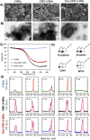
Representative (A) SEM and (B) TEM images of h-BNs, CMC-h-BNs, and Dox-CMC-h-BNs. (C) TGA results. (D) Western blot analysis for N-cadherin, beta-catenin, CD44, and NPTN proteins on Dox-CMC-h-BNs. (E) XPS analysis on h-BNs, CMC-h-BNs, and Dox-CMC-h-BNs.
Figure 2
Figure 2
Cumulative Dox release profile (in %) from Dox-CMCs-h-BNs at increasing time points (4, 24, and 72 h) and under different treatments (at pH 4.5, pH 4.5 + 100 μM H2O2, pH 7.4, and pH 7.4 + 100 μM H2O2).
Figure 3
Figure 3
Biocompatibility studies on HMC3, U87-MG, and astrocytes treated with Dox, CMC-h-BNs, and Dox-CMC-h-BNs for 24 and 72 h. The values are presented as the mean ± standard deviation of 3 different measurements (* p < 0.05).
Figure 4
Figure 4
Flow cytometry quantification of the percentage of Dox-CMC-h-BN-positive cells on HMC3, U87-MG, and astrocyte cultures after 24 and 72 h of incubation.
Figure 5
Figure 5
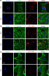
Representative confocal images of (A) HMC3, (B) U87-MG, and (C) astrocytes treated with Dox-CMC-h-BNs (red) for 24 and 72 h. Nuclei (blue) and f-actin (green) are also shown.
Figure 6
Figure 6
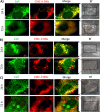
Confocal Raman imaging of (A) HMC3, (B) U87-MG, and (C) astrocytes treated with CMC-h-BNs for 24 and 72 h. Signal maps were obtained according to the signal of h-BNs (in red, Raman shift range: 1350–1380 cm–1) and of phenylalanine (in green, Raman shift range: 980–1020 cm–1).
Figure 7
Figure 7
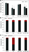
(A) Immunofluorescence analysis on Dox-, CMC-h-BNs-, and Dox-CMC-h-BNs-treated U87-MG cells for Ki-67 marker after 24 and 72 h of incubation (* p < 0.05). Flow cytometry analysis of apoptotic and live cells treated with Dox, CMC-h-BNs, and Dox-CMC-h-BNs after (B) 24 h and (C) 72 h.
Figure 8
Figure 8
Proteomic analysis. (A) Volcano plot and (B) heatmap showing over- and underexpression of proteins for Dox-CMC-h-BNs-treated HMC3 cells with respect to control cultures. (C) Complete list of the gene ontology terms related to the Dox-CMC-h-BN treatment.

References

    1. Rong L.; Li N.; Zhang Z. Emerging Therapies for Glioblastoma: Current State and Future Directions. J. Exp. Clin. Cancer Res. 2022, 41 (1), 1–18. 10.1186/s13046-022-02349-7. - DOI - PMC - PubMed
    1. Alcantara Llaguno S. R.; Parada L. F. Cell of Origin of Glioma: Biological and Clinical Implications. Br. J. Cancer 2016, 115 (12), 1445–1450. 10.1038/bjc.2016.354. - DOI - PMC - PubMed
    1. Baghban R.; Roshangar L.; Jahanban-Esfahlan R.; Seidi K.; Ebrahimi-Kalan A.; Jaymand M.; Kolahian S.; Javaheri T.; Zare P. Tumor Microenvironment Complexity and Therapeutic Implications at a Glance. Cell Commun. Signal. 2020, 18, 1–19. 10.1186/s12964-020-0530-4. - DOI - PMC - PubMed
    1. Wu S.-Y.; Watabe K. The Roles of Microglia/Macrophages in Tumor Progression of Brain Cancer and Metastatic Disease. Front. Biosci. (Landmark Ed.) 2017, 22, 1805.10.2741/4573. - DOI - PMC - PubMed
    1. Lanza M.; Casili G.; Campolo M.; Paterniti I.; Colarossi C.; Mare M.; Giuffrida R.; Caffo M.; Esposito E.; Cuzzocrea S. Immunomodulatory Effect of Microglia-Released Cytokines in Gliomas. Brain Sci. 2021, 11 (4), 466.10.3390/brainsci11040466. - DOI - PMC - PubMed