. 2023 Dec 12;120(50):e2122178120.
doi: 10.1073/pnas.2122178120. Epub 2023 Dec 5.

The TLR2/TLR6 ligand FSL-1 mitigates radiation-induced hematopoietic injury in mice and nonhuman primates

Affiliations

The TLR2/TLR6 ligand FSL-1 mitigates radiation-induced hematopoietic injury in mice and nonhuman primates

W June Brickey et al. Proc Natl Acad Sci U S A. .

Abstract

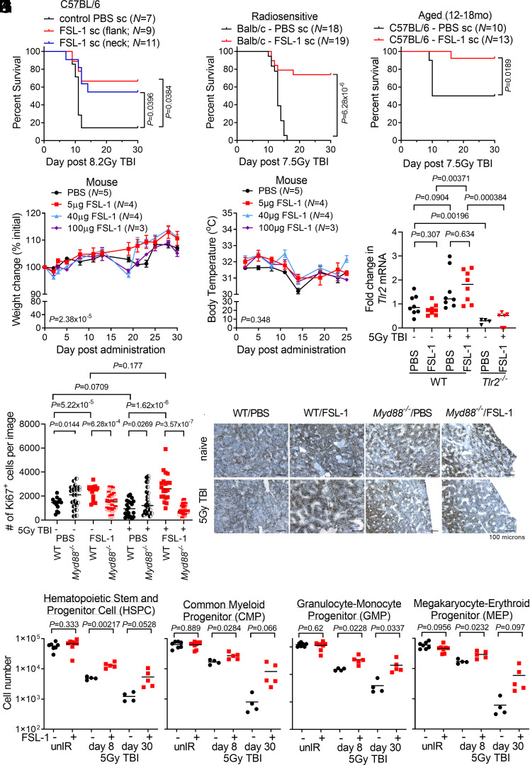
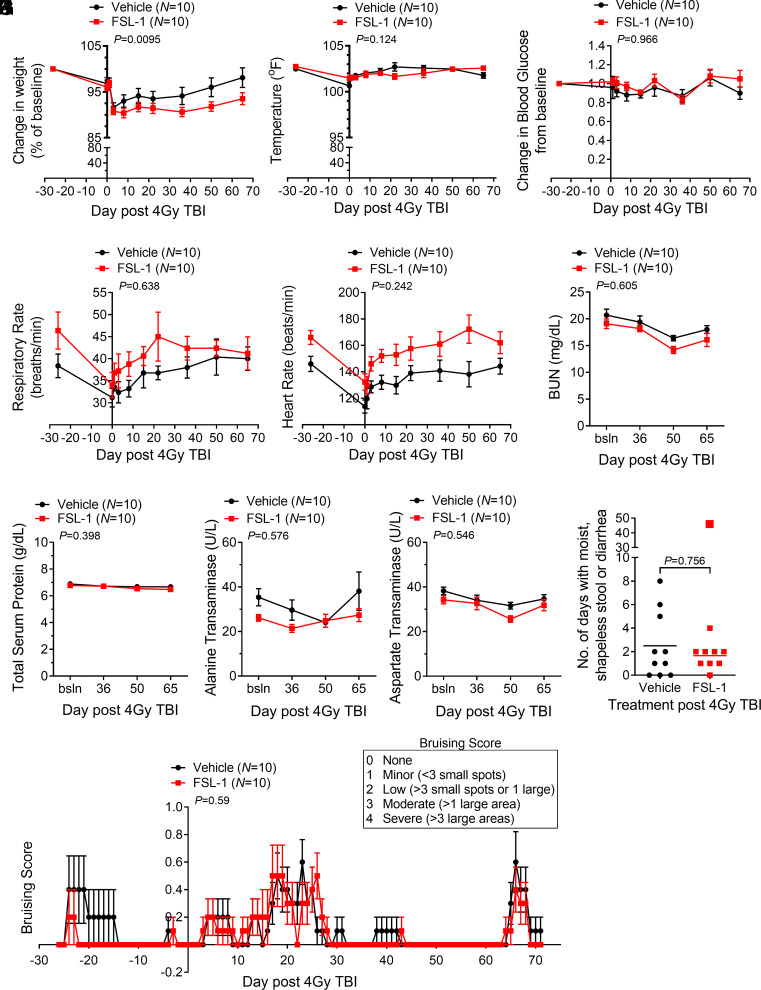
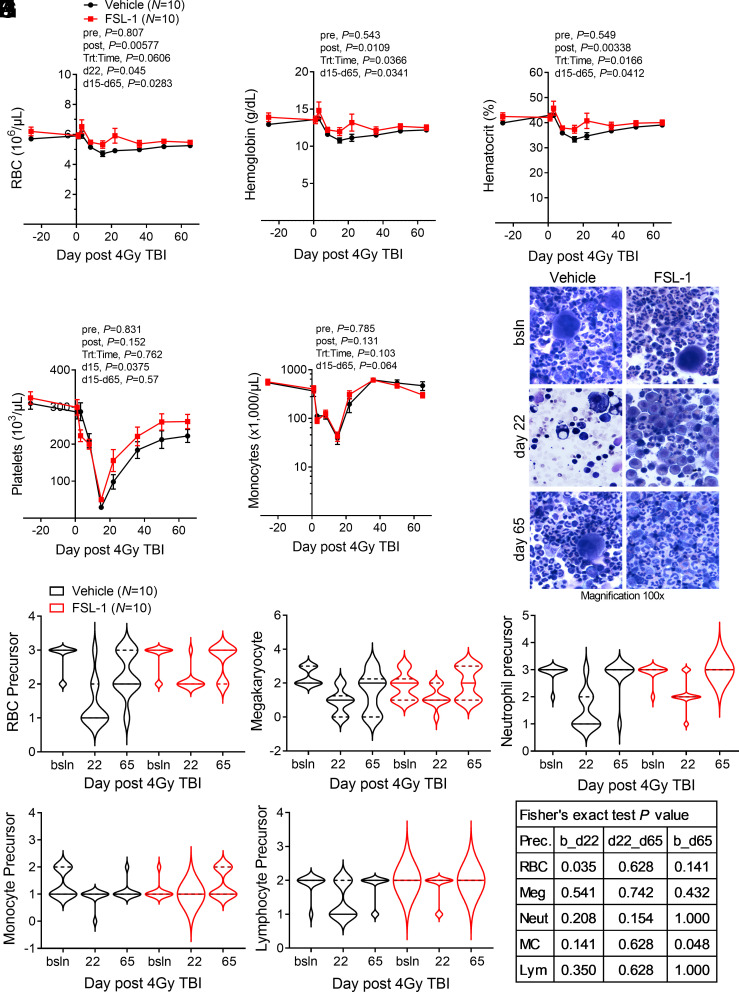
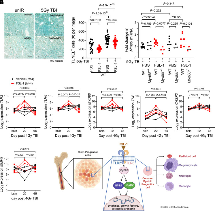
Thrombocytopenia, hemorrhage, anemia, and infection are life-threatening issues following accidental or intentional radiation exposure. Since few therapeutics are available, safe and efficacious small molecules to mitigate radiation-induced injury need to be developed. Our previous study showed the synthetic TLR2/TLR6 ligand fibroblast stimulating lipopeptide (FSL-1) prolonged survival and provided MyD88-dependent mitigation of hematopoietic acute radiation syndrome (H-ARS) in mice. Although mice and humans differ in TLR number, expression, and function, nonhuman primate (NHP) TLRs are like those of humans; therefore, studying both animal models is critical for drug development. The objectives of this study were to determine the efficacy of FSL-1 on hematopoietic recovery in small and large animal models subjected to sublethal total body irradiation and investigate its mechanism of action. In mice, we demonstrate a lack of adverse effects, an easy route of delivery (subcutaneous) and efficacy in promoting hematopoietic progenitor cell proliferation by FSL-1. NHP given radiation, followed a day later with a single subcutaneous administration of FSL-1, displayed no adversity but showed elevated hematopoietic cells. Our analyses revealed that FSL-1 promoted red blood cell development and induced soluble effectors following radiation exposure. Cytologic analysis of bone marrow aspirates revealed a striking enhancement of mononuclear progenitor cells in FSL-1-treated NHP. Combining the efficacy of FSL-1 in promoting hematopoietic cell recovery with the lack of adverse effects induced by a single administration supports the application of FSL-1 as a viable countermeasure against H-ARS.

Keywords: animal models; hematopoiesis; innate immunity; mitigator; radiation.


Conflict of interest statement

Competing interests statement:J.P.Y.T. is a cofounder of and stockholder in IMMvention Therapeutix, which is developing inflammasome inhibitors. US20200282006A1 United States Methods and treatments using toll-like receptor agonists to mitigate hematopoietic myeloid loss, increase gastrointestinal recovery and reduce tumor growth for J.P.Y.T., W.J.B., and H.G.

Figures

Fig. 1.
Fig. 1.
FSL-1 effectively mitigated acute radiation-induced lethality in mice by minimizing adverse effects and enhancing proliferation of hematopoietic progenitor cells. (A) C57BL/6 mice were administered FSL-1 subcutaneously (sc) in abdominal flank or scruffed neck 24 h after 8.2 Gy TBI. (B) Balb/c mice were administered FSL-1 (sc) at 24 h after 7.5 Gy TBI. (C) Aged C57BL/6 mice (12 to 18 mo) were given FSL-1 (sc) after 7.5 Gy TBI. Survival distributions in Kaplan–Meier plots and log-rank test P values are shown. Investigating adverse effects, naive mice were given a single FSL-1 sc injection (5 to 100 µg), and changes in weight (D) or body temperature (E) were monitored with mean ± SEM shown from a representative experiment of three replicate studies. Repeated measurements were assessed using linear mixed-effects modeling with P values for treatment to time interaction indicated. (F) qPCR studies were conducted to assess Tlr2 mRNA in bone marrow (BM) cells from WT and Tlr2−/− mice treated with radiation (5 Gy TBI) with (+) and without (−) FSL-1. (G and H) Femur tissue sections prepared at day 8 post treatment were probed with Ki67 antibodies, imaged and proliferating cells were quantified using ImageJ. Scale bar at 100 µm shown on representative images. Pairwise t tests (unpaired data, unequal variance, two-sided) were applied with P values shown and mean indicated by bar. (I) BM hematopoietic stem and progenitor cells (HSPCs), (J) common myeloid progenitors (CMPs), (K) granulocyte-macrophage progenitors (GMPs), and (L) megakaryocyte-erythroid progenitors (MEPs) in unirradiated (unIR) and irradiated mice BM harvested on days 8 and 30 after PBS or FSL-1 sc injections on day 1 were immunophenotyped by flow cytometry. Two-sample t tests were performed for unpaired BM cell data with Welch’s approximation assuming variance between treatment groups. P values are indicated. Each symbol represents an individual mouse (F and IL) or image of fixed bones (G).
Fig. 2.
Fig. 2.
Nonhuman primates (NHPs) given sublethal total body irradiation followed by one subcutaneous administration of FSL-1 showed few clinical signs of adversity. NHPs were subjected to 4 Gy TBI and given FSL-1 or Vehicle injections (sc) 24 h later. (A) Changes in body weight relative to baseline weight (%) and (B) body temperatures (°F) are indicated. The physiologic measures of blood glucose change (C), respiratory rate (D), and heart rate (E) are indicated. Injury markers in peripheral blood, including blood urea nitrogen (BUN) (F), total serum protein (G), alanine transaminase (H), and aspartate transaminase (I), are shown. Longitudinally repeated blood chemistry and physiologic measures were assessed by linear mixed-effects models. All pretreatment values were considered baseline and used as covariates in the mixed-effects models, with treatment over time P values shown (AE). For variables that indicated baseline categorically, a linear mixed-effects regression model was augmented with a fixed additive effect for the baseline value along with first- and second-order interaction terms (FI). (J) Softened stool or diarrhea over a span of 3 successive days was monitored. The outlier with 48 d of softened stool in the FSL-1 cohort is shown, but not included in statistical analysis. Since the assumption of normality was not appropriate based on the distribution, the Wilcoxon rank-sum test was applied to unpaired data, and asymptotic P value is reported. Incidences of bruising (K) are presented based on subjective scoring as shown in the box. The ordinal logistic regression model including additive fixed effects for time, treatment, and their interaction was applied to analyze treatment on bruising over time. Each Vehicle- and FSL-1-treated cohort are composed of N = 10 NHP with mean ± SEM shown (AI, and K), or each symbol represents an individual (J).
Fig. 3.
Fig. 3.
FSL-1 enhanced hematopoietic cell recovery in the periphery and bone marrow of irradiated NHP. Peripheral blood and BM samples were collected before radiation, at treatment, and at selected times for 65 d after radiation. Blood constituents were analyzed by hematologic assays, enumerating red blood cells (RBCs) (A), hemoglobin (B), hematocrit (C), platelets (D), and monocytes (E). For (AE), mean ± SEM is shown (N = 10/cohort), where lines connect means across time points. Repeated measurements were evaluated using linear mixed-effects models, except where the fitted linear mixed-effects model was singular, such that a Bayesian linear mixed-effects model was implemented pre and post the nadir (at day 15) (AE). As turning points were detected along the time, linear mixed-effects models were fit before (pre) and after (post) the detected turning time (nadir), with baseline as a fixed additive effect for prenadir models. P values for treatment pre/post nadir and for treatment to time interaction post nadir are indicated. Wilcoxon rank-sum testing was performed between Vehicle and FSL-1 treatment cohorts for measures on selected times (days d15 or d22) or for time period (d15 to d65), with indicated P values. (F) BM aspirates from FSL-1- or Vehicle-treated NHP at baseline (bsln) and days 22 and 65 after radiation were stained with Diff-Quik, with representative images at magnification of 100× under oil immersion for each cohort (N = 10) displayed. Abundance of cell types was tallied in a blinded fashion based on a scale from 0 (none) to 3 (most abundant). The violin plots reflect the abundance of precursor subpopulations with medians shown as solid bars and quartiles indicated by dashed bars for (G) RBC, (H) platelets (megakaryocytes), (I) neutrophils, (J) monocytes, and (K) lymphocytes. (L) Abundance changes between time points for Vehicle- vs. FSL-1-treated cohorts were evaluated using Fisher’s exact tests, with P values indicated (bsln or b to d22, d22 to d65 and b to d65).
Fig. 4.
Fig. 4.
FSL-1 stimulated TLR2/TLR6 signaling and activated downstream transcriptional regulation. (A) TUNEL immunohistochemistry of murine femur sections was conducted to study cell death. Femur tissue sections prepared from treated C57BL/6 mice on day 8 after radiation were probed with TUNEL HRP reagent. The scale bar is 100 µm. (B) Quantitation of number (#) of TUNEL+ cells per image was conducted using ImageJ, with each symbol representing an individual femur sample. Statistical significance was determined using pairwise t tests, with P values indicated. (C) Mmp9 mRNA from BM cells harvested at 8 d from FSL-1-treated control and irradiated mice was quantified by qPCR analysis. Each symbol represents an individual mouse with bar indicating the mean. Data (unpaired, unequal variance, two-sided) were evaluated using t tests (unpaired data), with P values indicated. (DI) Transcript profiling of NHP RNA was conducted on BM biopsies from Vehicle (N = 4) and FSL-1 (N = 4) treated irradiated NHP using NanoString technology. String plots for differentially expressed genes (TLR2, TLR6, MyD88, TNF, CASP3, and MMP9) are shown, where connected lines between symbols represent an individual NHP. The t test (unpaired data) was used to decipher changes in transcripts between FSL-1 and Vehicle treatments at bracketed time points between bsln to d22, d22 to d65 or bsln to d65. (J) A mechanism is proposed whereby FSL-1 binds TLR2/TLR6, resulting in activation of MyD88, NF-κB, and MAPK activities, and culminating in transcriptional regulation of cytokines, growth factors, and extracellular matrix components that promote proliferation of hematopoietic progenitors.

References

    1. Farese A. M., MacVittie T. J., Filgrastim for the treatment of hematopoietic acute radiation syndrome. Drugs Today (Barc) 51, 537–548 (2015). - PubMed
    1. McGettrick A. F., O’Neill L. A., Toll-like receptors: Key activators of leucocytes and regulator of haematopoiesis. Br J. Haematol. 139, 185–193 (2007). - PubMed
    1. Burdelya L. G., et al. , An agonist of toll-like receptor 5 has radioprotective activity in mouse and primate models. Science 320, 226–230 (2008). - PMC - PubMed
    1. Vijay-Kumar M., et al. , Flagellin treatment protects against chemicals, bacteria, viruses, and radiation. J. Immunol. 180, 8280–8285 (2008). - PubMed
    1. Gao F., et al. , A critical role of toll-like receptor 2 (TLR2) and its’ in vivo ligands in radio-resistance. Sci. Rep. 5, 13004 (2015). - PMC - PubMed

Publication types

LinkOut - more resources