. 2023 Dec 6;15(1):53.
doi: 10.1038/s41368-023-00259-8.

Dental impact of anti-fibroblast growth factor 23 therapy in X-linked hypophosphatemia

Affiliations

Dental impact of anti-fibroblast growth factor 23 therapy in X-linked hypophosphatemia

Elis J Lira Dos Santos et al. Int J Oral Sci. .

Abstract

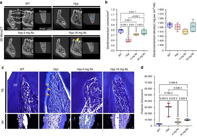
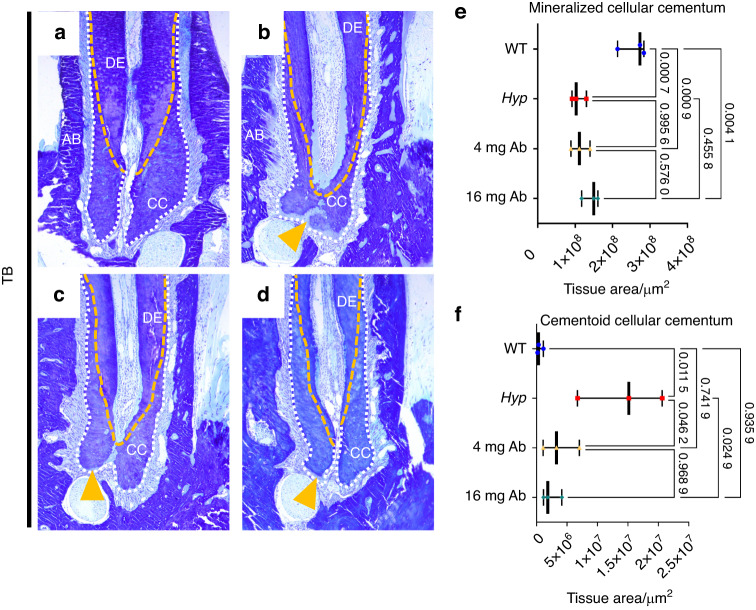
Elevated fibroblast growth factor 23 (FGF23) in X-linked hypophosphatemia (XLH) results in rickets and phosphate wasting, manifesting by severe bone and dental abnormalities. Burosumab, a FGF23-neutralizing antibody, an alternative to conventional treatment (phosphorus and active vitamin D analogs), showed significant improvement in the long bone phenotype. Here, we examined whether FGF23 antibody (FGF23-mAb) also improved the dentoalveolar features associated with XLH. Four-week-old male Hyp mice were injected weekly with 4 or 16 mg·kg-1 of FGF23-mAb for 2 months and compared to wild-type (WT) and vehicle (PBS) treated Hyp mice (n = 3-7 mice). Micro-CT analyses showed that both doses of FGF23-mAb restored dentin/cementum volume and corrected the enlarged pulp volume in Hyp mice, the higher concentration resulting in a rescue similar to WT levels. FGF23-mAb treatment also improved alveolar bone volume fraction and mineral density compared to vehicle-treated ones. Histology revealed improved mineralization of the dentoalveolar tissues, with a decreased amount of osteoid, predentin and cementoid. Better periodontal ligament attachment was also observed, evidenced by restoration of the acellular cementum. These preclinical data were consistent with the retrospective analysis of two patients with XLH showing that burosumab treatment improved oral features. Taken together, our data show that the dentoalveolar tissues are greatly improved by FGF23-mAb treatment, heralding its benefit in clinics for dental abnormalities.


Conflict of interest statement

K.N., A.H. and T.S. are industry scientists at Kyowa Kirin. A.L. reports a research grant from Kyowa Kirin, honoraria, and consulting fees from Pfizer, Novonordisk, Merck Serono and Sandoz, unrelated to this work. M.B.D. reports a research grant and honoraria from Kyowa Kirin. C.C. reports a research collaborative grant from Kyowa Kirin for URP2496 related to this work, and consulting fees from Novonordisk for URP2496 unrelated to this work. The other authors report no conflicts of interest.

Figures

Fig. 1
Fig. 1
Improved pulp chamber size and bone features in patients with XLH treated with burosumab. ad Representative periapical radiographs of a 20-y-old female with XLH treated with burosumab for 4 years. Blue dotted lines delimitate pulp chamber; Yellow dotted lines highlight reduced pulp chamber sizes after treatment. e Measurements of pulp chamber. f, g Representative images of CBCT in the mandibular premolar region from a 49-year-old male with XLH before and after treatment with burosumab for 6 years. h Bone fraction (BV/TV) at baseline and following treatment
Fig. 2
Fig. 2
Impact of FGF23-mAb therapy on dentin in 3-month-old Hyp mice. a Two-dimensional micro-computed tomography (mCT) shows mandibular first molar enamel (E), dentin (DE), pulp chamber (PC) and alveolar bone (AB). Hyp mice present enlarged pulp chamber (red asterisk), thin dentin and altered alveolar bone (blue hashtag). Treatment shows dentin formation (yellow arrow) with 4 or 16 mg·kg−1 of FGF23-mAb. b Both concentrations of FGF23-mAb restored dentin/cementum volume, the higher concentration resulting in a rescue similar to WT levels. c, d Toluidine blue (TD) staining (crown region, upper panel) reveals wide predentin (PD) (yellow *) and erratic PD-DE border with interglobular DE patterns in vehicle treated Hyp mice (yellow arrows). DE and PD are fully normalized by FGF23-mAb treatment at a dosage of 4 mg·kg−1 and 16 mg·kg−1. Von Kossa (VK) staining (root region) highlights the reduced predentin and improved calcospherite fusion during dentin mineralization in 3 months old FGF23 treated Hyp mice
Fig. 3
Fig. 3
Impact of FGF23-mAb therapy on pulp in Hyp mice. ae Three-dimensional micro-computed tomography (mCT) shows that enlargement of pulp chambers was also improved with a reduced pulp volume in treated Hyp mice
Fig. 4
Fig. 4
Histological analyses of the impact of FGF23-mAb therapy on the periodontal ligament in 3-month-old Hyp mice. ah Masson’s Trichrome (MT) staining revealed decreased periodontal ligament attachment in Hyp mice. fh Yellow arrowheads indicates defective Periodontal ligament (PDL) attachment. il Picrosirius red (PR) staining viewed under polarized light microscopy emphasizes highly organized periodontal ligament (PDL) fibers in wild-type (WT) mice (blue dotted line). m, n Hyp mice have a loss of PDL fiber organization and no alteration in PDL thickness, and higher dose treatment improved functional attachment between acellular cementum (AC) and alveolar bone (AB)
Fig. 5
Fig. 5
Histological analyses of the impact of FGF23-mAb therapy on cellular cementum in 3-month-old Hyp mice. ad Cementoid accumulation is observed in Hyp mice compared to WT mice (yellow arrow). ef Histomorphometry confirms significant effect of FGF23-mAb with complete rescue of cellular cementum mineralization
Fig. 6
Fig. 6
Impact of FGF23-mAb treatment on alveolar bone mineralization in Hyp mice. a Three-dimensional micro-computed tomography (mCT) present mandibular alveolar bone in the first molar region. b Treatment with both doses of FGF23-mAb improved alveolar bone volume fraction (BV/TV), bone mineral density (BMD) and total mineral density (TMD) in Hyp mice
Fig. 7
Fig. 7
Histological analyses of the impact of FGF23-mAb therapy on bone in 3-month-old Hyp mice. ad Von Kossa staining showed poor mineralization (in dark) of the collagen matrix revealing an increased amount of osteoid (yellow*) in Hyp mice alveolar bone. e Histomorphometry analysis demonstrated considerable osteoid reduction of alveolar bone after treatment with 4 mg or 16 mg·kg−1 of FGF23-mAb
Fig. 8
Fig. 8
Histomorphometrical analysis of alveolar bone in FGF23-mAb treated Hyp mice. ad Undecalcified sections automate segmented to measure osteoid in mice mandibles. e Histomorphometry analysis demonstrated a significant reduction of osteoid area/bone area (O.Ar/B.Ar) in alveolar bone after treatment with 4 mg or 16 mg·kg−1 of FGF23-mAb. f Alterations in osteocyte number (N.Ot) are observed in Hyp mice compared to WT mice, with full recovery by 16 mg·kg−1 FGF23-mAb treatment. gj Masson’s Trichrome (MT) staining demonstrated distribution of osteocytes (Ocy) in alveolar bone and the region of interest (ROI). CEJ Cementum enamel junction
Fig. 9
Fig. 9
Expression of bone and cementum markers in FGF23-mAb treated Hyp mice. ad Osteopontin (OPN) is a marker of acellular cementum (AC) and alveolar bone (AB), and all groups presented OPN staining on root surfaces, even though Hyp mice have a defective pattern (black arrows). Both treatments showed improved localization of OPN in acellular cementum. OPN is not similarly distributed in Hyp versus WT alveolar bone, and OPN is present in Hyp mice alveolar bone with both doses of treatment, showing a localization at the bone surface and around osteocytes. e–h Bone sialoprotein (BSP) is a marker of AC and AB. AC defects in Hyp mice are shown as irregular BSP staining (purple arrows), however, both 4 mg or 16 mg·kg−1 of FGF23-mAb improve BSP localization on root dentin surfaces

References

    1. Carpenter TO, Imel EA, Holm IA, Jan de Beur SM, Insogna KL. A clinician’s guide to X-linked hypophosphatemia. J. Bone Min. Res. 2011;26:1381–1388. doi: 10.1002/jbmr.340. - DOI - PMC - PubMed
    1. Hennig S, et al. A gene (PEX) with homologies to endopeptidases is mutated in patients with X-linked hypophosphatemic rickets. The HYP Consortium. Nat. Genet. 1995;11:130–136. doi: 10.1038/ng1095-130. - DOI - PubMed
    1. Zhukouskaya VV, et al. Increased prevalence of overweight and obesity in children with x-linked hypophosphatemia. Endocr. Connect. 2020;9:144–153. doi: 10.1530/EC-19-0481. - DOI - PMC - PubMed
    1. Chaussain-Miller C, et al. Dental abnormalities in patients with familial hypophosphatemic vitamin D-resistant rickets: prevention by early treatment with 1-hydroxyvitamin D. J. Pediatr. 2003;142:324–331. doi: 10.1067/mpd.2003.119. - DOI - PubMed
    1. Haffner D, et al. Clinical practice recommendations for the diagnosis and management of X-linked hypophosphataemia. Nat. Rev. Nephrol. 2019;15:435–455. doi: 10.1038/s41581-019-0152-5. - DOI - PMC - PubMed

Publication types

MeSH terms