. 2023 Dec 6;14(1):8064.
doi: 10.1038/s41467-023-43718-w.

Design and structural validation of peptide-drug conjugate ligands of the kappa-opioid receptor

Affiliations

Design and structural validation of peptide-drug conjugate ligands of the kappa-opioid receptor

Edin Muratspahić et al. Nat Commun. .

Abstract

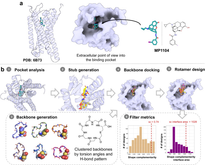
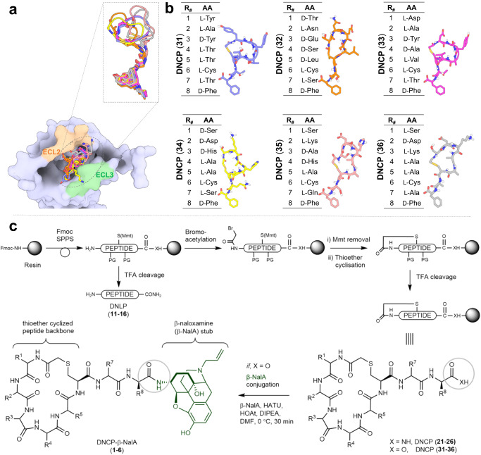
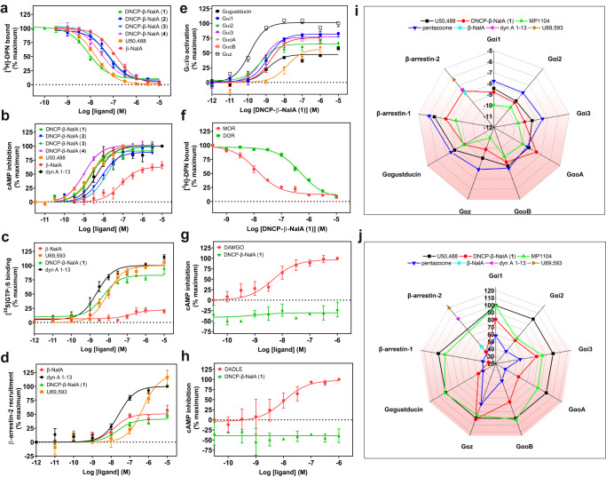
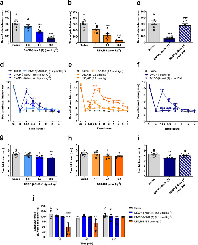
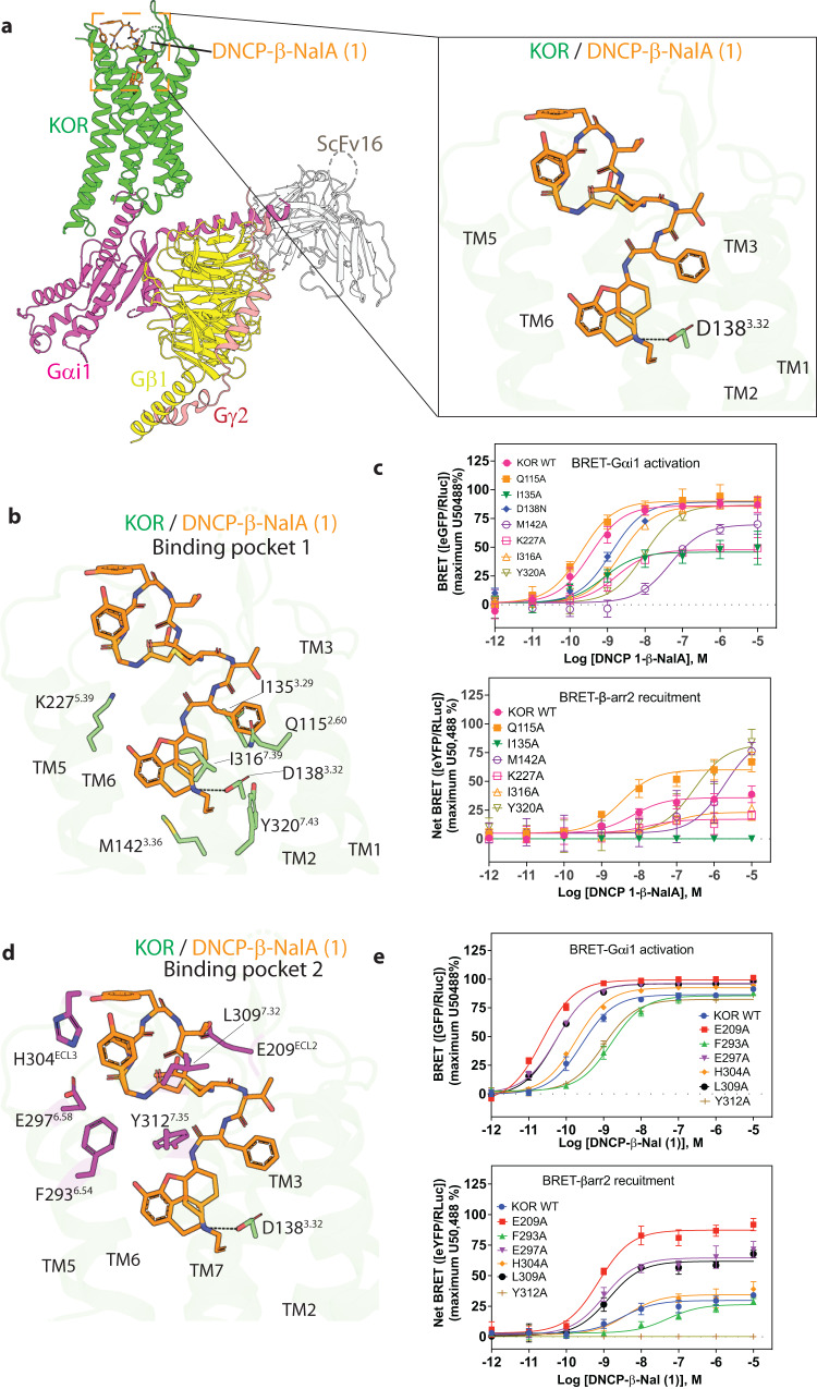
Despite the increasing number of GPCR structures and recent advances in peptide design, the development of efficient technologies allowing rational design of high-affinity peptide ligands for single GPCRs remains an unmet challenge. Here, we develop a computational approach for designing conjugates of lariat-shaped macrocyclized peptides and a small molecule opioid ligand. We demonstrate its feasibility by discovering chemical scaffolds for the kappa-opioid receptor (KOR) with desired pharmacological activities. The designed De Novo Cyclic Peptide (DNCP)-β-naloxamine (NalA) exhibit in vitro potent mixed KOR agonism/mu-opioid receptor (MOR) antagonism, nanomolar binding affinity, selectivity, and efficacy bias at KOR. Proof-of-concept in vivo efficacy studies demonstrate that DNCP-β-NalA(1) induces a potent KOR-mediated antinociception in male mice. The high-resolution cryo-EM structure (2.6 Å) of the DNCP-β-NalA-KOR-Gi1 complex and molecular dynamics simulations are harnessed to validate the computational design model. This reveals a network of residues in ECL2/3 and TM6/7 controlling the intrinsic efficacy of KOR. In general, our computational de novo platform overcomes extensive lead optimization encountered in ultra-large library docking and virtual small molecule screening campaigns and offers innovation for GPCR ligand discovery. This may drive the development of next-generation therapeutics for medical applications such as pain conditions.


Conflict of interest statement

S.M. is a co-founder of Sparian Biosciences. The other authors declare no competing interests.

Figures

Fig. 1
Fig. 1. Strategy for the computational design of thioether macrocyclized peptide–small molecule conjugates targeting KOR.
a 3D human KOR structure with small molecule agonist MP1104 was used as the starting template (PDB: 6B73). b Workflows for computational peptide–small molecule conjugate design: (1) Measurement of the pocket area (934 Å3) narrowed down the size of macrocycles to focus on 5- and 6-mer cyclic peptides. (2) Generation of small molecules with two additional amino acids, which were sampled and scored for optimal dimer sequence (select dipeptide modified small molecules: Gray: CVV-D-Phe-Thr; Yellow: CVV-D-Phe-Gln; Orange: CVV-D-Phe-Ser; CVV corresponds to N-cyclopropylmethyl-epoxy morphinan small molecule stub). (3) Generation of a comprehensive library of 5- and 6-mer thioether cyclized peptides clustered via torsion angle and hydrogen bond pattern. (4) Docked structure of thioether macrocyclized hexamers through coordinate-guided transformation of the backbone C-termini to the generated anchor N-termini. (5) Rotamer design to optimize the interface interactions of the backbones. (6) Design filtering based on shape complementarity and interface area as representative examples for interface metrics; dashed red line represents 90th percentile cut-off values.
Fig. 2
Fig. 2. Overview of the selected peptide macrocycle designs and the synthetic strategy to produce the peptide–small molecule conjugates and controls.
a Peptides have interactions with ECL2 and/or ECL3 of KOR with a subset overlay of the peptide backbone designs (aligned by the small molecule fragment anchor) showing shape diversity in the pocket. b Final selection of peptide sequences. Instead of focusing on multiple sequences for a single promising backbone, we sought to select designs across diverse shapes and sequences for experimental testing. c Synthetic scheme depicting solid phase synthesis of the de novo linear peptides (DNLP) (1116) and the de novo cyclic peptides (DNCP) (2126 and 3136) and the solution phase conjugation reaction with β-naloxamine (β-NalA) to generate the DNCP-β-NalA (16) conjugates. R# indicates a side chain of the respective amino acid. PG denotes protecting groups. Rink amide resin was used for synthesis of DNLP (1116) and DNCP (2126), whereas Fmoc-D-Phe preloaded Wang resin was used for DNCP (31–36) synthesis. The amino acids indicated by R# in Fig. 2b correspond to the identical side chain represented by R# in Fig. 2c.
Fig. 3
Fig. 3. In vitro receptor pharmacology of peptide–small molecule conjugates.
a, b Radioligand binding (n = 3) and functional cAMP assays (n = 3–4) of DNCP-β-NalA conjugates (14) were performed on HEK293T cell membranes stably expressing mouse KOR. Binding (a) was measured by displacing 1 nM of [3H]DPN whereas cAMP inhibition (b) was monitored after treatment with indicated concentrations of conjugates. U50,488 and β-NalA were positive controls. Final concentration of 10 µM of forskolin was used to stimulate cAMP production (Supplementary Table 3). c Concentration-dependent stimulation of [35S]GTPγS binding by the most potent DNCP-β-NalA(1) (n = 3), β-NalA (n = 3), U69,593 (n = 3) and dyn A1-13 (n = 4) in human KOR expressing CHO cell membranes (Supplementary Table 4). d β-arrestin-2 recruitment assay of DNCP-β-NalA(1), β-NalA and dynorphin (dyn) A1-13 was done in HEK293T cells transiently expressing mouse KOR-EGFP and β-arrestin-2-nano-luciferase (n = 3–6) (Supplementary Table 3). e α-Subtype screening of DNCP-β-NalA(1) at the mouse KOR in the TRUPATH assay (n = 8) (Supplementary Tables 5 and 6). f Selectivity of DNCP-β-NalA(1) was determined in a radioligand binding assay using HEK293T cell membrane preparations stably expressing mouse MOR and DOR and 1 nM of [3H]DPN, respectively (n = 3). g, hi-mediated cAMP inhibition of DNCP-β-NalA(1) at the mouse MOR (g) and DOR (h) was measured in stable HEK293T cells using DAMGO and DADLE as reference ligands, respectively (n = 3). i, j Spider plots from TRUPATH (n = 8 for each Gα), β-arrestin-1 (n = 4) and β-arrestin-2 (n = 3–6) recruitment assays represent potency (log EC50) (i) and normalized efficacy (j) of DNCP-β-NalA(1), U50,488, MP1104, pentazocine, β-NalA and dyn A1-13. Data were normalized to full KOR agonists U50,488, U69,593 or dyn A1-13, full MOR agonist DAMGO and full DOR agonist DADLE. All data are presented as mean values ± s.e.m. Source data are provided as a Source Data file.
Fig. 4
Fig. 4. In vivo pharmacology of DNCP-β-NalA(1) after s.c. administration in male mice.
a, b Formalin test, dose-dependent effect; groups: saline (n = 8 mice), DNCP-β-NalA(1) (0.8 µmol kg−1, n = 6 mice; 1.9 µmol kg−1, n = 6 mice; 3.8 µmol kg−1, n = 6 mice) and U50,488 (1.1 µmol kg−1, n = 6 mice; 2.1 µmol kg−1, n = 6 mice; 5.4 µmol kg−1, n = 6 mice); One-way ANOVA, F(3, 22) = 19.97, P < 0.0001 (a), and F(3, 22) = 27.10, P < 0.0001 (b). c Formalin test, antagonism by nor-BNI; groups: saline (n = 8 mice), DNCP-β-NalA(1) (3.8 µmol kg−1, n = 6 mice) and DNCP-β-NalA(1)+nor-BNI (3.8 µmol kg−1 + 13.6 µmol kg−1, n = 6 mice); One-way ANOVA, F(2, 17) = 29.67, P < 0.0001. d, e CFA-induced inflammatory hyperalgesia, dose- and time-dependent effect; groups: saline (n = 8 mice), DNCP-β-NalA (1) (0.4 µmol kg−1, n = 6 mice; 0.8 µmol kg−1, n = 6 mice; 1.9 µmol kg−1, n = 6 mice) and U50,488 (0.2 µmol kg−1, n = 6 mice; 0.6 µmol kg−1, n = 7 mice; 2.1 µmol kg−1, n = 8 mice); Two-way ANOVA, F(3, 176) = 46.10, P < 0.0001 (d), and F(3, 275) = 173.0, P < 0.0001 (e). f CFA-induced inflammatory hyperalgesia, antagonism by nor-BNI; groups: saline (n = 8 mice), DNCP-β-NalA(1) (1.9 µmol kg−1, n = 6 mice) and DNCP-β-NalA(1)+nor-BNI (1.9 µmol kg−1+13.6 µmol kg−1, n = 6 mice); Two-way ANOVA, F(2, 136) = 91.61, P < 0.0001. g, h Paw thickness, dose-dependent effect; groups: saline (n = 8 mice), DNCP-β-NalA(1) (0.8 µmol kg−1, n = 6 mice; 1.9 µmol kg−1, n = 6 mice; 3.8 µmol kg−1, n = 6 mice) and U50,488 (1.1 µmol kg−1, n = 6 mice; 2.1 µmol kg−1, n = 6 mice; 5.4 µmol kg−1, n = 6 mice); One-way ANOVA, F(3, 22) = 5.016, P = 0.0084 (g), and F(3, 22) = 1.770, P = 0.1823. i Paw thickness, antagonism by nor-BNI; groups: saline (n = 8 mice), DNCP-β-NalA(1) (3.8 µmol kg−1) and DNCP-β-NalA(1)+nor-BNI (3.8 µmol kg−1 + 13.6 µmol kg−1, n = 6 mice); One-way ANOVA, F(2, 17) = 7.239, P = 0.0053. j Rotarod test, motor coordination; groups: saline (n = 5 mice), DNCP-β-NalA(1) (3.8 µmol kg−1, n = 6 mice; 7.6 µmol kg−1, n = 5 mice) and U50,488 (5.4 µmol kg−1, n = 5 mice); Two-way ANOVA, F(3, 51) = 7.992, P = 0.0002. One-way ANOVA with Dunnett’s (a, b, g, h) and Tukey’s post hoc test (c, i); Two-way ANOVA with Bonferroni’s post-hoc test for (df, j). *P < 0.05, **P < 0.01, ***P < 0.001, drug vs. saline group; #P < 0.05, ##P < 0.001, DNCP-β-NalA(1) vs. DNCP-β-NalA(1)+nor-BNI. All data represent means ± s.e.m. Source data are provided as a Source Data file.
Fig. 5
Fig. 5. Cryo-EM structure of the KOR-DNCP-β-NalA(1)-Gi1 complex.
a Overall architecture of the active state human KOR bound to DNCP-β-NalA(1) and G-protein heterotrimer (Gαi1, Gβ1, Gγ2). The KOR-G-protein complex was further stabilized by a single-chain antibody scFv16. The right panel shows the binding pose of DNCP-β-NalA(1) at KOR. The highly conserved anchoring residue D138, as part of the orthosteric binding pocket of KOR is shown. b The interactions between the bottom half of DNCP-β-NalA(1) and the orthosteric site of KOR. c Effects of orthosteric residues on DNCP-β-NalA(1)-mediated Gαi1 protein and β-arrestin-2 signaling (n = 3). For KOR D138N mutant, the reference ligand is salvinorin A because U50,488 is inactive at this mutant. d The interactions between the peptide macrocycle of DNCP-β-NalA(1) and the extracellular binding pocket 2 of KOR. e Effects of binding pocket 2 residues on DNCP-β-NalA(1)-mediated Gαi1 protein and β-arrestin-2 signaling (n = 3). All data are presented as mean values ± s.e.m. Source data are provided as a Source Data file.

References

    1. Davenport AP, Scully CCG, de Graaf C, Brown AJH, Maguire JJ. Advances in therapeutic peptides targeting G protein-coupled receptors. Nat. Rev. Drug Discov. 2020;19:389–413. doi: 10.1038/s41573-020-0062-z. - DOI - PubMed
    1. Hauser AS, Attwood MM, Rask-Andersen M, Schioth HB, Gloriam DE. Trends in GPCR drug discovery: new agents, targets and indications. Nat. Rev. Drug Discov. 2017;16:829–842. doi: 10.1038/nrd.2017.178. - DOI - PMC - PubMed
    1. Muratspahic E, Freissmuth M, Gruber CW. Nature-derived peptides: a growing niche for GPCR ligand discovery. Trends Pharmacol. Sci. 2019;40:309–326. doi: 10.1016/j.tips.2019.03.004. - DOI - PMC - PubMed
    1. Muttenthaler M, King GF, Adams DJ, Alewood PF. Trends in peptide drug discovery. Nat. Rev. Drug Discov. 2021;20:309–325. doi: 10.1038/s41573-020-00135-8. - DOI - PubMed
    1. Volkow ND, Collins FS. The role of science in addressing the opioid crisis. N. Engl. J. Med. 2017;377:391–394. doi: 10.1056/NEJMsr1706626. - DOI - PubMed

Publication types