. 2023 Dec 5;14(1):8042.
doi: 10.1038/s41467-023-43798-8.

A lung-selective delivery of mRNA encoding broadly neutralizing antibody against SARS-CoV-2 infection

Affiliations

A lung-selective delivery of mRNA encoding broadly neutralizing antibody against SARS-CoV-2 infection

Wanbo Tai et al. Nat Commun. .

Abstract

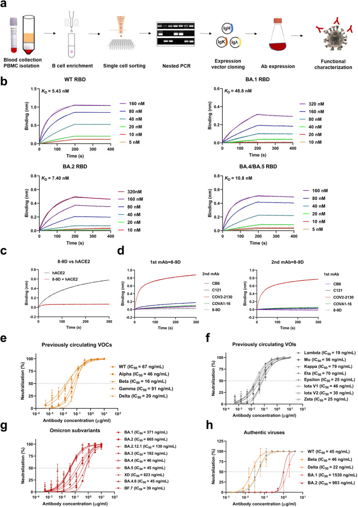
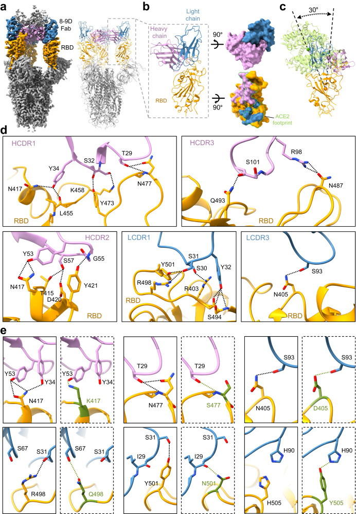
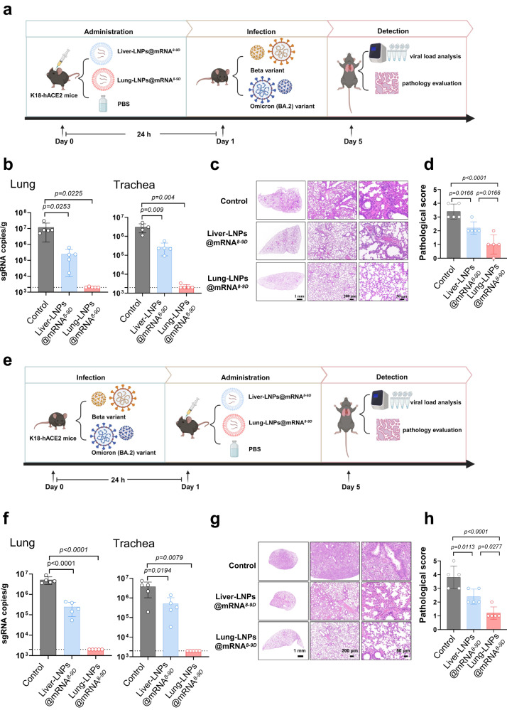
The respiratory system, especially the lung, is the key site of pathological injury induced by SARS-CoV-2 infection. Given the low feasibility of targeted delivery of antibodies into the lungs by intravenous administration and the short half-life period of antibodies in the lungs by intranasal or aerosolized immunization, mRNA encoding broadly neutralizing antibodies with lung-targeting capability can perfectly provide high-titer antibodies in lungs to prevent the SARS-CoV-2 infection. Here, we firstly identify a human monoclonal antibody, 8-9D, with broad neutralizing potency against SARS-CoV-2 variants. The neutralization mechanism of this antibody is explained by the structural characteristics of 8-9D Fabs in complex with the Omicron BA.5 spike. In addition, we evaluate the efficacy of 8-9D using a safe and robust mRNA delivery platform and compare the performance of 8-9D when its mRNA is and is not selectively delivered to the lungs. The lung-selective delivery of the 8-9D mRNA enables the expression of neutralizing antibodies in the lungs which blocks the invasion of the virus, thus effectively protecting female K18-hACE2 transgenic mice from challenge with the Beta or Omicron BA.1 variant. Our work underscores the potential application of lung-selective mRNA antibodies in the prevention and treatment of infections caused by circulating SARS-CoV-2 variants.


Conflict of interest statement

G.Y., G.C., W.T., K.Y., and Y.L. have applied for the patent (202310810894X) related to the lung-selective LNP formulation and its preparation. All other authors declare that they have no competing interests.

Figures

Fig. 1
Fig. 1. Characterization of SARS-CoV-2 targeting monoclonal antibody 8-9D.
a Schematic for the neutralizing monoclonal antibody selection and evaluation. b Binding affinity of 8-9D to RBDs of WT, Omicron BA.1, Omicron BA.2 and Omicron BA.4/BA.5 by biolayer interferometry (BLI). The association and dissociation of the response curves of 8-9D are shown. The gray lines represent the fitted curves based on the experimental data. Equilibrium dissociation constants (KD) are shown above each plot. c Antibody 8-9D and hACE2 competition for binding to SARS-CoV-2 RBD determined by BLI. The traces show the binding of the hACE2 to preformed 8-9D-RBD complexes. The level of reduction in shift of 8-9D + hACE2 compared to the hACE2-only control indicates the blocking capacity of the antibody. d Epitope competition measured by BLI. The antibody 8-9D was assayed for epitope specificity with 4 structurally defined monoclonal antibodies, CB6 (class 1), C121 (class 2), COV2-2130 (class 3), and COVA1-16 (class 4). The traces represent the binding of the second antibody (2nd Ab) to preformed first antibody (1st Ab)-RBD complexes in an in-tandem binning assay. eg Neutralization of monoclonal antibody 8-9D against pseudotyped SARS-CoV-2 wild type, previously circulating variants of concern (VOCs) (e), previously circulating variants of interest (VOIs) (f), and Omicron subvariants (g). The 50% inhibitory concentration (IC50) values are shown. The dashed line indicates a 50% reduction in viral infectivity. The curves were fitted by nonlinear regression (log [inhibitor] vs. normalized response, variable slope), n = 2 biologically independent samples, and the results are representative of 2 or more independent experiments with similar results. Data are mean ± SEM of the experiments. h Neutralizing activity of 8-9D against authentic SARS-CoV-2 wild type, Beta, Delta, Omicron BA.1, and Omicron BA.2 variants as in (eg). Source data are provided as a Source data file.
Fig. 2
Fig. 2. Structural characterization of monoclonal antibody 8-9D.
a Surface rendered representations and ribbon diagrams showing the structure of the Omicron BA.5 spike in complex with three 8-9D Fabs. Only the variable region of 8-9D Fab is displayed in the ribbon diagrams (right). The “up” RBDs are in orange, the heavy chain of 8-9D is colored pink, and the light chain is colored blue. The selected region in the dashed square shows one RBD with one bound Fab. b Ribbon diagrams showing the RBD interacting with the variable region of the 8-9D Fab (left). The rendered surface of 8-9D Fab and RBD in an open-up view (right). The epitopes on the RBD recognized by the 8-9D heavy chain and light chain are colored pink and blue, respectively. The ribbons of 8-9D Fab and RBD are colored the same as in (a). The ACE2 footprint on RBD is highlighted with green lines. c Ribbon diagrams showing the comparisons of the binding orientations of ACE2 and 8-9D on the RBD. The 8-9D Fab and RBD are colored the same as in (a) and ACE2 is colored green. The green, pink, and orange balls are the centroids of ACE2, 8-9D Fab, and RBD, respectively. d Detailed ribbon and stick diagrams showing the hydrogen bonds between the RBD and complementarity-determining region (CDR) of 8-9D heavy and light chains. RBD, 8-9D heavy and light chains are colored the same as in (a). Residues involved in hydrogen bond formation are shown in sticks with oxygen and nitrogen atoms colored red and blue, respectively. e Ribbon and stick diagrams comparing the corresponding residues in Omicron BA.5 RBD (in a solid rectangle) and in the wild-type RBD (in a dashed rectangle). The residues and hydrogen bonds in wild type RBD are colored green. The complex structure of 8-9D and the wild-type RBD was obtained through structural modeling with COOT and Foldit Standalone. RBD, 8-9D heavy and light chains are colored the same as in (a).
Fig. 3
Fig. 3. Characterization of organ-selective lipid nanoparticle systems.
a Structural formula and schematic diagram of each component of lung-selective and liver-selective LNPs. b Schematic diagram of lung-selective and liver-selective LNPs. c Transmission electron microscopy (TEM) results: upper image (Lung-LNPs) and lower image (Liver-LNPs). d, e Dynamic light scattering (DLS) results for liver-selective (d) and lung-selective LNPs (e). f Particle size change for two particles in PBS, data are shown as the mean ± SD, n = 3 biologically independent samples. g, h Zeta potential analysis of liver-selective (g) and lung-selective LNPs (h), n = 3 independent experiments. i, In vivo imaging of liver-selective and lung-selective LNPs@mRNALuci at different time points, n = 4 for each LNPs@mRNALuci at different time points. j, k Visceral imaging of liver-selective and lung-selective LNPs@mRNALuci. The organs are heart (h), lung (Lu), liver (Li), stomach (St), intestine (I), kidney (K), and spleen (Sp). l The statistical results of luminescence intensity from mice inoculated with liver-selective and lung-selective LNPs@mRNALuci at different time points, the ROI of liver-selective LNP statistics was the liver, and the ROI of lung-selective LNP statistics was the lungs, data are shown as the mean ± SD, n = 4 independent samples. m Imaging liver tissue or lung tissue of mice inoculated with liver-selective and lung-selective LNPs@mRNAeGFP at 24 h post injection. Source data are provided as a Source data file.
Fig. 4
Fig. 4. In vivo activity and protective efficacy of 8-9D mRNA.
a Schematic for distribution and kinetics analysis of 8-9D mRNA after injection. C57BL/6 mice were i.v. injected with 5 μg of 8-9D mRNA with Lung-LNPs or Liver-LNPs as the delivery system. The animals (n = 5) were euthanized at 24 h post injection. be The sera (b), BALF (c), livers (d), and lungs (e) were collected for 8-9D detection by ELISA. 8-9D kinetics levels in BALF (f) and lungs (g) were further measured for the samples (n = 3) collected on day 2, 4, 8, 15, and 21 post injection. IL-6, TNF-α and IL-1β levels in sera at 1 day, 2 days, 4 days, and 8 days post injection of Lung-LNPs@mRNA8-9D (hj). Data are presented as mean ± SD and representative of two independent experiments with similar results. P values were determined by one-way ANOVA with Tukey’s multiple comparison post-hoc test. Source data are provided as a Source data file.
Fig. 5
Fig. 5. 8-9D mRNA protective efficacy in SARS-CoV-2 animal model.
ad 8-9D mRNA protective efficacy as prophylaxis, the K18-hACE2 mice (n = 5) were i.v. injected with 5 μg of Liver-LNPs@mRNA8-9D or Lung-LNPs@mRNA8-9D. Twenty-four hours post LNP injection, the mice were challenged with 2 × 10⁴ TCID50 Beta variant. Four days post challenge, the mice were euthanized, and the lungs and tracheae were collected for viral load analysis by qRT–PCR, n = 5 biologically independent animals (b). The lungs were fixed for pathology evaluation (c), and pathological scores (d) were determined for significant comparison. PBS-injected mice were set as control, n = 5 biologically independent animals. eh 8-9D mRNA protective efficacy as treatment. The mice were first anesthetized and intranasally inoculated with 2 × 10⁴ TCID50 of authentic SARS-CoV-2 Beta variant, and after 24 h, the mice were intravenously administered one dose of 5 μg of Liver-LNPs@mRNA8-9D or Lung-LNPs@mRNA8-9D, or PBS alone as a control. The mice were euthanized 4 days post infection to harvest lung tissues and trachea tissues for viral load test (f) or histopathology evaluation (g, h). Data are presented as mean ± SD and representative of two independent experiments with similar results, n = 5 biologically independent animals. P values were determined by one-way ANOVA with Tukey’s multiple comparison post-hoc test. Source data are provided as a Source data file.

References

    1. WHO. WHO Coronavirus (COVID-19) Dashboard. https://covid19.who.int (2023).
    1. Menni C, et al. Vaccine side-effects and SARS-CoV-2 infection after vaccination in users of the COVID Symptom Study app in the UK: a prospective observational study. Lancet Infect. Dis. 2021;21:939–949. doi: 10.1016/S1473-3099(21)00224-3. - DOI - PMC - PubMed
    1. Baden LR, et al. Efficacy and Safety of the mRNA-1273 SARS-CoV-2 Vaccine. N. Engl. J. Med. 2021;384:403–416. doi: 10.1056/NEJMoa2035389. - DOI - PMC - PubMed
    1. Voysey M, et al. Safety and efficacy of the ChAdOx1 nCoV-19 vaccine (AZD1222) against SARS-CoV-2: an interim analysis of four randomised controlled trials in Brazil, South Africa, and the UK. Lancet. 2021;397:99–111. doi: 10.1016/S0140-6736(20)32661-1. - DOI - PMC - PubMed
    1. Logunov DY, et al. Safety and immunogenicity of an rAd26 and rAd5 vector-based heterologous prime-boost COVID-19 vaccine in two formulations: two open, non-randomised phase 1/2 studies from Russia. Lancet. 2020;396:887–897. doi: 10.1016/S0140-6736(20)31866-3. - DOI - PMC - PubMed

Publication types

Supplementary concepts