. 2023 Dec 5;24(1):279.
doi: 10.1186/s13059-023-03110-9.

Dengue and Zika RNA-RNA interactomes reveal pro- and anti-viral RNA in human cells

Affiliations

Dengue and Zika RNA-RNA interactomes reveal pro- and anti-viral RNA in human cells

Kuo-Chieh Liao et al. Genome Biol. .

Abstract

Background: Identifying host factors is key to understanding RNA virus pathogenicity. Besides proteins, RNAs can interact with virus genomes to impact replication.

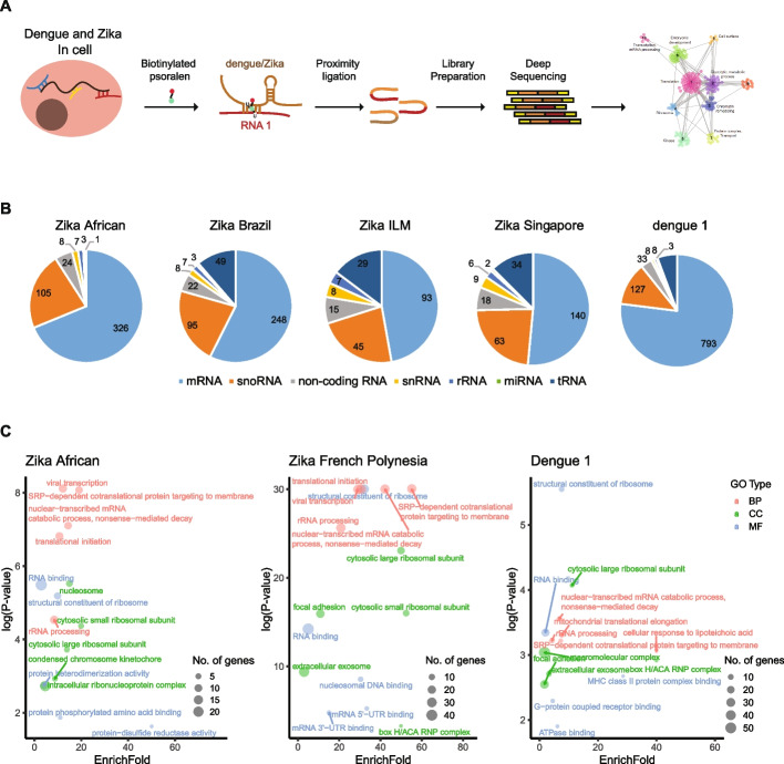
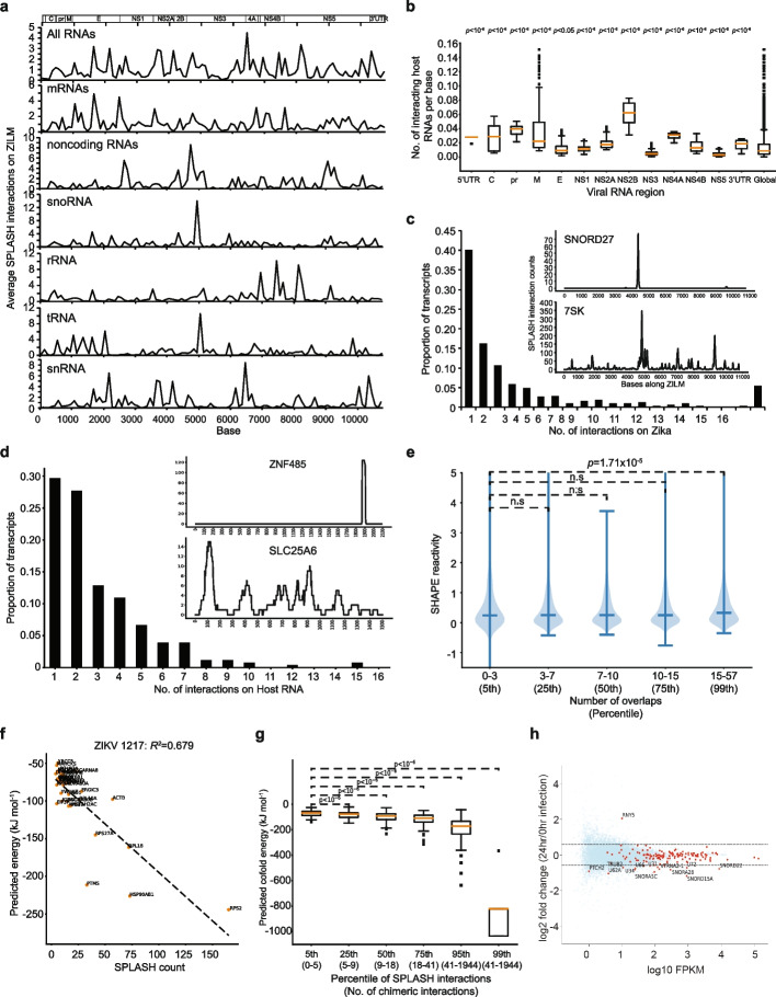
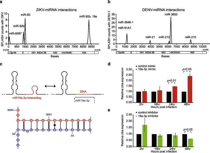
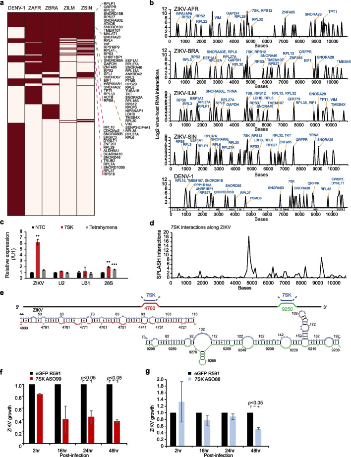
Results: Here, we use proximity ligation sequencing to identify virus-host RNA interactions for four strains of Zika virus (ZIKV) and one strain of dengue virus (DENV-1) in human cells. We find hundreds of coding and non-coding RNAs that bind to DENV and ZIKV viruses. Host RNAs tend to bind to single-stranded regions along the virus genomes according to hybridization energetics. Compared to SARS-CoV-2 interactors, ZIKV-interacting host RNAs tend to be downregulated upon virus infection. Knockdown of several short non-coding RNAs, including miR19a-3p, and 7SK RNA results in a decrease in viral replication, suggesting that they act as virus-permissive factors. In addition, the 3'UTR of DYNLT1 mRNA acts as a virus-restrictive factor by binding to the conserved dumbbell region on DENV and ZIKV 3'UTR to decrease virus replication. We also identify a conserved set of host RNAs that interacts with DENV, ZIKV, and SARS-CoV-2, suggesting that these RNAs are broadly important for RNA virus infection.

Conclusions: This study demonstrates that host RNAs can impact virus replication in permissive and restrictive ways, expanding our understanding of host factors and RNA-based gene regulation during viral pathogenesis.


Conflict of interest statement

The authors declare that they have no competing interests.

Figures

Fig. 1
Fig. 1
DENV and ZIKV genomes interact with hundreds of host RNAs in cells. a Experimental workflow to identify virus-host RNA interactions globally using biotinylated psoralen and proximity ligation sequencing. b Pie chart showing the number of RNAs from different RNA classes that interact with DENV and ZIKV viruses. c GO term enrichments of the host RNAs that interact with different strains of DENV and ZIKV viruses. The Y-axis indicates log (p-value) and the X-axis indicates the number of folds that the genes are enriched in binding to the virus genome as compared to the background
Fig. 2
Fig. 2
General properties of Zika-host RNA-RNA interactions. a Locations of interacting host RNAs on the viral genomes. Aggregate data and data classified by type of host RNA are shown. We observed differences in binding patterns by different classes of RNA along the genome. b The number of interactors in each coding region normalized by the length of the respective region. We observed the highest abundance of interactions in the NS2B-coding region. c Number of Zika genome binding sites for any specific host RNA (“Zika to host”). Most interactions bind at a specific, unique site. Promiscuous interactions of host RNA are present but considerably rarer. Inset, top: An example of a highly specific interaction between host RNA and virus genome, whereby SNORD27 only binds to a single region in ZIKV. Bottom: An example of promiscuous interaction between host RNA and virus genome whereby 7SK binds to many locations along ZIKV. d Number of interaction sites on host RNAs that bind to ZIKV genome (“host to Zika”). Data shows mostly unique interactions, but a higher propensity for 2 or more interaction sites. Inset, top: An example showing only 1 host region in ZNF485 interacting with the virus genome. Bottom: An example showing that many regions along SLC25A6 bind to the virus genome. e Sites along the Zika genome that show high numbers of interactions (99th percentile of interactions) show a significant preference for single-stranded, high SHAPE reactivity segments inside virion particles. f Host RNAs interacting at ZIKV position 1217 with low predicted interaction energy corresponding to low observed read counts and high interaction energies coinciding with high observed read counts, indicating interaction energetics is a significant factor driving specific host-virus interactions. g Aggregate interaction energy statistics for percentiles of observed interaction counts, again with high interaction counts corresponding to high predicted interaction energy and significant differences between all classes. h Volcano plot showing the fold change in gene expression of host RNAs after 24 h of ZIKV infection. ZIKV interactors are colored as red dots, while the non-interactors are in blue. The dotted lines indicate a 1.5-fold change in gene expression
Fig. 3
Fig. 3
DENV and ZIKV interact with host miRNAs. a, b Line plot showing the locations along ZIKV (a) and DENV (b) that interact with host miRNAs. Y-axis indicates the number of interaction counts between the virus genome and a specific miRNA. The X-axis indicates the position along the virus genome. c Top, secondary structure modeling of ZIKV genome before and after miR-19a-3p binding. Bottom: Predicted ZIKV: miR-19a-3p interactions using the program RNAcofold. The ZIKV interacting sequence is in red and the miRNA sequence is in blue. d, e Bar charts showing the amount of Zika virus detected inside Huh7 cells using qPCR, 2, 16, 24, and 48 h post-infection in cells that are transfected with miR-19a-3p or control (d), or in cells that are transfected with miR-19a-3p inhibitor or control (e). The data in cells with over-expression of miR-19a-3p or inhibitor is normalized to its control
Fig. 4
Fig. 4
Noncoding RNAs interact with DENV and ZIKV genomes and impact virus fitness. a Heatmap showing the host RNAs that interact with DENV-1 and 4 strains of Zika. Host RNAs that interact with more than 3 viruses are listed on the right of the heatmap. b Locations of top 50 virus-host interaction sites along DENV-1 and the 4 ZIKV strains. Many of the top virus-host interaction sites are conserved across the viruses (shown in red). c qPCR analysis of ZIKV, U2, U31 and 28S rRNA pulled down by 7SK and tetrahymena RNA. Tetrahymena RNA is used as a negative control. **, *** indicate p-values ≤ 0.01 and 0.001 respectively, using Student’s T-test. d Locations along ZIKV that bind to 7SK RNA. The Y-axis indicates the number of SPLASH interactions between ZIKV and 7SK at that position. e Top, schematic of the strongest ZIKV-7SK interactions along the ZIKV genome. Bottom, predicted pairing interactions between ZIKV and 7SK using the program RNAcofold. 7SK sequences are shown in blue and the two ZIKV interaction sequences are in red and green respectively. f, g Bar charts showing the amount of ZIKV inside Huh7 cells at 2,16, 24, and 48 h post-infection, using qPCR analysis. ZIKV amount inside cells is decreased upon knockdown of 7SK using ASO99 (f) and after its interaction with 7SK is blocked using a 2’O-methylated anti-sense oligo to 7SK (ASO88) (g)
Fig. 5
Fig. 5
DYNLT1 is an anti-ZIKV factor that binds to ZIKV 3′UTRs. a Top, Location along DYNLT1 that binds to ZIKV. Bottom, Location along ZIKV that binds to DYNLT1. b Schematic and predicted RNA structures on ZIKV before and after DYNLT1 binding to the 3′UTR. The DYNLT1 sequence is in blue and the ZIKV sequence is in black and red. c Structure model of DYNLT1-ZIKV interaction using RNAcofold program. DYNLT1 RNA is in blue while the ZIKV genome is in red. The mutations on DYNLT1 3′UTR RNA are indicated in purple above the original bases. d EMSA data showing ZIKV and DYNLT1 RNA-RNA interactions using different amounts of WT and MT DYNLT1 3′UTR RNA. e Barplots showing the effect of DYNLT1 knockdown on ZIKV replication. Huh7 cells were transfected with 20 nM of DYNLT1 siRNA or a control siRNA. At 24 h post-transfection, cells were infected with recombinant ZIKV strain PRVABC59 at a multiplicity of infection (MOI) 0.5. The infectivities of the supernatants 1 day after infection were determined by plaque assay. Means and standard deviations (SDs) from six independent experiments are presented. f DYNLT1 knockdown reduces the replication of the ZIKV African lineage. Huh7 cells were transfected with 20 nM of DYNLT1 siRNA or a control siRNA. At 24 h post-transfection, cells were infected with a ZIKV strain Dakar containing a nanoluciferase gene. At 24 h post-infection, luciferase signals were measured. The relative luciferase signals were obtained by normalizing the luciferase readouts of the DYNLT1 siRNA-treated groups to those of the siRNA control. Means and SDs from five independent experiments are presented. g Overexpression of DYNLT1 3′UTR suppresses ZIKV replication. Huh7 cells were transfected with DYNLT 3′UTR, DYNLT1 3′UTR with mutation, or pXJ vector (100, 200, or 400 ng per well in a 24-well plate). At 24 h post-transfection, cells were infected with recombinant ZIKV strain PRVABC59 containing a nanoluciferase gene at an MOI of 0.5. At 24 h post-infection, luciferase signals were measured. The relative luciferase signals were obtained by normalizing the luciferase readouts of each group to those of the pXJ vector control. Means and SDs from four independent experiments are presented. h DYNLT1 3′UTR inhibits ZIKV replication. Huh7 cells were transfected with 200 ng of DYNLT 3′UTR, DYNLT1 3′UTR with mutation, or pXJ vector. At 24 h post-transfection, cells were infected with recombinant ZIKV strain PRVABC59 containing a nanoluciferase gene at an MOI of 0.5. At given time points, cells were harvested. The relative luciferase signals were obtained by normalizing the luciferase readouts of each group to those of control at 2 h post-infection. Means and SDs from four independent experiments are presented. i Venn diagram showing the amount of overlap between RNAs that interact with all 5 DENV and ZIKV viruses and RNAs that interact with SARS-CoV-2

References

    1. Bhatt S, Gething PW, Brady OJ, Messina JP, Farlow AW, Moyes CL, et al. The global distribution and burden of dengue. Nature. 2013;496:504–507. doi: 10.1038/nature12060. - DOI - PMC - PubMed
    1. Musso D, Gubler DJ. Zika virus. Clin Microbiol Rev. 2016;29:487–524. doi: 10.1128/CMR.00072-15. - DOI - PMC - PubMed
    1. Barrows NJ, Campos RK, Liao K-C, Prasanth KR, Soto-Acosta R, Yeh S-C, et al. Biochemistry and molecular biology of flaviviruses. Chem Rev. 2018;118:4448–4482. doi: 10.1021/acs.chemrev.7b00719. - DOI - PMC - PubMed
    1. Zeidler JD, Fernandes-Siqueira LO, Barbosa GM, Da Poian AT. Non-canonical roles of dengue virus non-structural proteins. Viruses. 2017;9:42. doi: 10.3390/v9030042. - DOI - PMC - PubMed
    1. Sessions OM, Barrows NJ, Souza-Neto JA, Robinson TJ, Hershey CL, Rodgers MA, et al. Discovery of insect and human dengue virus host factors. Nature. 2009;458:1047–1050. doi: 10.1038/nature07967. - DOI - PMC - PubMed

Publication types