

Atrophy of specific amygdala subfields in subjects converting to mild cognitive impairment
- PMID: 38053753
- PMCID: PMC10694338
- DOI: 10.1002/trc2.12436
Atrophy of specific amygdala subfields in subjects converting to mild cognitive impairment
Abstract
Introduction: Accumulating evidence indicates that the amygdala exhibits early signs of Alzheimer's disease (AD) pathology. However, it is still unknown whether the atrophy of distinct subfields of the amygdala also participates in the transition from healthy cognition to mild cognitive impairment (MCI).
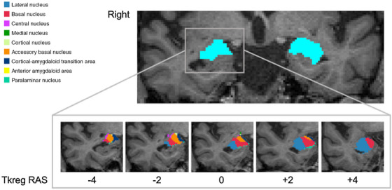
Methods: Our sample was derived from the AD Neuroimaging Initiative 3 and consisted of 97 cognitively healthy (HC) individuals, sorted into two groups based on their clinical follow-up: 75 who remained stable (s-HC) and 22 who converted to MCI within 48 months (c-HC). Anatomical magnetic resonance (MR) images were analyzed using a semi-automatic approach that combines probabilistic methods and a priori information from ex vivo MR images and histology to segment and obtain quantitative structural metrics for different amygdala subfields in each participant. Spearman's correlations were performed between MR measures and baseline and longitudinal neuropsychological measures. We also included anatomical measurements of the whole amygdala, the hippocampus, a key target of AD-related pathology, and the whole cortical thickness as a test of spatial specificity.
Results: Compared with s-HC individuals, c-HC subjects showed a reduced right amygdala volume, whereas no significant difference was observed for hippocampal volumes or changes in cortical thickness. In the amygdala subfields, we observed selected atrophy patterns in the basolateral nuclear complex, anterior amygdala area, and transitional area. Macro-structural alterations in these subfields correlated with variations of global indices of cognitive performance (measured at baseline and the 48-month follow-up), suggesting that amygdala changes shape the cognitive progression to MCI.
Discussion: Our results provide anatomical evidence for the early involvement of the amygdala in the preclinical stages of AD.
Highlights: Amygdala's atrophy marks elderly progression to mild cognitive impairment (MCI).Amygdala's was observed within the basolateral and amygdaloid complexes.Macro-structural alterations were associated with cognitive decline.No atrophy was found in the hippocampus and cortex.
Keywords: Alzheimer's disease (AD); amygdala; magnetic resonance imaging (MRI); mild cognitive impairment (MCI); preclinical; subfields.
© 2023 The Authors. Alzheimer's & Dementia: Translational Research & Clinical Interventions published by Wiley Periodicals LLC on behalf of Alzheimer's Association.
Conflict of interest statement
The authors declare no conflicts of interest. Author disclosures are available in the Supporting information.
Figures

References
-
- Hampel H, Lista S. The rising global tide of cognitive impairment. Nat Rev Neurol. 2016;12(3):131‐132. - PubMed
-
- Kepp KP, Robakis NK, Høilund‐Carlsen PF, Sensi SL, Vissel B. The amyloid cascade hypothesis: an updated critical review. Brain. 2023;15: awad159. - PubMed
-
- Delli Pizzi S, Granzotto A, Bomba M, Frazzini V, Onofrj M, Sensi SL. Acting before: a combined strategy to counteract the onset and progression of dementia. Curr Alzheimer Res. 2020;17(9):790‐804. - PubMed
-
- Delli Pizzi S, Punzi M, Sensi, SL , Alzheimer's Disease Neuroimaging Initiative. Functional signature of conversion of patients with mild cognitive impairment. Neurobiol Aging. 2019;74:21‐37. - PubMed
Grants and funding
LinkOut - more resources
Full Text Sources
Miscellaneous