. 2023 Oct 31:22:1135-1145.
doi: 10.17179/excli2023-6458. eCollection 2023.

Proteomic analysis of hepatic effects of okadaic acid in HepaRG human liver cells

Affiliations

Proteomic analysis of hepatic effects of okadaic acid in HepaRG human liver cells

Leonie T D Wuerger et al. EXCLI J. .

Abstract

The marine biotoxin okadaic acid (OA) is produced by dinoflagellates and enters the human food chain by accumulating in the fatty tissue of filter-feeding shellfish. Consumption of highly contaminated shellfish can lead to diarrheic shellfish poisoning. However, apart from the acute effects in the intestine, OA can also provoke toxic effects in the liver, as it is able to pass the intestinal barrier into the blood stream. However, molecular details of OA-induced hepatotoxicity are still insufficiently characterized, and especially at the proteomic level data are scarce. In this study, we used human HepaRG liver cells and exposed them to non-cytotoxic OA concentrations for 24 hours. Global changes in protein expression were analyzed using 2-dimensional gel electrophoresis in combination with mass-spectrometric protein identification. The results constitute the first proteomic analysis of OA effects in human liver cells and indicate, amongst others, that OA affects the energy homeostasis, induces oxidative stress, and induces cytoskeletal changes.

Keywords: HepaRG cells; liver proteome; okadaic acid.


Figures

Table 1
Table 1. The 15 most prominently upregulated proteins in all samples. Proteins were isolated from HepaRG cells incubated with 33 or 100 nM OA for 24 h. Proteins were then separated using 2D gel electrophoresis and deregulated proteins were identified using MALDI-MS.
Table 2
Table 2. The 15 most prominently downregulated proteins in all samples. Proteins were isolated from HepaRG cells incubated with 33 or 100 nM OA for 24 h. Proteins were then separated using 2D gel electrophoresis and deregulated proteins were identified using MALDI-MS.
Figure 1
Figure 1. Representative gels of separated proteins from HepaRG cells incubated with 33 or 100 nM OA for 24 h. HepaRG cells were lysed, and proteins were extracted. Proteins were first separated using isoelectric focusing and then further using SDS-PAGE. Gels were stained using Ruthenium II Tris solution, n=3.
Figure 2
Figure 2. Deregulated protein spots and identified deregulated proteins in OA-treated HepaRG cells. Proteins were isolated from HepaRG cells incubated with 33 or 100 nM OA for 24 h. Proteins were then separated using 2D gel electrophoresis and deregulated protein spots were identified using the Wilcoxon Rank sum test (p ≤ 0.01 and │log2 ratio│≥ 0.5). (A) Total numbers of up- and downregulated protein spots. (B) Venn diagram visualization of the overlap of deregulated protein spots between the treatment groups, presented in absolute numbers and percentages. (C) The proteins corresponding to each spot were identified using MALDI-MS; the diagram shows the numbers and percentages of identified proteins in the treatment groups and their overlap.
Figure 3
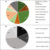
Figure 3. Grouping of identified OA-regulated proteins according to their cellular functions. (A) Distribution of the proteins between different cellular functions and processes according to the UniProt database. (B) Detailed functional grouping of the proteins related to energy homeostasis

References

    1. Alves-Bezerra M, Cohen DE. Triglyceride metabolism in the liver. Compr Physiol. 2017;8(1):1–8. doi: 10.1002/cphy.c170012. - DOI - PMC - PubMed
    1. Bialojan C, Takai A. Inhibitory effect of a marine-sponge toxin, okadaic acid, on protein phosphatases. Specificity and kinetics. Biochem J. 1988;256:283–290. doi: 10.1042/bj2560283. - DOI - PMC - PubMed
    1. Cordier S, Monfort C, Miossec L, Richardson S, Belin C. Ecological analysis of digestive cancer mortality related to contamination by diarrhetic shellfish poisoning toxins along the coasts of France. Environ Res. 2000;84:145–150. doi: 10.1006/enrs.2000.4103. - DOI - PubMed
    1. Del Campo M, Zhong TY, Tampe R, García L, Lagos N. Sublethal doses of dinophysistoxin-1 and okadaic acid stimulate secretion of inflammatory factors on innate immune cells: Negative health consequences. Toxicon. 2017;126:23–31. doi: 10.1016/j.toxicon.2016.12.005. - DOI - PubMed
    1. Dietrich J, Grass I, Günzel D, Herek S, Braeuning A, Lampen A, et al. The marine biotoxin okadaic acid affects intestinal tight junction proteins in human intestinal cells. Toxicol Vitro. 2019;58:150–160. doi: 10.1016/j.tiv.2019.03.033. - DOI - PubMed

LinkOut - more resources