. 2024 Jan 23;98(1):e0166423.
doi: 10.1128/jvi.01664-23. Epub 2023 Dec 6.

Pseudorabies virus upregulates low-density lipoprotein receptors to facilitate viral entry

Affiliations

Pseudorabies virus upregulates low-density lipoprotein receptors to facilitate viral entry

Ying-Xian Ma et al. J Virol. .

Abstract

Pseudorabies virus (PRV) is the causative agent of Aujeszky's disease in pigs. The low-density lipoprotein receptor (LDLR) is a transcriptional target of the sterol-regulatory element-binding proteins (SREBPs) and participates in the uptake of LDL-derived cholesterol. However, the involvement of LDLR in PRV infection has not been well characterized. We observed an increased expression level of LDLR mRNA in PRV-infected 3D4/21, PK-15, HeLa, RAW264.7, and L929 cells. The LDLR protein level was also upregulated by PRV infection in PK-15 cells and in murine lung and brain. The treatment of cells with the SREBP inhibitor, fatostatin, or with SREBP2-specific small interfering RNA prevented the PRV-induced upregulation of LDLR expression as well as viral protein expression and progeny virus production. This suggested that PRV activated SREBPs to induce LDLR expression. Furthermore, interference in LDLR expression affected PRV proliferation, while LDLR overexpression promoted it. This indicated that LDLR was involved in PRV infection. The study also demonstrated that LDLR participated in PRV invasions. The overexpression of LDLR or inhibition of proprotein convertase subtilisin/kexin type 9 (PCSK9), which binds to LDLR and targets it for lysosomal degradation, significantly enhanced PRV attachment and entry. Mechanistically, LDLR interacted with PRV on the plasma membrane, and pretreatment of cells with LDLR antibodies was able to neutralize viral entry. An in vivo study indicated that the treatment of mice with the PCSK9 inhibitor SBC-115076 promoted PRV proliferation. The data from the study indicate that PRV hijacks LDLR for viral entry through the activation of SREBPs.IMPORTANCEPseudorabies virus (PRV) is a herpesvirus that primarily manifests as fever, pruritus, and encephalomyelitis in various domestic and wild animals. Owing to its lifelong latent infection characteristics, PRV outbreaks have led to significant financial setbacks in the global pig industry. There is evidence that PRV variant strains can infect humans, thereby crossing the species barrier. Therefore, gaining deeper insights into PRV pathogenesis and developing updated strategies to contain its spread are critical. This study posits that the low-density lipoprotein receptor (LDLR) could be a co-receptor for PRV infection. Hence, strategies targeting LDLR may provide a promising avenue for the development of effective PRV vaccines and therapeutic interventions.

Keywords: LDLR; PCSK9; SREBPs; pseudorabies virus; viral entry.


Conflict of interest statement

The authors declare no conflict of interest.

Figures
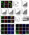
FIG 1
FIG 1
PRV stimulates LDLR mRNA and protein expression. (A) PK-15 cells infected with PRV-GFP (multiplicity of infection, MOI = 1) for 12 h were cultured in serum-free medium containing Dil-LDL (10 µg/mL) for 5 h. Dil-LDL uptake was analyzed using fluorescence microscopy. White dotted lines represent PRV-infected cells (left). Scale bar: 10 µm. Quantitative analysis of Dil-LDL fluorescence intensity in mock- or PRV-infected cells was analyzed using ImageJ (right, n = 15). ***P < 0.001. (B) 3D4/21, (C) PK-15, (D) HeLa, (E) RAW264.7, and (F) L929 cells were infected with PRV-QXX (MOI = 1) for 0–36 h. Relative mRNA expression of LDLR was evaluated using quantitative RT-PCR analysis. *P < 0.05, **P < 0.01, ***P < 0.001. (G) PK-15 cells were infected with PRV-GFP (MOI = 1) for 12 h. PRV-GFP (green) and LDLR (red) were examined using immunofluorescence analysis. The asterisk indicates mock-infected cells. Scale bar: 10 µm. The relative fluorescence intensity of LDLR in mock- or PRV-infected cells was quantified using ImageJ (right, n = 15). ***P < 0.001. (H) PK-15 cells were mock infected or infected with PRV-QXX (MOI = 1) for 0–36 h. LDLR and gE expression were analyzed using immunoblotting assays. (I and J) Mice were infected with PRV-QXX (2 × 10⁵ TCID50) for 166 h. Immunofluorescence analysis of prepared lung and brain sections was conducted with anti-gE (green) and anti-LDLR (red) antibodies. Scale bar: 50 µm.
FIG 2
FIG 2
PRV-induced transcriptional upregulation of LDLR is mediated by SREBP. (A) PK-15 cells were infected with PRV-QXX (multiplicity of infection, MOI = 1) or UV-inactivated PRV-QXX (MOI = 1) for 24 h. Relative mRNA expression of LDLR was evaluated using quantitative reverse transcription PCR (qRT-PCR) analysis. *P < 0.05. (B–C) PK-15 cells were infected with PRV-QXX (MOI = 1) for 0–36 h. Relative mRNA expression of (B) HMGCR and (C) HMGCS1 was evaluated using qRT-PCR analysis. *P < 0.05, **P < 0.01, ***P < 0.001. (D–F) PK-15 cells were infected with PRV-QXX (MOI = 1) and simultaneously treated with fatostatin (3 µM) for 0–36 h. Relative mRNA expression of (D) LDLR, (E) HMGCR, and (F) HMGCS1 was evaluated using qRT-PCR analysis. *P < 0.05, **P < 0.01, ***P < 0.001. (G) PK-15 cells were transfected with NC, siSREBP2-1, and siSREBP2-2 for 48 h. SREBP2 was analyzed using immunoblotting assays. (H) PK-15 cells were transfected with NC, siSREBP2-1, and siSREBP2-2 for 24 h and infected with PRV-QXX (MOI = 1) for 24 h. Relative mRNA expression of LDLR was evaluated using qRT-PCR analysis. ***P < 0.001. (I) PK-15 cells were mock infected or infected with PRV-QXX (MOI = 1) and simultaneously treated with fatostatin (0–3 µM) for 36 h. LDLR and gE expression were analyzed using immunoblotting assays. (J) PK-15 cells were transfected with NC, siSREBP2-1, and siSREBP2-2 for 24 h, and mock infected or infected with PRV-QXX (MOI = 1) for 24 h. PRV gE, SREBP2, and LDLR expression were analyzed using immunoblotting assays. (K) PK-15 cells were infected with PRV-QXX (MOI = 1) and simultaneously treated with fatostatin (0–3 µM) for 36 h. The viral titer was determined using a TCID50 assay. *P < 0.05, **P < 0.01, ***P < 0.001. (L) PK-15 cells were transfected with NC, siSREBP2-1, and siSREBP2-2 for 24 h and infected with PRV-QXX (MOI = 1) for 24 h. The viral titer was determined using a TCID50 assay. **P < 0.01.
FIG 3
FIG 3
LDLR is responsible for PRV proliferation. (A) Relative mRNA expression of LDLR in scramble and shLDLR PK-15 cells was evaluated using qRT-PCR analysis. ***P < 0.001. (B) LDLR protein in scramble and shLDLR PK-15 cells was evaluated using immunoblotting assays. (C) Proliferation of scramble and shLDLR PK-15 cells was determined using the CCK-8 assay. ns, no significance. (D) Scramble and shLDLR PK-15 cells were mock infected or infected with PRV-QXX (multiplicity of infection, MOI = 1) for 24 h. LDLR and gE expression were analyzed using immunoblotting assays. (E) Scramble and shLDLR PK-15 cells were infected with PRV-QXX (MOI = 1) for 24 h. The viral titer was determined using a TCID50 assay. ***P < 0.001. (F) Scramble and shLDLR PK-15 cells were infected with PRV-QXX (MOI = 1). A one step growth curve of the progeny virus was determined using a TCID50 assay for 2–24 h. **P < 0.01. (G) PK-15 cells were transfected with LDLR-FLAG (0–8 µg) and mock infected or infected with PRV-QXX (MOI = 1) for 24 h. LDLR-FLAG and gE expression were analyzed using an immunoblotting assays. (H) PK-15 cells were transfected with LDLR-FLAG (0–2 µg) and infected with PRV-QXX (MOI = 1) for 24 h. The viral titer was determined using a TCID50 assay. *P < 0.05, **P < 0.01, ***P < 0.001.
FIG 4
FIG 4
PCSK9 inhibition promotes PRV proliferation. (A) PK-15 cells were treated with SBC-115076 (0–300 nM) for the indicated times. Cell proliferation was determined using the CCK-8 assay. (B) PK-15 cells were pre-treated with SBC-115076 (0–300 nM) for 5 h and mock infected or infected with PRV-QXX (multiplicity of infection, MOI = 1) for 24 h. PRV gE, LDLR, and PCSK9 were evaluated using immunoblotting assays. (C) PK-15 cells were pre-treated with SBC-115076 (0–300 nM) for 5 h and then infected with PRV-GFP (MOI = 0.01) for 36 h. PRV-GFP proliferation was analyzed using fluorescence microscopy (left). Scale bar: 50 µm. Quantitative analysis of GFP-positive cells was determined using flow cytometry (right). **P < 0.01. (D) PK-15 cells were treated as in B. The viral titer was determined using a TCID50 assay. *P < 0.05, **P < 0.01. (E) PK-15 cells were transfected with vector or PSCK9 variants (WT, D374Y, and C679X) for 24 h and then infected with PRV-GFP (MOI = 0.01) for 36 h. PRV-GFP proliferation was analyzed using fluorescence microscopy (left). Scale bar: 50 µm. Quantitative analysis of GFP-positive cells was performed using flow cytometry (right). **P < 0.01. ns, no significance. (F) PK-15 cells were transfected with vector or PSCK9 variants (WT, D374Y, and C679X) for 24 h and then infected with PRV-QXX (MOI = 1) for 24 h. The viral titer was determined using a TCID50 assay. *P < 0.05. ns, no significance.
FIG 5
FIG 5
LDLR promotes PRV attachment and entry. (A) PRV attachment to PM of scramble and shLDLR PK-15 cells was evaluated using immunofluorescence analysis of PRV gE. Scale bar: 10 µm. (B) Quantification of gE puncta number per cell from (A) using ImageJ (n = 20). ***P < 0.001. (C) PRV entry into scramble and shLDLR PK-15 cells was evaluated using immunoblotting assays of PRV gE. (D) PRV entry into scramble and shLDLR PK-15 cells was evaluated using qRT-PCR analysis of viral genome copy numbers. ***P < 0.001. (E) PRV attachment to PM of control and LDLR-FLAG-expressing PK-15 cells was evaluated using immunofluorescence analysis of PRV gE. Scale bar: 10 µm. (F) Quantification of gE puncta number per cell from (F) using ImageJ (n = 20). ***P < 0.001. (G) PRV entry into control and LDLR-FLAG-expressing PK-15 cells was evaluated using immunoblotting assays of PRV gE. (H) PRV entry into control and LDLR-FLAG-expressing PK-15 cells was evaluated using qRT-PCR analysis of viral genome copy numbers. **P < 0.01.
FIG 6
FIG 6
LDLR interacts with PRV during viral entry. (A) PK-15 cells were transfected with the LDLR-FLAG plasmid for 24 h and then infected with PRV-QXX (multiplicity of infection, MOI = 100) at 4°C for 1 h. The interaction of PRV (gE) with LDLR-FLAG was evaluated using immunofluorescence analysis. Scale bar: 10 µm. (B) PK-15 cells were infected with PRV-QXX (MOI = 100) at 4°C for 1 h. Then, cells were cultured at 37°C for 0–30 min. Interactions of LDLR with PRV gE on PM were detected using a cell surface biotinylation assay. (C) PK-15 cells were incubated with indicated concentrations of anti-LDLR antibody at 4°C for 30 min and then infected with PRV-GFP (MOI = 0.01) for 36 h. PRV-GFP proliferation was analyzed using fluorescence microscopy and flow cytometry. Scale bar: 50 µm. (D) Quantitative analysis of GFP-positive cells from C was performed using flow cytometry. *P < 0.05, ***P < 0.001. (E) PK-15 cells were incubated with indicated concentrations of anti-LDLR antibody at 4°C for 30 min and then infected with PRV-QXX (MOI = 1) for 24 h. The viral titer was determined using a TCID50 assay. *P < 0.05, **P < 0.01, ***P < 0.001.
FIG 7
FIG 7
Inhibition of PCSK9 promotes PRV infection in vivo. (A) Experimental strategy for SBC-115076 treatment and PRV-QXX challenge. (B) PCSK9, LDLR, and PRV gE in the lung were evaluated using immunoblotting assays 3 days post-infection (n = 3). (C and D) The mRNA levels of LDLR (C) and IL-1β (D) in the lung were evaluated using qRT-PCR analysis 0–3 days post-infection (n = 4). *P < 0.05, **P < 0.01. (E) Serum IL-1β was analyzed using ELISA 0–3 days post-infection (n = 4). *P < 0.05. ns, no significance. (F) PRV genome copy numbers in the lung were determined using qRT-PCR analysis 3 days post-infection (n = 4). *P < 0.05. (G) Lung injury was assessed using hematoxylin and eosin staining 3 days post-infection (n = 4). Scale bar: 100 µm. (H) Survival rate was monitored daily for 6 days (n = 12). *P < 0.05.

References

    1. Pomeranz LE, Reynolds AE, Hengartner CJ. 2005. Molecular biology of Pseudorabies virus: impact on neurovirology and veterinary medicine. Microbiol Mol Biol Rev 69:462–500. doi:10.1128/MMBR.69.3.462-500.2005 - DOI - PMC - PubMed
    1. Laval K, Vernejoul JB, Van Cleemput J, Koyuncu OO, Enquist LW, Longnecker RM. 2018. Virulent pseudorabies virus infection induces a specific and lethal systemic inflammatory response in mice. J Virol 92. doi:10.1128/JVI.01614-18 - DOI - PMC - PubMed
    1. Avak S, Bienzle U, Feldmeier H, Hampl H, Habermehl K-O. 1987. Pseudorabies in man. Lancet 329:501–502. doi:10.1016/S0140-6736(87)92105-2 - DOI - PubMed
    1. Ai JW, Weng SS, Cheng Q, Cui P, Li YJ, Wu HL, Zhu YM, Xu B, Zhang WH. 2018. Human endophthalmitis caused by pseudorabies virus infection, China, 2017. Emerg. Infect. Dis 24:1087–1090. doi:10.3201/eid2406.171612 - DOI - PMC - PubMed
    1. Zheng L, Liu X, Yuan D, Li R, Lu J, Li X, Tian K, Dai E. 2019. Dynamic cerebrospinal fluid analyses of severe Pseudorabies encephalitis. Transbound Emerg Dis 66:2562–2565. doi:10.1111/tbed.13297 - DOI - PubMed

Publication types

LinkOut - more resources