. 2023 Dec 21;89(12):e0063023.
doi: 10.1128/aem.00630-23. Epub 2023 Dec 6.

Genetic dissection of the degradation pathways for the mycotoxin fusaric acid in Burkholderia ambifaria T16

Affiliations

Genetic dissection of the degradation pathways for the mycotoxin fusaric acid in Burkholderia ambifaria T16

Matias Vinacour et al. Appl Environ Microbiol. .

Abstract

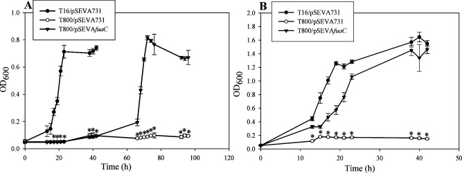
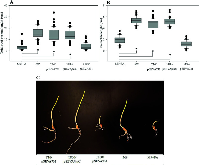
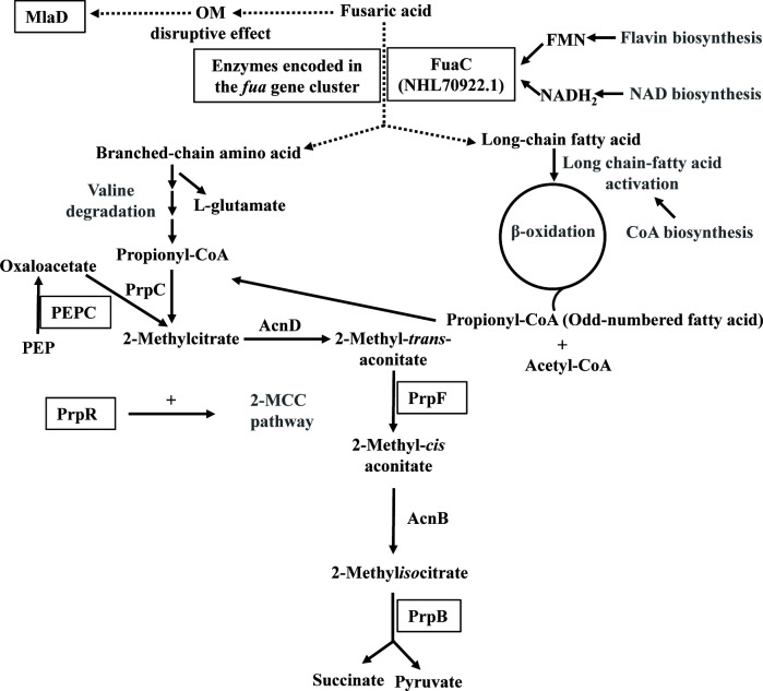
Fusaric acid (FA) is an important virulence factor produced by several Fusarium species. These fungi are responsible for wilt and rot diseases in a diverse range of crops. FA is toxic for animals, humans and soil-borne microorganisms. This mycotoxin reduces the survival and competition abilities of bacterial species able to antagonize Fusarium spp., due to its negative effects on viability and the production of antibiotics effective against these fungi. FA biodegradation is not a common characteristic among bacteria, and the determinants of FA catabolism have not been identified so far in any microorganism. In this study, we identified genes, enzymes, and metabolic pathways involved in the degradation of FA in the soil bacterium Burkholderia ambifaria T16. Our results provide insights into the catabolism of a pyridine-derivative involved in plant pathogenesis by a rhizosphere bacterium.

Keywords: 2-methylcitrate cycle; Burkholderia ambifaria T16; catabolism; detoxification; fusaric acid; two-component flavin-dependent monooxygenase.


Conflict of interest statement

The authors declare no conflict of interest.

Figures

Fig 1
Fig 1
(A) Genomic organization of the prp locus in B. ambifaria T16 and localization of mini-Tn5 insertions in the mutants B. ambifaria FA2, FA4, FA92, and FA91. Genes are represented by arrows and black inverted triangles represent the localization of the mini-Tn5 insertions in each mutant. (B) 2-Methylcitrate cycle in B. ambifaria T16. Enzymes are shaded in gray rectangles. PrpC: methylcitrate synthase, AcnD: methylcitrate dehydratase, PrpF: methylaconitate cis-trans isomerase, AcnB: aconitase, PrpB: methylisocitrate lyase. The AcnB enzyme is not encoded in the prp locus.
Fig 2
Fig 2
Growth of B. ambifaria strains in M9 minimal medium with 0.2% (wt/vol) sodium citrate (A), 0.2% (wt/vol) sodium propionate (B) or 450 µg/mL fusaric acid (C) as sole carbon and energy source. The graphs show the means ± SD obtained from three biological replicates. The OD600 values obtained were subjected to Kruskal-Wallis analysis of variance followed by Dunnett’s test. The ΔprpB strain bearing the vector plasmid (T42/pSEVA234) was used as control in the test. P values were considered significant when they were lower than 0.05. Asterisks indicate that there is a statistically significant difference between OD600 values of strains T16/pSEVA234, T42/pSEVAicl or T42/pSEVAprpB and OD600 values of T42/pSEVA234.
Fig 3
Fig 3
Differential protein abundance in B. ambifaria T16 grown in M9 minimal medium with citrate (M9 + CA) or fusaric acid (M9 + FA) as sole carbon source. Red dots represent some upregulated proteins during growth with FA. Proteins IDs NHL69607.1 to NHL69612.1 correspond to proteins involved in valine degradation. Proteins NHL70913.1 to 70915.1: 2-oxo acid dehydrogenase complex. NHL70916.1: predicted BdkR transcriptional regulator involved in the regulation of isoleucine and valine catabolism. Proteins NHL70920.1 to 70924.1 are encoded in a gene cluster of unknown function.
Fig 4
Fig 4
Organization of the gene cluster EIB72_31580-EIB72_31610 (fua cluster) encoding proteins NHL70920.1 to 70924. 1. Genes are represented by arrows. Locus IDs are shown above the genes. FR, flavin reductase; LLM, luciferase-like monooxygenase.
Fig 5
Fig 5
Growth of B. ambifaria strains with fusaric acid as the sole carbon and energy source (A) or as the sole nitrogen source (B). The graphs show the means ± SD obtained from three biological replicates. OD600 values were subjected to Kruskal-Wallis analysis of variance, followed by Dunnett’s test. B. ambifaria T800/pSEVA731 was used as a control in the test. P values were considered significant when they were lower than 0.05. Asterisks indicate that there is a statistically significant difference with B. ambifaria T800/pSEVA731.
Fig 6
Fig 6
FA toxicity assay using barley seedlings. Box plots of median, 1st and 3rd quartiles (boxes), and 1.5x quartiles (whiskers) corresponding to total root lenght (A) and coleoptile length (B) of seedlings (n=60) treated with cell-free supernatants from stationary phase-cultures of B. ambifaria strains, M9 minimal medium (M9), or M9 supplemented with 450 μg/mL FA (M9+FA). Three biological replicates were used. Dots denote observations outside the range of adjacent values. Data were subjected to Kruskal-Wallis analysis of variance and compared by Dunnet’s test. Seeds treated with M9 + FA were used as control. Asterisks indicate significant differences at P < 0.05 between values from the barley seedlings treated with bacterial supernatants or M9, and the barley seedlings treated with M9 + FA (control). (C) Photographic image showing barley seedlings treated with culture supernatants of B. ambifaria strains, M9 minimal medium (M9), or M9 supplemented with 450 μg/mL FA (M9 + FA).
Fig 7
Fig 7
Schematic representation of key proteins and metabolic pathways likely required for fusaric acid catabolism in B. ambifaria T16. Proteins belonging to metabolic pathways written in gray were significantly more abundant during growth on fusaric acid compared to sodium citrate. Proteins essential for growth in the presence of fusaric acid are black-framed. OM, outer membrane; PEP, phosphoenolpyruvate.

References

    1. Bacon CW, Porter JK, Norred WP, Leslie JF. 1996. Production of fusaric acid by Fusarium species. Appl Environ Microbiol 62:4039–4043. doi:10.1128/aem.62.11.4039-4043.1996 - DOI - PMC - PubMed
    1. Ma L-J, Geiser DM, Proctor RH, Rooney AP, O’Donnell K, Trail F, Gardiner DM, Manners JM, Kazan K. 2013. Fusarium pathogenomics. Annu Rev Microbiol 67:399–416. doi:10.1146/annurev-micro-092412-155650 - DOI - PubMed
    1. Pegg GF. 1981. Biochemistry and physiology of pathogenesis, p 193–246. In Mace ME, Bell AA, Beckman CH (ed), Fungal wilt diseases of plants. Academic Press Inc, New York.
    1. El-Hassan KI, El-Saman MG, Mosa AA, Mostafa M 2. 2007. Variation among Fusarium spp., the causal of potato tuber dry rot in their pathogenicity and mycotoxins production. Egypt J Phytopathol 35:53–68.
    1. Liu S, Li J, Zhang Y, Liu N, Viljoen A, Mostert D, Zuo C, Hu C, Bi F, Gao H, Sheng O, Deng G, Yang Q, Dong T, Dou T, Yi G, Ma LJ, Li C 2. 2020. Fusaric acid Instigates the invasion of banana by Fusarium oxysporum f. sp. cubense TR4. New Phytol 225:913–929. doi:10.1111/nph.16193 - DOI - PMC - PubMed

Publication types

Supplementary concepts

LinkOut - more resources