. 2023 Dec 6:12:RP86678.
doi: 10.7554/eLife.86678.

Discovery and characterization of cross-reactive intrahepatic antibodies in severe alcoholic hepatitis

Affiliations

Discovery and characterization of cross-reactive intrahepatic antibodies in severe alcoholic hepatitis

Ali Reza Ahmadi et al. Elife. .

Abstract

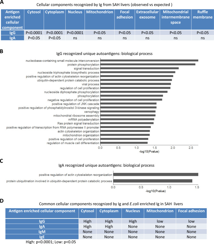
The pathogenesis of antibodies in severe alcoholic hepatitis (SAH) remains unknown. We analyzed immunoglobulins (Ig) in explanted livers from SAH patients (n=45) undergoing liver transplantation and tissues from corresponding healthy donors (HD, n=10) and found massive deposition of IgG and IgA isotype antibodies associated with complement fragment C3d and C4d staining in ballooned hepatocytes in SAH livers. Ig extracted from SAH livers, but not patient serum exhibited hepatocyte killing efficacy. Employing human and Escherichia coli K12 proteome arrays, we profiled the antibodies extracted from explanted SAH, livers with other diseases, and HD livers. Compared with their counterparts extracted from livers with other diseases and HD, antibodies of IgG and IgA isotypes were highly accumulated in SAH and recognized a unique set of human proteins and E. coli antigens. Further, both Ig- and E. coli-captured Ig from SAH livers recognized common autoantigens enriched in several cellular components including cytosol and cytoplasm (IgG and IgA), nucleus, mitochondrion, and focal adhesion (IgG). Except IgM from primary biliary cholangitis livers, no common autoantigen was recognized by Ig- and E. coli-captured Ig from livers with other diseases. These findings demonstrate the presence of cross-reacting anti-bacterial IgG and IgA autoantibodies in SAH livers.

Keywords: E. coli; E. coli arrays; IgA; IgG; alcoholic hepatitis; autoantibodies; human; immunology; inflammation.


Conflict of interest statement

AA, GS, TG, JM, XH, MH, AC, RW, BP, SO, EK, AG, LQ, BP, JB, RA, ZZ, HL, DF, CC, JQ, BG, HZ, ZS No competing interests declared

Figures

Figure 1.
Figure 1.. Immunoglobulin (Ig) deposition in ballooned hepatocytes of explanted livers from severe alcoholic hepatitis (SAH) patients.
(A) Liver tissue sections with H&E staining showed histologic features of SAH. (b, c) Immunohistochemistry staining by using anti-human IgA (B) or IgG (C) antibodies demonstrated IgA and IgG deposition in ballooned hepatocytes in SAH livers. Representative tissue sections from 45 SAH or 10 healthy donor (HD) livers. (D–E) Ig levels in liver tissue homogenates from SAH or HD (n=7/group) were quantified by western blot analysis (D). Western blot analysis demonstrated that the levels of IgA, IgG, and IgM were significantly increased in SAH livers as compared with the HD livers (E). (F–G) Ig isotypes (f) and IgG subclass levels (G) were quantified by ELISA (n=7/group). (H) IgG-positive hepatocytes in tissue sections from 45 SAH patients and 10 HD were quantified by immunohistochemistry staining and using HALO Image Analysis Software.
Figure 1—figure supplement 1.
Figure 1—figure supplement 1.. IgG deposition.
HALO image analysis of IgG-positive hepatocytes in severe alcoholic hepatitis livers. Representative images of liver tissue sections stained with anti-human IgG antibody. Positive cells were reported as percentage stained surface area of total annotated area by digital analysis (Hallo, Indicalabs, Corrales, NM, USA).
Figure 2.
Figure 2.. Immunoglobulin (Ig) deposition is associated with activation of complement in ballooned hepatocytes and Ig extracted from severe alcoholic hepatitis (SAH) livers exhibits hepatocyte killing efficacy in vitro.
To determine if immunoglobulin in ballooning hepatocytes induces activation of complement, complement fragments C3d and C4d were analyzed in SAH livers. (A–B) Immunohistochemistry staining showed the presence of both C3d (A) and C4d (B) in ballooning hepatocytes in SAH livers but not in the donor livers. (C) Double staining for IgG and complement fragments C3d or C4d showed IgG co-stained with C3d or C4d in ballooning hepatocytes. Representative tissue sections from seven samples per group. (E–F) C3d and C4d levels in SAH livers were quantified by western blot analysis (n=7). (G) Ig extracted from SAH livers but not serum exhibit hepatocyte killing efficacy in antibody-dependent cell-mediated cytotoxicity (ADCC) assay. Representative data from three independent experiments.
Figure 3.
Figure 3.. To determine if immunoglobulin (Ig) recognize cell surface antigens of ballooned hepatocytes, multiplex immunofluorescence staining was performed in liver tissue sections.
(A) Images of confocal microscopy showed the presence of both IgG (red) and IgA (green) in ballooning hepatocytes (blue) in severe alcoholic hepatitis (SAH) livers (lower panels), while only hepatic sinusoid endothelial cells (CD32+, purple) stained with IgG and IgA in donor livers (upper panels). (B) Co-staining with β-catenin (blue) demonstrated IgG (red) and IgA (green) deposition on membrane of ballooning hepatocytes in SAH livers. (C) Triple staining for IgG (red), IgA (green), and C4d (blue) showed both IgG and IgA co-stained with C4d on the surface of hepatocyte. Representative tissue sections from six samples per group.
Figure 4.
Figure 4.. Human and Escherichia coli proteome arrays that identify a group of unique antibodies in the severe alcoholic hepatitis (SAH) liver recognize both human and bacterial antigens.
(A) Each liver tissue piece was ground up and treated under low pH to release tissue-deposited Ig. After neutralization, the extracted Ig from each liver sample were separately probed to the HuProt or E. coli protein arrays, followed by incubation with the isotype-specific secondary antibodies to obtain the Ig isotype immune signatures of the same liver sample. (B) Representative images of HuProt arrays. (C) Venn diagram analysis to identify shared autoantigens of each liver disease. 346 autoantigens were shared by the IgG antibodies in all five SAH livers (left panel), 95 autoantigens were shared by the IgG antibodies in all five healthy donor (HD) livers (middle panel), and a large fraction (i.e., 267 proteins) of the SAH-shared IgG autoantigens was not found in the HD livers (right panel), suggesting existence of an SAH-specific autoimmune signature. (D) A seven-way Venn diagram analysis showed that 45 autoantigens were commonly recognized by IgG isotype autoantibodies in liver tissue homogenates extracted from different liver diseases, while 188 unique IgG autoantigens were recognized by tissue homogenates from the SAH livers. (E) Representative images of E. coli protein arrays. (F) Venn diagram analysis to identify shared E. coli antigens recognized by each liver disease. 435 E. coli antigens were commonly recognized by IgG antibodies in the five SAH livers (left panel), while only 1 E. coli antigen was commonly recognized by IgG antibodies in the five HD livers (middle panel). 434 out of 435 E. coli antigens were uniquely recognized by IgG antibodies in SAH livers but not HD livers (right panel). (G) A seven-way Venn diagram analysis showed that unique IgG bacterial antigens were only identified by using liver tissue homogenates from SAH or AC.
Figure 4—figure supplement 1.
Figure 4—figure supplement 1.. Numbers of autoantibodies shared by all five severe alcoholic hepatitis (SAH) livers.
Venn diagram analysis of autoantigens recognized by antibodies extracted from the five SAH liver tissues. After individual elution of antibodies from the five SAH liver tissues (i.e., SAH1–5), they were individually profiled on the HuProt arrays. The identified autoantigens were grouped on the basis of anti-IgG, -IgA, -IgM, and -IgE isotypes, and the shared and unique autoantigens were analyzed using the Venn diagram analysis. As illustrated in each isotype panel, the shared autoantigens recognized by the anti-IgG, -IgA, -IgM, and -IgE antibodies among the five SAH samples are 346, 319, 194, and 10, respectively.
Figure 4—figure supplement 2.
Figure 4—figure supplement 2.. Venn diagram analysis of all liver samples.
Summary of the Venn diagram analysis of autoantigens recognized by antibodies extracted from the five donor, five AC, five AIH, five HBV, five HCV, five NASH, five primary biliary cholangitis (PBC), and five severe alcoholic hepatitis (SAH) liver tissues. Using the same approach as described in Figure 4—figure supplement 1, the shared and disease-specific autoantibodies were identified. The Ig isotypes are designated in each roll of the Venn diagrams, and the tissue types are shown on the top of each column of the Venn diagrams.
Figure 4—figure supplement 3.
Figure 4—figure supplement 3.. Unique autoantigens recognized by immunoglobulin (Ig) from diseased livers on the HuProt arrays.
A seven-member Venn diagram analysis of the shared autoantigens recognized by antibodies extracted from the diseased liver tissues. The shared autoantigens identified by the antibodies extracted from the seven diseased liver tissues were subjected to a seven-member Venn diagram analysis. The Ig isotypes are shown on the top of each Venn diagram.
Figure 4—figure supplement 4.
Figure 4—figure supplement 4.. Venn diagram analysis of antigens identified on the E. coli proteome arrays.
Summary of the Venn diagram analysis of bacterial antigens recognized by human antibodies extracted from the five donor, five AC, five AIH, five HBV, five HCV, five NASH, five primary biliary cholangitis (PBC), and five severe alcoholic hepatitis (SAH) liver tissues. Using the same approach as described in Figure 4—figure supplement 1, the shared and disease-specific antibodies were identified. The immunoglobulin (Ig) isotypes are designated in each roll of the Venn diagrams, and the tissue types are shown on the top of each column of the Venn diagrams.
Figure 4—figure supplement 5.
Figure 4—figure supplement 5.. Unique bacterial antigens recognized by immunoglobulin (Ig) from diseased livers on the E. coli proteome arrays.
Venn diagram analysis of the shared bacterial antigens recognized by human antibodies extracted from the diseased liver tissues. The shared bacterial antigens identified by the antibodies extracted from the seven diseased liver tissues were subjected to a seven-member Venn diagram analysis. The Ig isotypes are shown on the top of each Venn diagram.
Figure 5.
Figure 5.. E. coli antigens-captured immunoglobulins (Ig) from severe alcoholic hepatitis (SAH) livers recognize human protein antigens.
(A) To determine if anti-bacterial antibodies cross-react with human proteins in the liver, total proteins from E. coli (strain K12) were extracted and immobilized on NHS-activated magnetic beads to capture Ig pooled from the five SAH livers. E. coli protein-captured antibodies (E. coli-Ig) were then released and incubated on the HuProt arrays. (B) Representative images of E. coli-Ig on HuProt arrays. (C) 937, 1041, 932, and 85 human proteins were reproducibly identified by E. coli protein-captured IgG, IgA, IgM, and IgE antibodies from the five SAH livers. Venn diagram analysis showed many of these proteins (694/937, 796/1041, 451/932, and 42/95, respectively) were also found to be the autoantigens recognized by Ig recovered directly from the five SAH livers.
Figure 6.
Figure 6.. Gene ontology (GO) enrichment analysis of proteome arrays identifies autoantigen-enriched common cellular components recognized by both immunoglobulin (Ig)- and E. coli-captured Ig in severe alcoholic hepatitis (SAH) livers.
(A) Cellular components recognized by IgG and IgA antibodies in SAH livers. (B–C) Biological processes are involved by IgG autoantigens (B) and IgA autoantigens (C). (D) Common cellular components recognized by both Ig- and E. coli antigens-captured Ig in SAH livers.
Figure 7.
Figure 7.. Increased bacterial antigens, B and plasma cell infiltration, and immunoglobulin gene expression in severe alcoholic hepatitis (SAH) livers.
(A) Immunohistochemistry staining for the gram-negative bacterial product lipopolysaccharides (LPS) and gram-positive bacteria antigen lipoteichoic acid (LTA) in SAH livers. (B) LPS levels in SAH liver tissues were quantified by using Pierce Chromogenic Endotoxin Quant Kit (n=7/group). (C) Immunohistochemistry staining for CD20+ B cells and CD138+ plasma cells in SAH livers. Representative images from 20 SAH or 10 healthy donor (HD) livers (A–C). (D) Double immunofluorescence staining with CD20 (green) and LTA (red) showed colocalization of B cells and bacteria antigens in the inflammatory areas. Representative tissue sections from six samples per group. (E) RNA-sequencing analysis identified differentially expressed genes representing immunoglobulin fragments in SAH livers. Purple dots represented 91 differentially expressed immunoglobulin genes (log2FC >1 and adjusted p-value <0.01) in the comparison of SAH patients between normal donors. Among them, 87 were upregulated and 4 downregulated. Gray dots were the remaining immunoglobulin genes that did not meet the thresholds. n=7 per group. (F) Schematic representation of mechanism of cross-reacting anti-bacterial autoantibody-mediated hepatocyte damage in SAH.

Update of

References

    1. Amano K, Tsukada K, Takeuchi T, Fukuda Y, Nagura H. IgA deposition in alcoholic liver disease. An immunoelectron microscopic study. American Journal of Clinical Pathology. 1988;89:728–734. doi: 10.1093/ajcp/89.6.728. - DOI - PubMed
    1. Bajaj JS. Alcohol, liver disease and the gut microbiota. Nature Reviews. Gastroenterology & Hepatology. 2019;16:235–246. doi: 10.1038/s41575-018-0099-1. - DOI - PubMed
    1. Bjorneboe M, Prytz H, Orskov F. Antibodies to intestinal microbes in serum of patients with cirrhosis of the liver. Lancet. 1972;1:58–60. doi: 10.1016/s0140-6736(72)90060-8. - DOI - PubMed
    1. Bogdanos DP, Baum H, Grasso A, Okamoto M, Butler P, Ma Y, Rigopoulou E, Montalto P, Davies ET, Burroughs AK, Vergani D. Microbial mimics are major targets of crossreactivity with human pyruvate dehydrogenase in primary biliary cirrhosis. Journal of Hepatology. 2004;40:31–39. doi: 10.1016/s0168-8278(03)00501-4. - DOI - PubMed
    1. Chen CS, Sullivan S, Anderson T, Tan AC, Alex PJ, Brant SR, Cuffari C, Bayless TM, Talor MV, Burek CL, Wang H, Li R, Datta LW, Wu Y, Winslow RL, Zhu H, Li X. Identification of novel serological biomarkers for inflammatory bowel disease using Escherichia coli proteome chip. Molecular & Cellular Proteomics. 2009;8:1765–1776. doi: 10.1074/mcp.M800593-MCP200. - DOI - PMC - PubMed

Associated data