. 2023 Dec 7;7(12):e0337.
doi: 10.1097/HC9.0000000000000337. eCollection 2023 Dec 1.

IL-2 produced by HBV-specific T cells as a biomarker of viral control and predictor of response to PD-1 therapy across clinical phases of chronic hepatitis B

Affiliations

IL-2 produced by HBV-specific T cells as a biomarker of viral control and predictor of response to PD-1 therapy across clinical phases of chronic hepatitis B

Conan Chua et al. Hepatol Commun. .

Abstract

Background: There are no immunological biomarkers that predict control of chronic hepatitis B (CHB). The lack of immune biomarkers raises concerns for therapies targeting PD-1/PD-L1 because they have the potential for immune-related adverse events. Defining specific immune functions associated with control of HBV replication could identify patients likely to respond to anti-PD-1/PD-L1 therapies and achieve a durable functional cure.

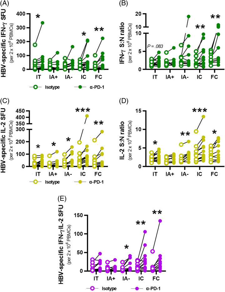
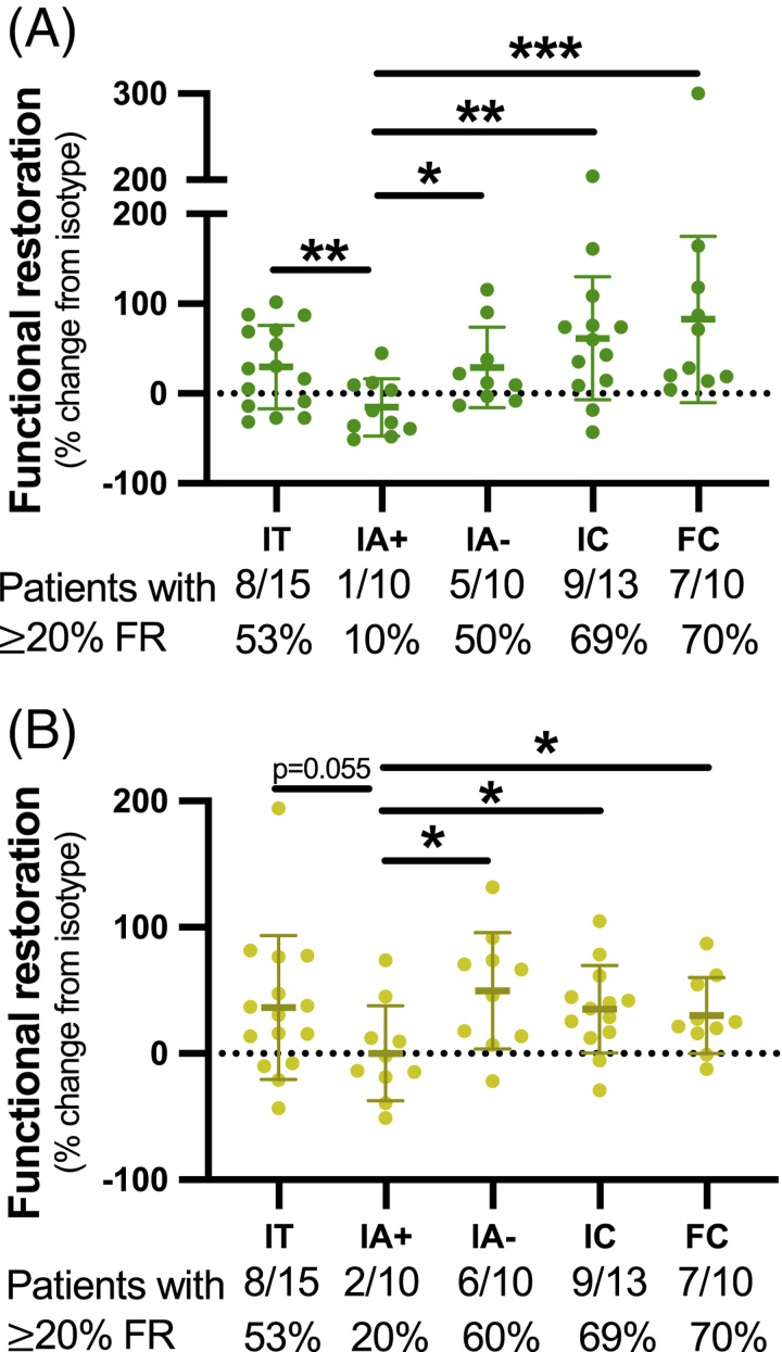
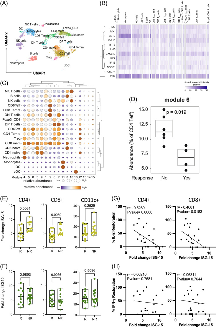
Methods: We enrolled immunotolerant, HBeAg+ immune-active (IA+), HBeAg- immune-active (IA-), inactive carriers, and functionally cured patients to test ex vivo PD-1 blockade on HBV-specific T cell functionality. Peripheral blood mononuclear cells were stimulated with overlapping peptides covering HBV proteins +/-α-PD-1 blockade. Functional T cells were measured using a 2-color FluoroSpot assay for interferon-γ and IL-2. Ex vivo functional restoration was compared to the interferon response capacity assay, which predicts overall survival in cancer patients receiving checkpoint inhibitors.

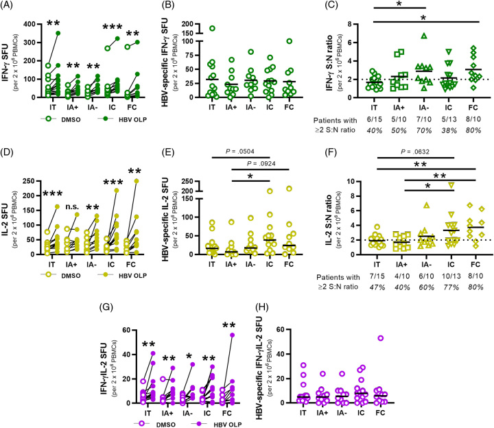
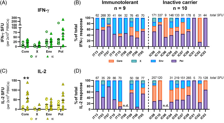
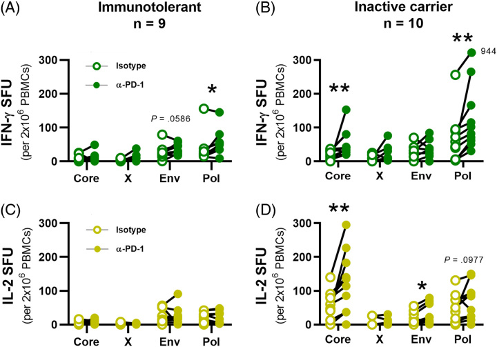
Results: Ex vivo interferon-γ+ responses did not differ across clinical phases. IL-2+ responses were significantly higher in patients with better viral control and preferentially restored with PD-1 blockade. Inactive carrier patients displayed the greatest increase in IL-2 production, which was dominated by CD4 T cell and response to the HBcAg. The interferon response capacity assay significantly correlated with the degree of HBV-specific T cell restoration.

Conclusions: IL-2 production was associated with better HBV control and superior to interferon-γ as a marker of T cell restoration following ex vivo PD-1 blockade. Our study suggests that responsiveness to ex vivo PD-1 blockade, or the interferon response capacity assay, may support stratification for α-PD-1 therapies.


Conflict of interest statement

The authors declare that this study was funded in part by AstraZeneca. Authors Sara Ferrando-Martinez and Scott H Robins were employed by AstraZeneca. Jordan J Feld receives research funding by Abbvie, Arbutus Biopharma, Gilead Sciences Inc., Janssen Pharmaceuticals, Eiger Biopharmaceuticals, and Enanta Pharmaceuticals; and reports compensation from consulting/scientific advising for Abbvie, Arbutus Biopharma, Gilead Sciences Inc., and GlaxoSmithKline. Scott Fung receives research funding by Gilead Sciences Inc.; and reports compensation from consulting/scientific advising for Gilead Sciences Inc., Abbvie, Janssen Pharmaceuticals, Assembly Biosciences. Harry L.A. Janssen receives research funding by Abbvie, Gilead Sciences Inc., GlaxoSmithKline, Janssen Pharmaceuticals, Roche, Vir Biotechnology; and reports compensation from consulting/scientific advising for ALIGOS Therapeutics, Antios Therapeutics, Arbutus Biopharma, Eiger Biopharmaceuticals, Gilead Sciences Inc., GlaxoSmithKline, Janssen Pharmaceuticals, Merck, Roche, VBI Vaccines Inc., Vir Biotechnology, and Viroclinics Biosciences. Adam J. Gehring. receives research funding from Janssen Pharmaceuticals, GlaxoSmithKline, and Gilead Sciences Inc. and reports compensation from consulting/scientific advising: Janssen Pharmaceuticals, Roche, GlaxoSmithKline, Vir Biotechnology. David G. Brooks holds the US Patent (PCT/CA2022/051519) for the Interferon response capacity assay. The remaining authors have no conflicts to report.

Figures

FIGURE 1
FIGURE 1
Ex vivo detection of HBV-specific T cells in patient cohorts with chronic hepatitis B by means of multianalyte FluoroSpot assays. (A) IFN-γ+ SFU for HBV OLP (filled circles) and DMSO (empty circles) conditions were linked per patient in each cohort. (B) IFN-γ+ HBV-specific SFU for each patient were calculated by subtracting DMSO SFU from HBV OLP SFU. (C) IFN-γ+ signal:noise (S:N) ratios were calculated for each patient by dividing HBV OLP SFU by DMSO SFU. Ratios ≥2 were considered positive responses. Fractions beneath each cohort denote the number of patients with detectable responses. Black lines indicate sample means. (D) IL-2 + SFU for HBV OLP and DMSO conditions linked per patient in each cohort. (E) IL-2+ HBV-specific SFU for each patient from subtracting DMSO SFU from HBV OLP SFU. (F) IL-2+ S:N ratios for each patient by dividing HBV OLP SFU by DMSO SFU in each patient. (G) Multifunctional IFN-γ+IL-2+ SFU for HBV OLP and DMSO were linked per patient. (H) HBV-specific IFN-γ+IL-2+ SFU for each patient from subtracting DMSO SFU from HBV OLP SFU. Wilcoxon tests were conducted to compare SFU between HBV OLP and DMSO conditions (A, D, G). Mann-Whitney tests were used to compare HBV-specific SFU and S:N ratios between patient cohorts (B, C, E, F, H) (*p<0.05, **p<0.01, ***p<0.001). Abbreviations: FC, functionally cured; IA, immune-active; IC, inactive carrier; IFN-γ, interferon; IT, immunotolerant; OLP, overlapping peptide pool; PBMC, peripheral blood mononuclear cells; S:N ratio, signal-to-noise ratio; SFU, spot-forming unit.
FIGURE 2
FIGURE 2
Ex vivo α-PD-1 blocking leads to functional restoration of HBV-specific T cells among patients with chronic hepatitis B. (A) HBV-specific IFN-γ+ SFU for each patient were calculated by subtracting DMSO SFU from HBV OLP SFU for isotype- (empty circles) and α-PD-1 blocked (filled circles) conditions. (B) IFN-γ+ S:N ratios were calculated for each patient by dividing HBV OLP SFU by DMSO SFU for each blocking condition. (C) HBV-specific IL-2+ SFU for each patient from subtracting DMSO SFU from HBV OLP SFU per blocking condition. (D) IL-2+ S:N ratios for each patient by dividing HBV OLP SFU by DMSO SFU per blocking condition. (E) Multifunctional IFN-γ+IL-2+ HBV-specific SFU for each patient following background subtraction for each blocking condition. Wilcoxon tests were conducted to compare HBV-specific SFU and S:N ratios between isotype- and α-PD-1 blocked conditions in each cohort (A–E) (*p<0.05, **p<0.01, ***p<0.001). Abbreviations: FC, functionally cured; IA, immune-active; IC, inactive carrier; IFN-γ, interferon; IT, immunotolerant; PBMC, peripheral blood mononuclear cells; S:N ratio, signal-to-noise ratio; SFU, spot-forming unit; PD-1, programmed cell death ligand protein 1.
FIGURE 3
FIGURE 3
Functional restoration from ex vivo PD-1 blockade across patient cohorts with chronic hepatitis B. Functional restoration was calculated and tabulated per patient for (A) IFN-γ and (B) IL-2 responses. The dotted line represents 0%, while full black lines indicate means with SDs. Fractions beneath each cohort indicate the number of patients having a functional restoration percentage of ≥20% and considered treatment-responsive to ex vivo PD-1 blockade. Mann-Whitney tests were used to compare functional restoration between patient cohorts (A, B) (* p<0.05, ** p<0.01, *** p<0.001). Abbreviations: FC, functionally cured; FR, functional restoration; IA, immune-active; IC, inactive carrier; IFN-γ, interferon; IT, immunotolerant PD-1, programmed cell death protein 1.
FIGURE 4
FIGURE 4
Liver biopsy stains for PD-1/PD-L1 across patient cohorts with chronic hepatitis B. (A) Area of PD-1+ expression among intrahepatic immune cells across chronic hepatitis B cohorts. (B) Area of PD-L1+ expression among intrahepatic immune cells across chronic hepatitis B cohorts. (C) Correlation between PD-L1+ area among intrahepatic immune cells and PD-L1 scoring on hepatocytes among IC patients. (D) Representative PD-L1 staining on hepatocytes to demonstrate weak (left) and diffuse (right) stains, black scale bars represent specified lengths. Unpaired parametric t tests were conducted to compare marker expression between cohorts (A, B). Pearson correlation coefficient and p-values are listed (C). (*p<0.05). Abbreviations: IA, immune-active; IC, inactive carrier; IT, immunotolerant; PD-1, programmed cell death protein 1; PD-L1, programmed cell death ligand 1; Tx, treatment.
FIGURE 5
FIGURE 5
Ex vivo HBV antigen hierarchies are distinct between chronic hepatitis B phase. (A) IFN-γ+ SFU for IT (circles) and IC patients (triangles) were calculated by subtracting DMSO SFU from each HBV overlapping peptide pool antigen SFU. Black lines indicate sample means. (B) Total IFN-γ SFU are listed above each patient and the proportions of IFN-γ responses were calculated for each HBV antigen. (C) IL-2+ SFU for IT and IC patients were calculated by subtracting DMSO SFU from each HBV overlapping peptide pool antigen SFU. (D) Total IL-2 SFU are listed for each patient and the proportions of IL-2 responses were calculated for each HBV antigen. Mann-Whitney tests were used to compare antigen-specific SFU between IT and IC patients (A, C) (**p<0.01). Abbreviations: IC, inactive carrier; IFN-γ, interferon; IT, immunotolerant; n.d., not detected; SFU, spot-forming unit.
FIGURE 6
FIGURE 6
Differential functional restoration of HBV antigen-specific T cell responses by clinical phase and cytokine response. Ag-specific IFN-γ+ SFU for (A) IT and (B) IC patients were each calculated for isotype- (empty circles) and α-PD-1 blocked (filled circles) conditions. Ag-specific IL-2+ SFU for (C) immunotolerant and (D) inactive carrier patients between isotype- and α-PD-1 blocked conditions. Wilcoxon tests were conducted to compare Ag-specific SFU between isotype- and α-PD-1 blocked conditions (*p<0.05, **p<0.01). Abbreviations: IFN-γ, interferon; PBMCs, peripheral blood mononuclear cells; PD-1, programmed cell death protein 1; SFU, spot-forming unit.
FIGURE 7
FIGURE 7
IFN response capacity correlates with IL-2 restoration after PD-1 blockade. (A) UMAP projection of immune cell populations identified in the cytof staining panel. (B) Single-cell heatmap showing the expression pattern of 12 interferon-stimulated proteins across the immune populations defined in (A). The transformed expression of each protein is scaled as a percentage of maximum expression. (C) Fast-Phenograph was used to define distinct modules of interferon-stimulated proteins. The relative abundance of each module within a given cell subset (size of dots) and the enrichment of the cell subset within a module (color of dots) are shown for the 16 modules. (D) Frequency of CD4 Teff cells positive for interferon-stimulated protein module 6 between anti-PD1, IL-2+IFN-γ+ responder (n=6) and nonresponder (n=4) patients. Boxes show the median, upper, and lower quartile and whiskers extend to 1.5X the interquartile range. Patients were stratified to responder/nonresponders to anti-PD-1 based on their (E) IL-2 (yellow) (R n=13; NR n=12 ) or (F) IFN-γ (green) (R n=11 ; NR n=14 ) HBV-specific T cell FluoroSpot results. Fold change in ISG15 expression in CD4 T cells, CD8 T cells, and myeloid cells was calculated by dividing the mean fluorescent intensity of IFN-β-stimulated cells by that of the unstimulated sample. Nonparametric Mann-Whitney t test was used for the statistical analysis. (G) Correlation between the percent increase in IL-2 production after PD-1 blockade and fold change in ISG15 expression. (H) Correlation between the percent increase in IFN-γ production after PD-1 blockade and fold change in ISG15 expression. Abbreviations: BST2, bone marrow stromal cell antigen 2; CD274, cluster of differentiation 274; CXCL-10, C-X-C motif chemokine 10; DC, dendritic cell; DN, double negative; DP, double positive; IDO, Indoleamine 2,3-dioxygenase; IFI16, interferon gamma inducible protein 16; IFIT3, interferon induced protein with tetratricopeptide repeats 3; IFN-γ, interferon; IRF7, interferon regulatory factor 7; ISG15, interferon stimulated gene 15; MX1, Myxovirus resistance protein 1; NK, natural killer cell; NR, nonresponder; PD-1, programmed cell death protein 1; pDC, plasmacytoid dendritic cell; PKR, protein kinase R; R, responder SOCS1, suppressor of cytokine signaling 1; Treg, regulatory CD4 T cell; UMAP, uniform manifold approximation and projection.

References

    1. Alatrakchi N, Koziel MJ. Antiviral T-cell responses and therapy in chronic hepatitis B. J Hepatol. 2003;39:631–4. - PubMed
    1. Rivino L, Bert N Le, Gill US, Kunasegaran K, Cheng Y, Tan DZM, et al. . Hepatitis B virus–specific T cells associate with viral control upon nucleos(t)ide-analogue therapy discontinuation. J Clin Invest. 2018;128:668–81. - PMC - PubMed
    1. Asabe S, Wieland SF, Chattopadhyay PK, Roederer M, Engle RE, Purcell RH, et al. . The size of the viral inoculum contributes to the outcome of hepatitis B virus infection. J Virol. 2009;83:9652–62. - PMC - PubMed
    1. Thimme R, Wieland S, Steiger C, Ghrayeb J, Reimann KA, Purcell RH, et al. . CD8+ T cells mediate viral clearance and disease pathogenesis during acute hepatitis B virus infection. J Virol. 2003;77:68–76. - PMC - PubMed
    1. Boni C, Laccabue D, Lampertico P, Giuberti T, Viganò M, Schivazappa S, et al. . Restored function of HBV-specific T cells after long-term effective therapy with nucleos(t)ide analogues. Gastroenterology. 2012;143:963–73.e9. - PubMed

Publication types