. 2023 Dec 6;19(12):e1011849.
doi: 10.1371/journal.ppat.1011849. eCollection 2023 Dec.

Citrullination profile analysis reveals peptidylarginine deaminase 3 as an HSV-1 target to dampen the activity of candidate antiviral restriction factors

Affiliations

Citrullination profile analysis reveals peptidylarginine deaminase 3 as an HSV-1 target to dampen the activity of candidate antiviral restriction factors

Selina Pasquero et al. PLoS Pathog. .

Abstract

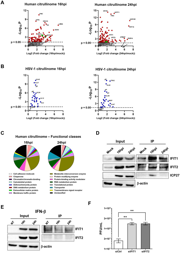
Herpes simplex virus 1 (HSV-1) is a neurotropic virus that remains latent in neuronal cell bodies but reactivates throughout an individual's life, causing severe adverse reactions, such as herpes simplex encephalitis (HSE). Recently, it has also been implicated in the etiology of Alzheimer's disease (AD). The absence of an effective vaccine and the emergence of numerous drug-resistant variants have called for the development of new antiviral agents that can tackle HSV-1 infection. Host-targeting antivirals (HTAs) have recently emerged as promising antiviral compounds that act on host-cell factors essential for viral replication. Here we show that a new class of HTAs targeting peptidylarginine deiminases (PADs), a family of calcium-dependent enzymes catalyzing protein citrullination, exhibits a marked inhibitory activity against HSV-1. Furthermore, we show that HSV-1 infection leads to enhanced protein citrullination through transcriptional activation of three PAD isoforms: PAD2, PAD3, and PAD4. Interestingly, PAD3-depletion by specific drugs or siRNAs dramatically inhibits HSV-1 replication. Finally, an analysis of the citrullinome reveals significant changes in the deimination levels of both cellular and viral proteins, with the interferon (IFN)-inducible proteins IFIT1 and IFIT2 being among the most heavily deiminated ones. As genetic depletion of IFIT1 and IFIT2 strongly enhances HSV-1 growth, we propose that viral-induced citrullination of IFIT1 and 2 is a highly efficient HSV-1 evasion mechanism from host antiviral resistance. Overall, our findings point to a crucial role of citrullination in subverting cellular responses to viral infection and demonstrate that PAD inhibitors efficiently suppress HSV-1 infection in vitro, which may provide the rationale for their repurposing as HSV-1 antiviral drugs.


Conflict of interest statement

The authors have declared that no competing interests exist.

Figures

Fig 1
Fig 1. The pan-PAD inhibitors Cl-A and BB-Cl inhibit HSV-1 replication in human fibroblasts.
(A) Protein lysates from HFFs infected with HSV-1 (MOI 1 PFU/cell) at different hours post-infection (hpi) or from uninfected HFFs (Mock) were exposed to an Rh-PG citrulline-specific probe (left panel) and subjected to gel electrophoresis to detect citrullinated proteins. An anti-gD antibody was used to assess HSV-1 infection, while β-actin cellular expression was used for protein loading control. Equal loading was also assessed by Coomassie blue staining (right panel). One representative gel of three independent experiments is shown. (B, C) Dose-response curves of the cell-permeable pan-PAD inhibitors Cl-A (B) and BB-Cl (C) in HFFs infected with HSV-1 (MOI 1). Inhibitors were given 1 h prior to virus adsorption and kept throughout the whole experiment. 24 hpi viral plaques were microscopically counted and the number of plaques was plotted as a function of inhibitor concentration. Values are expressed as mean ± SEM of three independent experiments, *P < 0.05, **P < 0.01, ***P < 0.001; one-way ANOVA followed by Bonferroni’s post test. (D) Protein lysates from uninfected (mock) or infected HFFs (24 hpi) at an MOI of 1 PFU/cell treated with Cl-A (100 μM), BB-Cl (2.5 μM), or vehicle (DMSO) were analyzed by immunoblotting for viral expression (gD) and normalized to β-actin. (E) To determine the number of viral DNA genomes in HSV-1-infected HFFs, viral DNA was isolated at 24 hpi and analyzed by qPCR using primers amplifying a region of the gE gene. GAPDH was used to normalize HSV-1 genome counts. Values are expressed as mean ± SEM of three independent experiments. HFFs were infected with HSV-1 (MOI 1 PFU/cell) and then treated with Cl-A (100 μM), BB-Cl (2.5 μM), or vehicle (DMSO), which were given at four different time points as indicated (F). At 24 hpi, viral plaques were microscopically counted and expressed as PFU/mL (G). Values are expressed as mean ± SEM of three independent experiments, *P < 0.05, **P < 0.01; one-way ANOVA followed by Bonferroni’s post test.
Fig 2
Fig 2. HSV-1 infection upregulates PAD expression in human fibroblasts.
(A) mRNA expression levels of PADI isoforms by RT-qPCR of HSV-1-infected (8 and 16 hpi) vs uninfected (mock) HFFs were normalized to the housekeeping gene glyceraldehyde-3-phosphate dehydrogenase (GAPDH) and expressed as mean fold change ± SEM over mock-infected cells. *P < 0.05, **P < 0.01, ***P < 0.001; one-way ANOVA followed by Bonferroni’s post test. (B) Western blot analysis of protein lysates from uninfected (mock) or infected HFFs (MOI 1) at different time points using antibodies against PAD2, PAD3, and PAD4. An anti-gD antibody was used to assess HSV-1 infection, while β-actin cellular expression was used for protein loading control. (C) Western blot analysis of protein lysates from untreated (NT) or IFN-β-treated (500 U/mL) HFFs using antibodies against PAD2, PAD3, and PAD4. An anti-ICP27 antibody was used to assess HSV-1 infection, while β-actin cellular expression was used for protein loading control. (D) Western blot analysis of protein lysates from uninfected (mock) or HSV-1-infected (MOI 1) cells at different time points using antibody against PAD3. An anti-ICP27 antibody was used to assess HSV-1 infection, while β-actin cellular expression was used for protein loading control. Representative blots of three independent experiments are shown.
Fig 3
Fig 3. PAD3 induction by HSV-1 early mechanism.
(A) HFFs were transiently transfected with luciferase plasmid encoding the wild-type PADI3 promoter region (pGL4.20-PADI3) or the pGL4.20 empty vector. Twenty-four h later, the cells were mock-infected or infected with HSV-1 at an MOI of 1. At 24 hpi, firefly and Renilla luciferase activities were measured. Luciferase activity in whole-cell lysates was normalized to Renilla luciferase activity and plotted as folds induction relative to infected HFFs carrying the pGL4.20 empty vector (set at 1). Results are shown as means of fold change ± SEM (error bars) of three independent experiments. (B) Western blot analysis of protein lysates from uninfected (mock) or infected HFFs with HSV-1 wild-type (WT) or UV-inactivated HSV1 (UV), at 24 hpi (MOI 1). Analysis was performed using antibodies against PAD3, ICP27, or β-actin. One representative blot of three independent experiments is shown. (C) Western blot and Rh-PG analysis of protein lysates from uninfected (mock) or HSV1-infected HFFs (MOI 1), treated with 150 μg/ml CHX or left untreated. Analysis was performed using antibodies against PAD2, PAD3, PAD4, ICP27, or β-actin to assess for equal loading. One representative blot of three independent experiments is shown. (D) Protein lysates from uninfected (mock) or infected HFFs (16 hpi) at an MOI of 1 PFU/cell treated with (PFA 250 μM) or vehicle were analyzed by immunoblotting for PAD3, viral protein gD, or β-actin. (E) PAD3 and ATF-6 mRNA expression levels by RT-qPCR of HSV-1-infected (16 hpi, MOI 1) or mock-infected HFFs treated or not with the indicated amounts of thapsigargin (TG). The results were normalized to the housekeeping gene glyceraldehyde-3-phosphate dehydrogenase (GAPDH) and expressed as mean fold change ± SEM over mock-infected cells. *P < 0.05, **P < 0.01, ***P < 0.001; one-way ANOVA followed by Bonferroni’s post test. (F) Western blot analysis of protein lysates from uninfected (mock), HSV-1-infected (16 hpi, MOI 1) or mock-infected HFFs treated with TG for 16 h. Analysis was performed using antibodies against PAD3, ICP27, or β-actin. Representative blots of three independent experiments are shown.
Fig 4
Fig 4. PAD3 targeting impairs HSV-1 replication in human cells.
HFFs were infected with HSV-1 (MOI 1) and then treated with increasing concentrations of HF4 (A) or CAY10727 (B), two PAD3-specific inhibitors, which was given 1 h prior to virus adsorption and kept throughout the whole experiment. At 24 hpi, viral plaques were microscopically counted, and the number of plaques was plotted as a function of inhibitor concentration. Values are expressed as means ± SEM (error bars) of three independent experiments. Values are expressed as mean ± SEM of three independent experiments, *P < 0.05, **P < 0.01, ***P < 0.001; one-way ANOVA followed by Bonferroni’s post test. (C) Representative images of infected HFFs (24 hpi) at an MOI of 1 PFU/cell and treated with HF4 (5 μM), CAY10727 (1 μM), or vehicle (DMSO). (D) Protein lysates from uninfected (mock) or infected HFFs (24 hpi) at an MOI of 1 PFU/cell treated with AFM30a (20 μM), HF4 (5 μM), GSK199 (20 μM) or vehicle (DMSO) were analyzed by immunoblotting to assess for viral expression with an anti-gD antibody; β-actin cellular expression was used for protein loading control. SH-SY5Y, ARPE-19, and HEK293 cells were infected with HSV-1 (MOI 1 PFU/cell) and then treated with increasing concentrations of HF4 (E) or CAY10727 (F), which were given 1 h prior to virus adsorption and kept throughout the whole experiment. At 24 hpi, viral plaques were microscopically counted, and the number of plaques was plotted as a function of inhibitor concentration. Values are expressed as means ± SEM (error bars) of four independent experiments, *P < 0.05, **P < 0.01, ***P < 0.001; one-way ANOVA followed by Bonferroni’s post test. (G) HFFs were silenced for PAD3 using specific siRNAs (siPAD3), as negative control cells were also similarly transfected with scrambled siRNA (siCTRL). At 24 h post-treatment (hpt), cells were infected with HSV-1 at an MOI of 1 PFU/cell. The efficiency of PAD3 protein depletion at 24 hpi was assessed by immunoblotting using antibodies against PAD3 or β-actin for equal loading. An anti-gD antibody was used to verify HSV-1 infection. Representative blots of three independent experiments are shown. (H) PAD3-silenced cells were infected with HSV-1 at an MOI of 1 PFU/cell. Viral supernatants were collected at 24 hpi and analyzed by standard plaque assay. Values are expressed as means ± SEM. Values are expressed as mean ± SEM of three independent experiments, ***P < 0.001; one-way ANOVA followed by Bonferroni’s post test. (I) Representative images of infected HFFs (24 hpi) at an MOI of 1 PFU/cell and transfected with the same siCTRL and siPAD3 described in the legend to Fig 3D.
Fig 5
Fig 5. Citrullinome analysis reveals IFIT proteins as restriction factors for HSV-1 replication.
(A-B) Volcano plot depicting the host (A, red dots) and viral (B, blue dots) citrullinated proteins of infected cells vs mock-infected cells at 16 hpi (left panels) and 24 hpi (right panels). Cell lysates from uninfected (mock) or HSV-1-infected HFFs (MOI 1) were exposed to a biotin-PG to isolate citrullinated proteins on streptavidin agarose. Bound proteins were then subjected to on-bead tryptic digestion and analyzed by LC-MS/MS—in the graph, every identified citrullinated protein corresponds to a dot. The x-axis represents the ratio of citrullination between mock and infected cells at the indicated time points, while the y-axis indicates the statistical significance. Both variables were plotted on a logarithmic scale (n = 3). IFIT1 and IFIT2 proteins are reported as green dots, while grey dots represent proteins that do not reach statistical significance. (C) The pie charts show the classification of citrullinated cellular proteins at 16 hpi (left) and 24 hpi (right) based on protein classes. Only classes with a representation exceeding 1% are reported. (D) Immunoprecipitation (IP) of total cell extracts (Input) from mock or infected HFFs at 24 hpi using an anti-peptidylcitrulline antibody. The IP complexes were analyzed by Western blotting using antibodies against IFIT1 and IFIT2. An anti-ICP27 antibody was used to assess HSV-1 infection, while β-actin cellular expression was used for protein loading control. The blot shown is representative of three independent experiments. (E) Immunoprecipitation (IP) of total cell extracts (input) from untreated (NT) or IFN-β-treated (500U/ml) HFFs using an anti-peptidylcitrulline antibody. The IP complexes were analyzed by Western blot using antibodies against IFIT1 and IFIT2. Equal loading was assessed by β-actin immunoblotting. The blot shown is representative of three independent experiments. (F) HFFs were silenced for IFIT1 and IFIT2 using specific siRNAs (siIFIT1 and siIFIT2, respectively). As negative control cells were also similarly transfected with scrambled siRNA (siCtrl. At 24 hpt, cells were infected with HSV-1 at an MOI of 1 PFU/cell. Viral supernatants were collected at 24 hpi and analyzed by standard plaque assay. Values are expressed as means ± SEM of three independent experiments. Representative blots of three independent experiments are shown.

References

    1. Yousuf W, Ibrahim H, Harfouche M, Abu Hijleh F, Abu-Raddad L. Herpes simplex virus type 1 in Europe: Systematic review, meta-analyses and meta-regressions. BMJ Global Health. 2020;5: 1–15. doi: 10.1136/bmjgh-2020-002388 - DOI - PMC - PubMed
    1. Rechenchoski DZ, Faccin-Galhardi LC, Linhares REC, Nozawa C. Herpesvirus: an underestimated virus. Folia Microbiologica. 2017;62: 151–156. doi: 10.1007/s12223-016-0482-7 - DOI - PubMed
    1. Itzhaki RF. Herpes and Alzheimer’s Disease: Subversion in the Central Nervous System and How It Might Be Halted. Journal of Alzheimer’s Disease. 2016;54: 1273–1281. doi: 10.3233/JAD-160607 - DOI - PubMed
    1. De Chiara G, Piacentini R, Fabiani M, Mastrodonato A, Marcocci ME, Limongi D, et al. Recurrent herpes simplex virus-1 infection induces hallmarks of neurodegeneration and cognitive deficits in mice. PLoS Pathogens. 2019;15: 1–30. doi: 10.1371/journal.ppat.1007617 - DOI - PMC - PubMed
    1. Krishnan R, Stuart PM. Developments in Vaccination for Herpes Simplex Virus. Frontiers in Microbiology. 2021;12. doi: 10.3389/fmicb.2021.798927 - DOI - PMC - PubMed