. 2023 Dec 8;9(49):eadi6681.
doi: 10.1126/sciadv.adi6681. Epub 2023 Dec 6.

RAD54L2-mediated DNA damage avoidance pathway specifically preserves genome integrity in response to topoisomerase 2 poisons

Affiliations

RAD54L2-mediated DNA damage avoidance pathway specifically preserves genome integrity in response to topoisomerase 2 poisons

Huimin Zhang et al. Sci Adv. .

Abstract

Type II topoisomerases (TOP2) form transient TOP2 cleavage complexes (TOP2ccs) during their catalytic cycle to relieve topological stress. TOP2ccs are covalently linked TOP2-DNA intermediates that are reversible but can be trapped by TOP2 poisons. Trapped TOP2ccs block transactions on DNA and generate genotoxic stress, which are the mechanisms of action of TOP2 poisons. How cells avoid TOP2cc accumulation remains largely unknown. In this study, we uncovered RAD54 like 2 (RAD54L2) as a key factor that mediates a TOP2-specific DNA damage avoidance pathway. RAD54L2 deficiency conferred unique sensitivity to treatment with TOP2 poisons. RAD54L2 interacted with TOP2A/TOP2B and ZATT/ZNF451 and promoted the turnover of TOP2 from DNA with or without TOP2 poisons. Additionally, inhibition of proteasome activity enhanced the chromatin binding of RAD54L2, which in turn led to the removal of TOP2 from chromatin. In conclusion, we propose that RAD54L2-mediated TOP2 turnover is critically important for the avoidance of potential TOP2-linked DNA damage under physiological conditions and in response to TOP2 poisons.


Figures
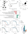
Fig. 1.
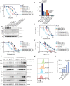
Fig. 1.. Suppression of RNF4 and proteasome-mediated ubiquitination and degradation processes confers cellular resistance to TOP2 poison.
(A) Schematic of the whole-genome CRISPR-Cas9 screens performed in HEK293A WT and TDP2-KO cells with Toronto Knock Out Library v3 (TKOv3). ETO (40 nM) was used for screening. NGS, next-generation sequencing. NT, not-treated. (B) Ranking of ETO coessential genes on the basis of drug Z analysis of the results of CRISPR/Cas9-based screening in HEK293A TDP2-KO cells. The z-score was used to define a possible synthetic lethal interaction with ETO. All genes targeted by the TKOv3 were scored according to the fold change of levels of their sgRNAs. The ETO-treated group and NT group in WT cells were compared. Genes whose loss of function led to ETO sensitivity appear on the left side, with a minus z-score, and genes whose loss of function led to ETO resistance appear on the right side, with a positive z-score. (C) Combinational comparison of ETO coessential genes between HEK293A WT and TDP2-KO cells. The z-scores from screening in HEK293A WT (29) and TDP2-KO cells were used. (D) Gene ontology clustering analysis of genes whose loss of function led to ETO sensitivity or resistance (P < 0.01) in WT and TDP2-KO cells was performed with DAVID Bioinformatics Resources 6.8, NIAID/NIH (https://david.ncifcrf.gov/summary.jsp). The top five enriched biological process were presented. (E) WT and TDP2-KO cells were transfected with siNC (nontargeting control) or siRNAs targeting RNF4. Cell proliferation was measured using a CellTiter-Glo assay after 4 days in the presence of the indicated concentrations of ETO, CPT, HU, or cisplatin. Data are presented as the mean ± SD (n = 3). (F) A two-tailed unpaired t test was used for statistical analysis of the IC50 of ETO treatment of each cell line in (E).
Fig. 2.
Fig. 2.. Depletion of RAD54L2 endows unique cellular sensitivity to treatment with TOP2 poisons.
(A) Proliferation of WT, TDP2-KO, RAD54L2-KO, and TDP2/RAD54L2-DKO cells were measured using a CellTiter-Glo assay after 4 days in the presence of indicated concentration of ETO. Data are presented as the mean ± SD (n = 3). (B) A two-tailed unpaired t test was used for statistical analysis of the IC50 of ETO treatment of each cell line in (A). (C) Western blotting showing TDP2 and RAD54L2 depletion in HEK293A cells. (D to F) Proliferation of WT, TDP2-KO, RAD54L2-KO, and TDP2/RAD54L2-DKO cells were measured using a CellTiter-Glo assay after 4 days in the presence of indicated concentration of doxorubicin (D), pyridostatin (E), and ICRF-187 (F). Data are presented as the mean ± SD (n = 3). (G) WT and RAD54L2-KO cells were treated with 10 μM ETO for the indicated times. Whole-cell extracts (WCEs) were prepared and subjected to Western blotting with the indicated antibodies. (H) A flow cytometry analysis of pH2AX-S139 intensity in WT and RAD54L2-KO cells that were either NT or treated with 4 μM ETO for 2 hours. (I) Quantification of (H). Mean pH2AX-S139 intensity from three independent experiments were shown in a bar chart (mean ± SD, n = 3). An unpaired t test with Welch’s correction was used for statistical analysis.
Fig. 3.
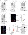
Fig. 3.. RAD54L2 interacts with TOP2A/TOP2B and ZNF451.
(A to D) HEK293A WT cells were either transfected with constructs encoding RAD54L2-SFB (A), SFB-ZNF451 (B), SFB-TOP2A (C), SFB-TOP2B (D), or empty vector as control and were subjected to immunoprecipitation (IP) with S beads. Western blotting was conducted with antibodies as indicated. (E) Representative confocal microscopy images from a TOP2A/RAD54L2-SFB (FLAG) PLA experiment. Scale bar, 10 μm. Quantification of indicated PLA foci in per nucleus was shown. The median number was indicated. (F) Representative confocal microscopy images from a ZNF451/RAD54L2-SFB (FLAG) PLA experiment. Scale bar, 10 μm. Quantification of indicated PLA foci in per nucleus was shown. The median number was indicated. (G) Coomassie Blue staining of purified RAD54L2 protein from insect cells was shown. Insect cell expressed 3FLAG-TOP2A was bound to FLAG-beads and subjected to washes with buffer that containing benzonuclease or high salt to eliminate contaminations from DNA or nonspecific proteins. Purified RAD54L2 (1 μg) was then mixed with FLAG-beads only or FLAG-beads bound with 3FLAG-TOP2A in NETN-100 lysis buffer for 2 hours. Beads were washed with NETN-100 lysis buffer three times and subjected to Western blotting with RAD54L2 antibody. Coomassie Blue staining was presented to show the bound 3FLAG-TOP2A on beads.
Fig. 4.
Fig. 4.. RAD54L2 interacts with SUMOylated proteins.
(A) HEK293A WT cells were transfected with constructs encoding RAD54L2-SFB or its deletion variants and were subjected to pull down with S beads. Western blotting was conducted with antibodies as indicated. (B) HEK293A WT cells were either transfected with constructs encoding RAD54L2-SFB or empty vector as control and were subjected to immunoprecipitation with S beads. Western blotting was conducted with antibodies as indicated. (C) HEK293A WT cells were either transfected with constructs encoding RAD54L2-SFB or empty vector as control. Cells were then treated with 10 μM MG132 or 10 μM ML-792 for 1 hour before subjected to immunoprecipitation with S beads. Western blotting was conducted with antibodies as indicated. The gray line indicates modified forms of TOP2A, TOP2B, or ZNF451. (D) Insect cell expressing 3FLAG-TOP2A, 3FLAG-TOP2A-3xSUMO2, and 3FLAG-TOP2AY805F-3xSUMO2 was bound to FLAG-beads and subjected to washes with buffer that containing benzonuclease or high salt to eliminate contaminations from DNA or nonspecific proteins. Purified RAD54L2 (1 μg) was then mixed with FLAG-beads only or FLAG-beads bound with 3FLAG-TOP2A, 3FLAG-TOP2A-3xSUMO2, or 3FLAG-TOP2AY805F-3xSUMO2 in NETN-100 lysis buffer for 2 hours. Beads were washed with NETN-100 lysis buffer three times and subjected to Western blotting with RAD54L2 antibody. Coomassie Blue staining was presented to show the bound 3FLAG-TOP2A, 3FLAG-TOP2A-3xSUMO2, or 3FLAG-TOP2AY805F-3xSUMO2 on beads.
Fig. 5.
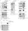
Fig. 5.. RAD54L2 reduces chromatin-bound TOP2 with or without ETO treatment, and this function requires its ATPase activity.
(A) RAD54L2-KO cells were infected with virus expressing RAD54L2WT-SFB or RAD54L2K310A-SFB. Cell proliferation was measured using a CellTiter-Glo assay after 4 days in the presence of the indicated concentrations of ETO. Data are presented as the mean ± SD (n = 3). (B) TDP2/RAD54L2-DKO cells were infected with virus expressing RAD54L2WT-SFB or RAD54L2K310A-SFB. Cell proliferation was measured using a CellTiter-Glo assay after 4 days in the presence of the indicated concentrations of ETO. Data are presented as the mean ± SD (n = 3). (C) WCE and TurboNuclease-mediated chromatin fraction of WT and RAD54L2-KO cells were prepared and subjected to Western blotting with the indicated antibodies. (D) Quantification of (C) from three independent experiments. Mean ± SD are shown. (E) WT and RAD54L2-KO cells were pretreated with 10 μM MG132 for 1 hour and then treated with 100 μM ETO for indicated times. Samples were collected for DUST assay to detect SUMO-2/3, and total TOP2A-DPCs. dsDNA is used as a loading control. dsDNA was run at the same gel but cut for presentation as shown. (F) In vitro TOP2 cleavage assay was performed with indicated amounts of proteins. Reactions were conducted at 37°C for 20 min. Western blotting was conducted for the detection of TOP2Acc and free TOP2A with anti-TOP2A antibody. Proteinase K was used to digest all proteins before running agarose gel. (G) A model showing that RAD54L2 prevents the excessive accumulation of TOP2 on chromatin with or without ETO treatment.
Fig. 6.
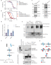
Fig. 6.. RAD54L2, ZNF451, and TDP2 constitute overlapping pathways involved in cellular resistance to ETO.
(A) Representative images of RAD54L2-SFB localization to chromatin with the indicated treatment are presented. Scale bar, 10 μm. Quantification of RAD54L2-SFB intensity per cell were shown. The median intensity was indicated. (B) Chromatin association of indicated proteins were determined with Western blotting with indicated treatment. Histone H3 served as loading control. (C) WT and RAD54L2-KO cells were transfected with siNC (nontargeting control) or siRNAs targeting ZNF451. Cell proliferation was measured using a CellTiter-Glo assay after 4 days in the presence of the indicated concentrations of ETO. Data are presented as the mean ± SD (n = 3). (D) A two-tailed unpaired t test was used for statistical analysis of the IC50 of ETO treatment of each cell line in (C). (E) WT and RAD54L2-KO cells were transfected with constructs encoding RAD54L2-SFB or infected with siRNAs targeting ZNF451. Cells were treated with 100 μM ETO for 20 min. Samples were collected for DUST assay to detect SUMO-2/3, and total TOP2-DPCs. dsDNA is used as a loading control. (F) TDP2-KO and TDP2/RAD54L2-DKO cells were transfected with siNC or siRNAs targeting ZNF451. Cell proliferation was measured using a CellTiter-Glo assay after 4 days in the presence of the indicated concentrations of ETO. Data are presented as the mean ± SD (n = 3). (G) A two-tailed unpaired t test was used for statistical analysis of the median inhibitory concentration (IC50) of ETO treatment of each cell line in (F). (H) WT and TDP2/ZNF451/RAD54L2-TKO cells were transfected with siNC or siRNAs targeting RNF4. Cell proliferation was measured using a CellTiter-Glo assay after 4 days in the presence of the indicated concentrations of ETO. Data are presented as the mean ± SD (n = 3).
Fig. 7.
Fig. 7.. A model of multiple mechanisms involved in the resolution of TOP2 and TOP2-linked DNA damage.
TOP2 forms transient TOP2cc during its catalytic cycle, which can be trapped by TOP2 poisons such as ETO. Here, we propose that RAD54L2 directly remove excess TOP2 on chromatin and thus reduce TOP2cc formation, especially when cells are treated with TOP2 poisons. This conserved RAD54L2-dependent TOP2-specific DNA damage avoidance pathway acts to ensure genome stability. Once the trapped TOP2cc forms, it can also be resolved via several distinct mechanisms. First, it can be precisely removed by the coordinated actions of TDP2 and ZATT/ZNF45. Second, TOP2cc can be targeted by RNF4-mediated pathway for ubiquitination and degradation. Residual peptides after proteasome-mediated degradation can be further processed by TDP2 or other repair enzymes such as MRN/CtIP. Third, MRN/CtIP or other repair enzymes can cleave the DNA strands in the vicinity of intact trapped TOP2cc or proteolytically processed TOP2cc with residual peptides. The RNF4/proteasome-mediated and/or MRN/CtIP-dependent processing of TOP2cc lead to more complex DNA repair events and activate DNA damage responses.

References

    1. J. J. Champoux, DNA topoisomerases: Structure, function, and mechanism. Annu. Rev. Biochem. 70, 369–413 (2001). - PubMed
    1. Y. Pommier, Y. Sun, S. N. Huang, J. L. Nitiss, Roles of eukaryotic topoisomerases in transcription, replication and genomic stability. Nat. Rev. Mol. Cell Biol. 17, 703–721 (2016). - PMC - PubMed
    1. L. Uuskula-Reimand, M. D. Wilson, Untangling the roles of TOP2A and TOP2B in transcription and cancer. Sci. Adv. 8, eadd4920 (2022). - PMC - PubMed
    1. J. L. Nitiss, Targeting DNA topoisomerase II in cancer chemotherapy. Nat. Rev. Cancer 9, 338–350 (2009). - PMC - PubMed
    1. Y. Pommier, E. Leo, H. L. Zhang, C. Marchand, DNA topoisomerases and their poisoning by anticancer and antibacterial drugs. Chem. Biol. 17, 421–433 (2010). - PMC - PubMed