. 2024 Jan:238:109741.
doi: 10.1016/j.exer.2023.109741. Epub 2023 Dec 8.

Visualization of choroidal vasculature in pigmented mouse eyes from experimental models of AMD

Affiliations

Visualization of choroidal vasculature in pigmented mouse eyes from experimental models of AMD

Imran A Bhutto et al. Exp Eye Res. 2024 Jan.

Abstract

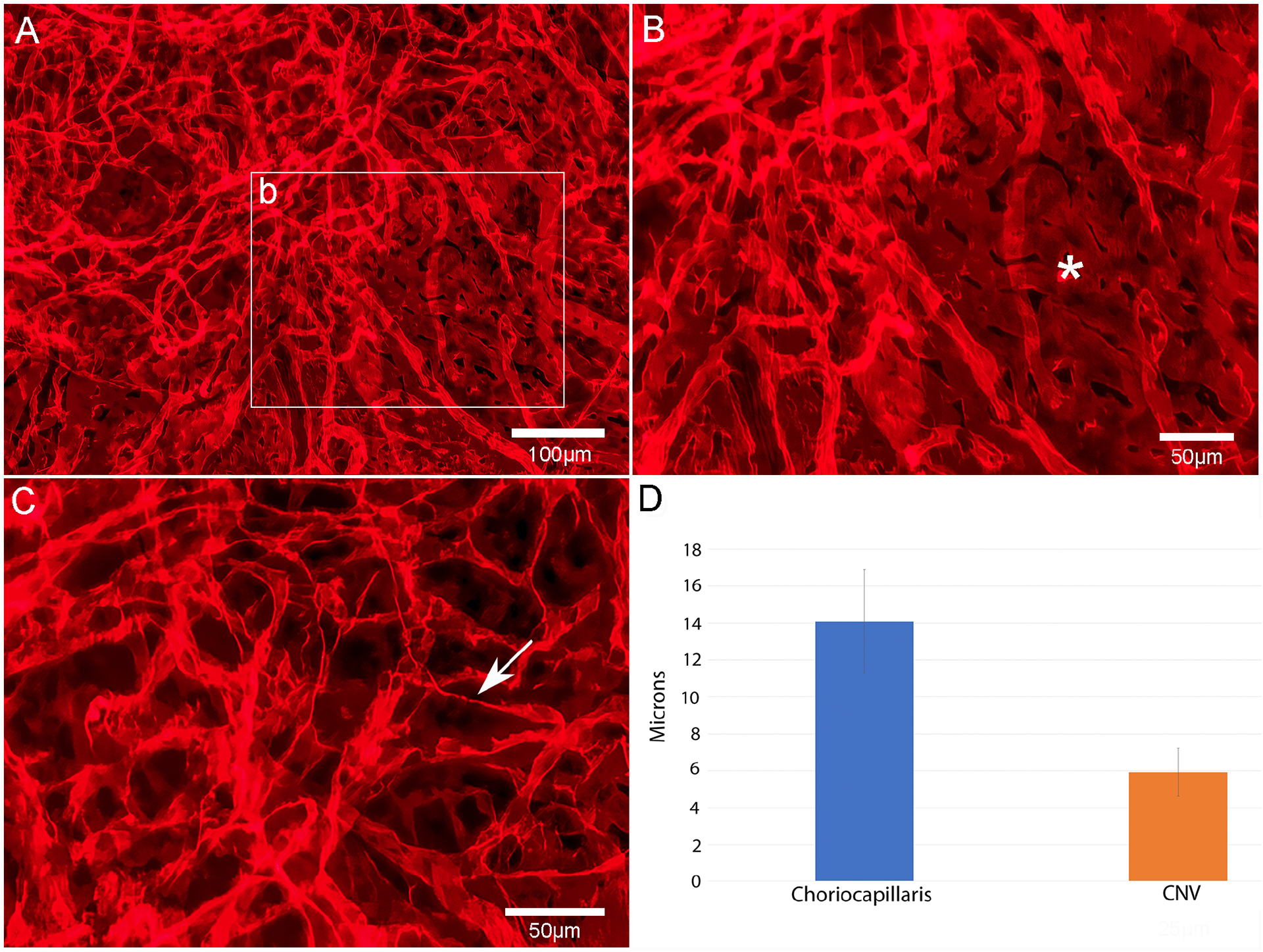
A variety of techniques exist to investigate retinal and choroidal vascular changes in experimental mouse models of human ocular diseases. While all have specific advantages, a method for evaluating the choroidal vasculature in pigmented mouse eyes has been more challenging especially for whole mount visualization and morphometric analysis. Here we report a simple, reliable technique involving bleaching pigment prior to immunostaining the vasculature in whole mounts of pigmented mouse choroids. Eyes from healthy adult pigmented C57BL/6J mice were used to establish the methodology. The retina and anterior segment were separated from the choroid. The choroid with retinal pigment epithelial cells (RPE) and sclera was soaked in 1% ethylenediaminetetraacetic acid (EDTA) to remove the RPE. Tissues were fixed in 2% paraformaldehyde (PFA) in phosphate-buffered saline (PBS). Choroids were subjected to melanin bleaching with 10% hydrogen peroxide (H2O2) at 55 °C for 90 min, washed in PBS and then immunostained with anti-podocalyxin antibody to label vascular endothelium followed by Cy3-AffiniPure donkey anti-goat IgG at 4 °C overnight. Images of immunostained bleached choroids were captured using a Zeiss 710 confocal microscope. In addition to control eyes, this method was used to analyze the choroids from subretinal sodium iodate (NaIO3) RPE atrophy and laser-induced choroidal neovascularization (CNV) mouse models. The H2O2 pretreatment effectively bleached the melanin, resulting in a transparent choroid. Immunolabeling with podocalyxin antibody following bleaching provided excellent visualization of choroidal vasculature in the flat perspective. In control choroids, the choriocapillaris (CC) displayed different anatomical patterns in peripapillary (PP), mid peripheral (MP) and far peripheral (FP) choroid. Morphometric analysis of the vascular area (VA) revealed that the CC was most dense in the PP region (87.4 ± 4.3% VA) and least dense in FP (79.9 ± 6.7% VA). CC diameters also varied depending on location from 11.4 ± 1.97 mm in PP to 15.1 ± 3.15 mm in FP. In the NaIO3-injected eyes, CC density was significantly reduced in the RPE atrophic regions (50.7 ± 5.8% VA in PP and 45.8 ± 6.17% VA in MP) compared to the far peripheral non-atrophic regions (82.8 ± 3.8% VA). CC diameters were significantly reduced in atrophic regions (6.35 ± 1.02 mm in PP and 6.5 ± 1.2 mm in MP) compared to non-atrophic regions (14.16 ± 2.12 mm). In the laser-induced CNV model, CNV area was 0.26 ± 0.09 mm2 and luminal diameters of CNV vessels were 4.7 ± 0.9 mm. Immunostaining on bleached choroids with anti-podocalyxin antibody provides a simple and reliable tool for visualizing normal and pathologic choroidal vasculature in pigmented mouse eyes for quantitative morphometric analysis. This method will be beneficial for examining and evaluating the effects of various treatment modalities on the choroidal vasculature in mouse models of ocular diseases such as age-related macular degeneration, and degenerative genetic diseases.

Keywords: Choroidal blood vessels; Immunostaining; Laser CNV; Mouse; Podocalyxin; Sodium iodate.


Conflict of interest statement

Declaration of competing interest The authors declare no relevant conflicts of interest.

Figures

Figure 1:
Figure 1:
Representative fundus photograph of a normal control mouse (A). Pigmented C57BL/6J mouse choroid before (B) and after 10% H2O2 bleaching (C). Flat mounted control mouse choroid immunolabeled with anti-podocalyxin antibody showing staining of the choroidal vasculature at low magnification (D). Morphology of the choriocapillaris (CC) in the peripapillary region (E) showing a dense freely interanastomosing honeycomb-like meshwork of capillaries. In the midperiphery (F), CC had a more linear arrangement with fewer interanastomosing connections and elongated intercapillary septa. In far periphery (G), choroidal vessels had a palm-shaped pattern and consisted of arterioles, venules and capillaries in the same plane. Scale bars = 1 mm in B–C, 500 μm in D and 50 μm in E–G.
Figure 2:
Figure 2:
Representative fundus photograph from a subretinal NalO3 injected mouse after 3 weeks showing extensive RPE degeneration and pigment clumping (A). Pigmented NaIO3-injected C57BL/6J mouse choroid before (B) and after 10% H2O2 bleaching (C). Flat mounted mouse choroid immunolabeled with anti-podocalyxin antibody three weeks after NalO3 injection showing the extent of CC degeneration (arrowheads) at low magnification (D). Higher magnification (E), shows the border of attenuated CC (arrowheads). CC degeneration in the bleb region (F) and unaffected CC outside the bleb in far periphery (G). Scale bars= 1 mm in B–C, 500 μm in A, 200 μm in D and 50 μm in E–G.
Figure 3:
Figure 3:
Flat mounted choroids from a control mouse (A) and NalO3 injected mice (B–F) showing area of CC degeneration delineated in tracings. The average size of the degenerative area was 7.0±1.56mm2 and the pathology was generally confined to the peripapillary and midperipheral choroid. Scale bar = 500 μm for A–F.
Figure 4:
Figure 4:
Comparison of CC morphology in three distinct regions of choroid in a representative control eye and three NalO3 injected mouse eyes. Three separate images (0.5 mm2 in area) from each region of choroid (control eyes n=3 and NalO3 eyes n=5) were used for morphometric analysis. Compared to control choroids (A, E and I), there was marked uniformity of CC degeneration in peripapillary (B–D) and mid peripheral choroid (F–H) of NalO3 injected eyes while the far peripheral CC was unaffected (J–L). Scale bar = 20 μm in all.
Figure 5:
Figure 5:
Morphometric analysis of control (blue bars) versus NalO3 injected eyes (orange bars) showing percent vascular area (A) and choriocapillaris luminal diameters (B) in peripapillary, mid peripheral and far peripheral choroid. There was a statistically significant decrease in %VA in peripapillary (*p<0.0001) and midperipheral (*p<0.0001) choroid of NalO3 injected eyes compared to controls. CC luminal diameters in NalO3 injected eyes were reduced by 44.3% in peripapillary (*p<0.0001), and 54.3% in mid peripheral (*p<0.0001) choroid. Far peripheral %VA and CC luminal diameters were not significantly different between control and NalO3 injected eyes.
Figure 6:
Figure 6:
Comparison of choroidal vascular attenuation in the mouse NalO3 model immunostained with podocalyxin (A) and in a 92-year-old human with GA immunostained with Ulex europaeus agglutinin (UEA) lectin. The region of complete RPE atrophy (asterisks) and border of RPE atrophy are indicated (arrows). Scale bars = 100 μm.
Figure 7:
Figure 7:
Fundus photo (A) and fluorescein angiogram (B) of a representative laser induced CNV treated mouse eye. Lesion (arrows in A&B) shows scarring of retina/choroid and leakage of dye at the site of CNV.
Figure 8:
Figure 8:
Whole bleached choroids of laser induced CNV mouse eyes immunostained with anti-podocalyxin antibody (A–C) showing CNV networks (arrows) adjacent to the optic nerve head. Higher magnification of CNV (D–F) shows thin diameter capillary networks (arrows) which have connections to the underlying choroidal vessels. Detail of laser induced CNV (arrowheads) and adjacent CC in “G”, at the edge of CNV (H) and adjacent CC (I). Blind ends of CNV at the neovascular front (arrow in H). Normal density and luminal diameters of CC in choroid nearest the CNV (I). Boxed areas in “G” represent higher magnification images shown in “H & I”. Scale bars = 500 μm in A, C & E, 100 μm in B, D, F, & G, and 50 μm in H & I .
Figure 9:
Figure 9:
Edge of podocalyxin stained laser induced CNV shows thin diameter capillaries of neovascularization (A), broad diameter lumen of CC adjacent to CNV (asterisk in B) and individual cells within the CNV with fine processes (arrow) representative of proliferating endothelial cells (C). Morphometric measurements of luminal diameters in CC vs CNV (D). Scale bars= 100 μm in A & 50 μm in B & C.
Figure 10:
Figure 10:
Comparison of CNV (arrowheads) in the mouse laser-induced CNV model immunostained with podocalyxin (A) and in a 92-year-old Caucasian male with neovascular AMD immunostained with Ulex europaeus agglutinin (UEA) lectin (B). Note the attenuated CC in advance of CNV in the human choroid (asterisks). Scale bars = 100 μm in A & 1mm in B.

References

    1. Baba T, Bhutto IA, Merges C, Grebe R, Emmert D, McLeod DS, Armstrong D, Lutty GA, 2010. A rat model for choroidal neovascularization using subretinal lipid hydroperoxide injection. Am. J. Pathol 176(6), 3085–3097. - PMC - PubMed
    1. Bhutto IA, Amemiya T, 2001. Microvascular Architecture of the Rat Choroid: Corrosion Cast Study. Anat. Rec 264, 63–71. - PubMed
    1. Bhutto IA, Tiwari A, Thomson BR, Edwards MM, Lutty GA, 2020. Visualization of choroidal vasculature in pigmented mouse eyes. Invest Ophthalmol Vis Sci. 61, 2235.
    1. Bhutto IA, Ogura S, Baldeosingh R, McLeod DS, Lutty GA, Edwards MM, 2018. An acute injury model for the phenotypic characteristics of geographic atrophy. Invest. Ophthalmol. Vis. Sci 59(4), AMD143–AMD151. 10.1167/iovs.18-24245 - DOI - PMC - PubMed
    1. Chowers G, Cohen M, Marks-Ohana D, Stika S, Eijzenberg A, Banin E, Obolensky A, 2017. Course of sodium iodate-induced retinal degeneration in albino and pigmented mice. Invest. Ophthalmol. Vis. Sci 58(4), 2239–2249. doi: 10.1167/iovs.16-21255 - DOI - PubMed

Publication types