Review
. 2024 May;42(5):544-559.
doi: 10.1016/j.tibtech.2023.10.009. Epub 2023 Dec 5.

Advances in 3D bioprinting for urethral tissue reconstruction

Affiliations
Review

Advances in 3D bioprinting for urethral tissue reconstruction

Daniel Booth et al. Trends Biotechnol. 2024 May.

Abstract

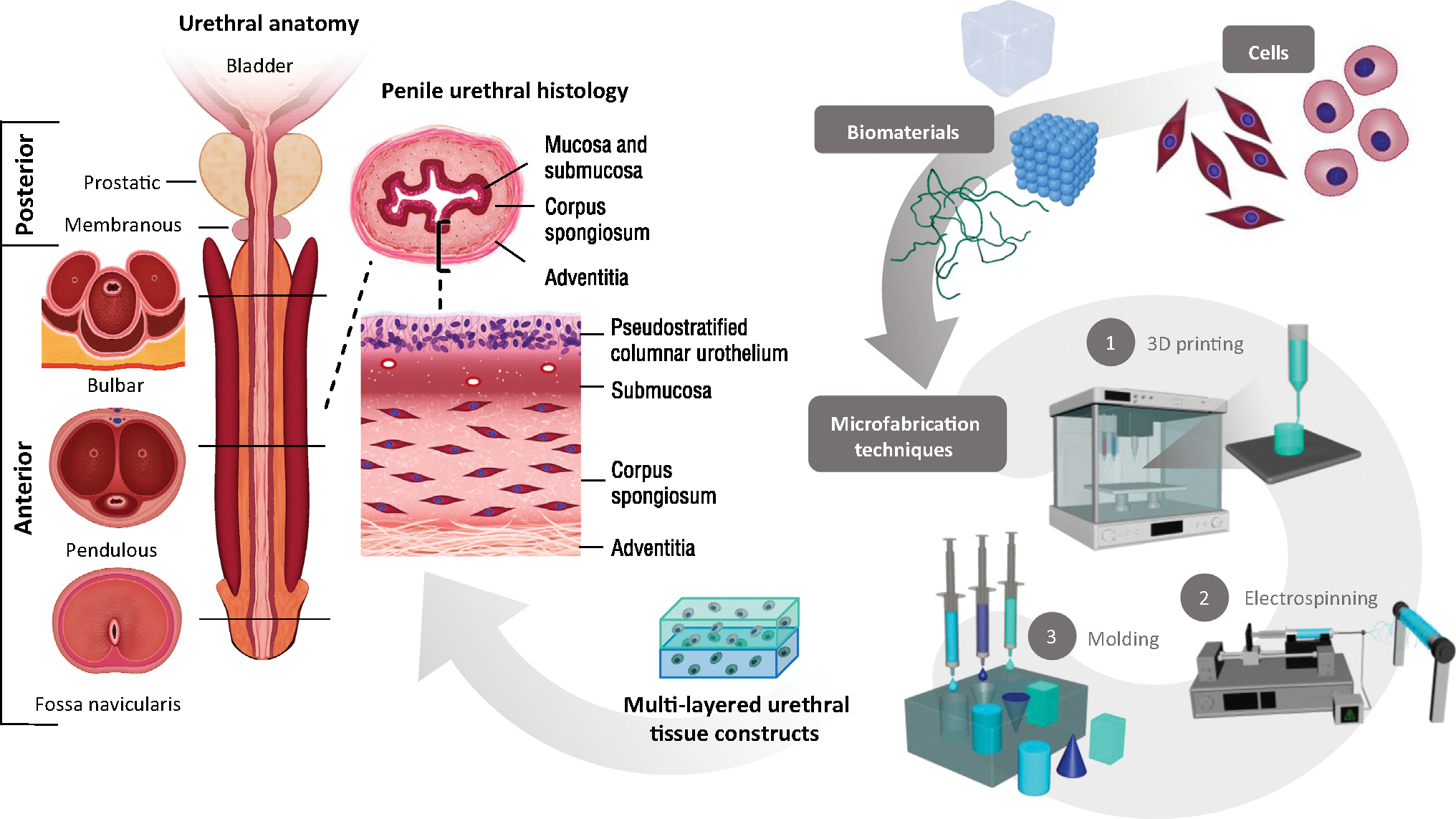
Urethral conditions affect children and adults, increasing the risk of urinary tract infections, voiding and sexual dysfunction, and renal failure. Current tissue replacements differ from healthy urethral tissues in structural and mechanical characteristics, causing high risk of postoperative complications. 3D bioprinting can overcome these limitations through the creation of complex, layered architectures using materials with location-specific biomechanical properties. This review highlights prior research and describes the potential for these emerging technologies to address ongoing challenges in urethral tissue engineering, including biomechanical and structural mismatch, lack of individualized repair solutions, and inadequate wound healing and vascularization. In the future, the integration of 3D bioprinting technology with advanced biomaterials, computational modeling, and 3D imaging could transform personalized urethral surgical procedures.

Keywords: 3D bioprinting; biomaterials; tissue engineering; urethra; urological disease.


Conflict of interest statement

Declaration of interests N.A., is a member of the journal’s advisory board and affirms that this affiliation did not influence the idea, execution, or interpretation of the article. N.A. holds equity in GelMEDIX Inc.; R.S. holds equity in Protean Surgical Instruments. The remaining authors declare no interests.

Figures

Figure 1.
Figure 1.. Anatomic, structural, and pathologic features of the male urethra.
These are critical features to guide the application of novel biomaterials, cell seeding, and microfabrication techniques in the development of personalized multilayered tissue-engineered constructs.
Figure 2.
Figure 2.. Common 3D bioprinting methods for engineering urethral tissue constructs.
(A) Direct inkjet-based bioprinting; (B) coaxial extrusion; (C) droplet-based bioprinting; (D) indirect inkjet-based bioprinting; (E) laser-based forward transfer bioprinting; (F) laser-based photolithographic bioprinting.
Figure 3.
Figure 3.. Star-shaped scaffold with radial elasticity for hollow organ regeneration.
Macroscopic view of the scaffold in (A) closed and (B) partially open position; (C) expansion and relaxation (folding, unfolding) of the scaffold after injection and removal of water; (D) burst pressure of round scaffolds without star-shaped compression, star-shaped scaffolds before and after fatigue test (both n = 8), and of native pig urethras (n = 5). Tubes were closed at both ends, and water was pumped into the lumen until rupture while continuously monitoring the pressure. Bars represent the mean ± standard deviation. One-way ANOVA with Bonferroni post hoc test, ***P < 0.0001; (E) hematoxylin and eosin staining (H&E) staining of cross-sections of cell-seeded star scaffold. (F) Immunostaining of dynamically cultured scaffold stained for cell nuclei with DAPI (blue), type I collagen (green), and cytokeratin 18 (red). Reproduced, with permission, from [57]. Copyright 2017, Elsevier.
Figure 4.
Figure 4.. 3D bioprinted PCL/PLCL urethral constructs with structural modification to tubularized design.
(A) CT scan of a male rabbit urethra. (B) PCL/PLCL (50:50) scaffold with spiral design. (C) Final structure of the bioprinted urethra. (D) The viability and proliferation of UCs and SMCs in the bioprinted urethra. (E) UCs (labeled with PKH67 green fluorescent dye) as seen in the hydrogel component of the bioprinted urethral construct after 7 days of culture. (F) SMCs (labeled with PKH26 red fluorescent dye) in the hydrogel component of the bioprinted urethral construct after 7 days of cell culture. Reproduced, with permission, from [50]. Copyright 2017, Elsevier. Abbreviations: PCL, poly(ε-caprolactone); PLCL, poly(lactide-co-caprolactone); SMC, smooth muscle cell; UC, urothelial cell.
Figure 5.
Figure 5.. Complex 3D printed structures with in situ crosslinking approach.
(A) Schematic and representative fluorescence images for printing filaments with core-shell structure using (B) two inks labeled with different fluorophores or (C) two inks containing cells labeled with different dyes. (D) Schematic and representative fluorescence images for printing heterogeneous filaments with intermittent structures using two inks labeled with (E) different fluorophores or (F) two inks containing cells labeled with different dyes. (G) Schematic for printing hollow filaments using a longer core coaxial nozzle and representative images of printed hollow tubes (H) either before or after perfusion with a dye solution or (I) with cells in the printed tubes. Scale bars are 500 μm. Reproduced, with permission, from [52]. Copyright 2017, Wiley.

References

    1. Pathak H et al. (2023) Surgical management of stricture urethra in patients with chronic renal failure: ten years’ experience at a tertiary center. Urol. Ann. 15, 22–26 - PMC - PubMed
    1. Baskin LS and Ebbers MB (2006) Hypospadias: anatomy, etiology, and technique. J. Pediatr. Surg. 41, 463–472 - PubMed
    1. Santucci RA et al. (2007) Male urethral stricture disease. J. Urol. 177, 1667–1674 - PubMed
    1. Alwaal A et al. (2014) Epidemiology of urethral strictures. Transl. Androl. Urol. 3, 209–213 - PMC - PubMed
    1. Barbagli G et al. (2010) Morbidity of oral mucosa graft harvesting from a single cheek. Eur. Urol. 58, 33–41 - PubMed

Publication types

Substances