. 2024 Apr;45(4):844-856.
doi: 10.1038/s41401-023-01199-z. Epub 2023 Dec 6.

SMAD4 endows TGF-β1-induced highly invasive tumor cells with ferroptosis vulnerability in pancreatic cancer

Affiliations

SMAD4 endows TGF-β1-induced highly invasive tumor cells with ferroptosis vulnerability in pancreatic cancer

Hai-di Chen et al. Acta Pharmacol Sin. 2024 Apr.

Abstract

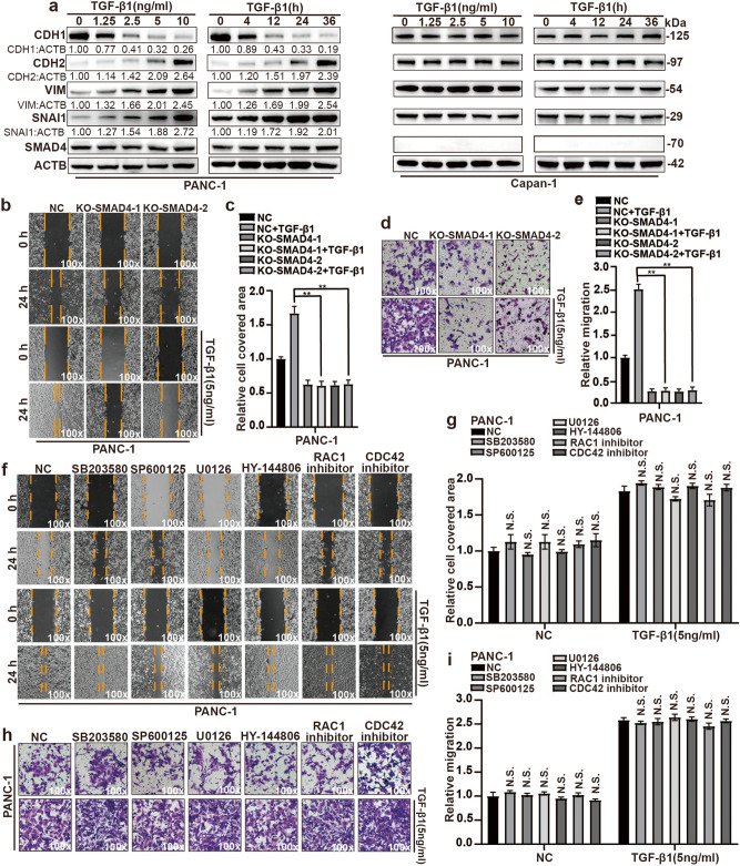
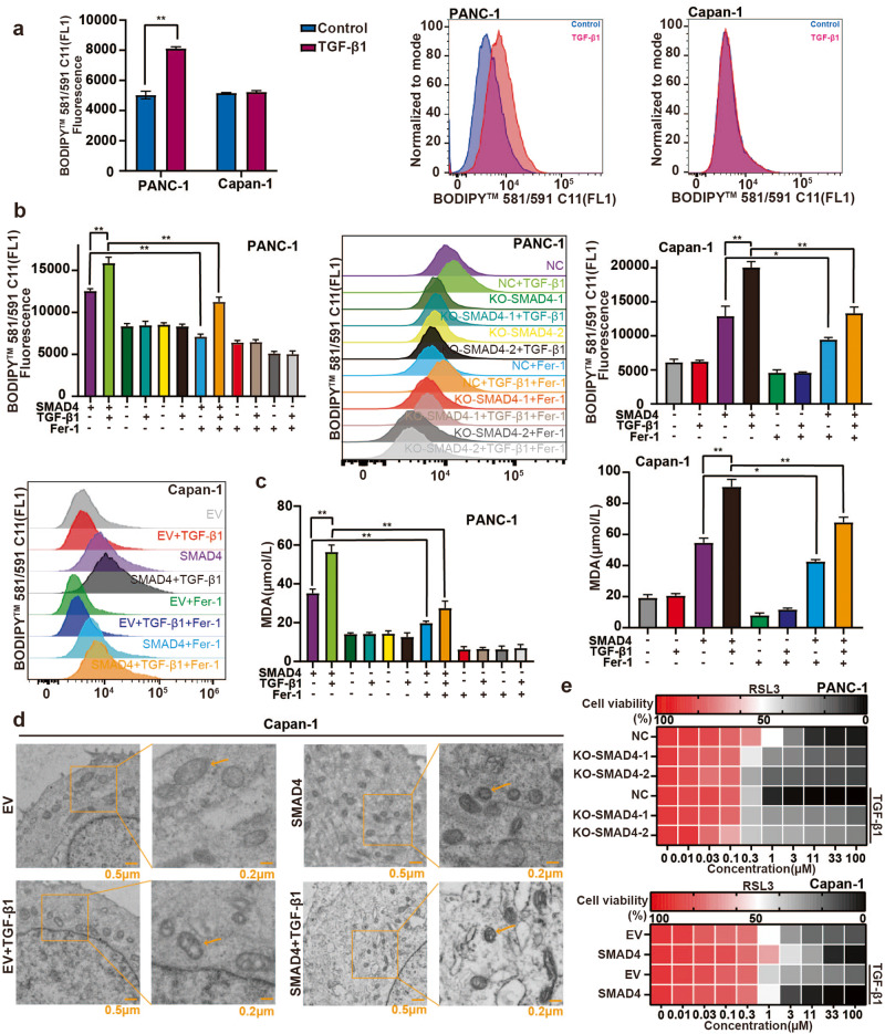
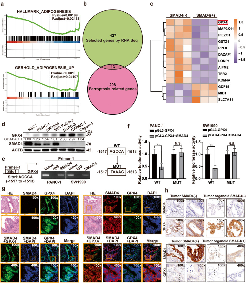
Pancreatic ductal adenocarcinoma (PDAC) is an extremely aggressive malignancy prone to recurrence and metastasis. Studies show that tumor cells with increased invasive and metastatic potential are more likely to undergo ferroptosis. SMAD4 is a critical molecule in the transforming growth factor β (TGF-β) pathway, which affects the TGF-β-induced epithelial-mesenchymal transition (EMT) status. SMAD4 loss is observed in more than half of patients with PDAC. In this study, we investigated whether SMAD4-positive PDAC cells were prone to ferroptosis because of their high invasiveness. We showed that SMAD4 status almost determined the orientation of transforming growth factor β1 (TGF-β1)-induced EMT via the SMAD4-dependent canonical pathway in PDAC, which altered ferroptosis vulnerability. We identified glutathione peroxidase 4 (GPX4), which inhibited ferroptosis, as a SMAD4 down-regulated gene by RNA sequencing. We found that SMAD4 bound to the promoter of GPX4 and decreased GPX4 transcription in PDAC. Furthermore, TGF-β1-induced high invasiveness enhanced sensitivity of SMAD4-positive organoids and pancreas xenograft models to the ferroptosis inducer RAS-selective lethal 3 (RSL3). Moreover, SMAD4 enhanced the cytotoxic effect of gemcitabine combined with RSL3 in highly invasive PDAC cells. This study provides new ideas for the treatment of PDAC, especially SMAD4-positive PDAC.

Keywords: EMT; GPX4; RSL3; SMAD4; ferroptosis; pancreatic cancer.


Conflict of interest statement

The authors declare no competing interests.

Figures

Fig. 1
Fig. 1. SMAD4 determines highly invasive status induced by TGF-β1 in PDAC.
a PANC-1 and Capan-1 cells were treated with various concentrations of TGF-β1 for 24 h or TGF-β1 at 5 ng/ml for various times, and the expressions of EMT marker (CDH1, CDH2, VIM and SNAI1) were evaluated. The grayscale values of the bands were calculated by ImageJ software and are annotated below the bands. b, c A wound healing assay was used to evaluate cell motility in PANC-1 cell lines with SMAD4 silencing treated with TGF-β1 (5 ng/ml, 24 h) or not (n = 3, independent t test, **P < 0.01) (original magnification, ×100). d, e The effect of SMAD4 expression on invasiveness was evaluated after treatment with TGF-β1 (5 ng/ml, 24 h) or not (n = 3, independent t test, **P < 0.01) (original magnification, ×100). fi Wound healing assay and transwell invasion were used to evaluate cell motility and invasion, respectively, in PANC-1 cell lines treated with TGF-β1 (5 ng/ml, 24 h) and a MAPK1/ERK2-MAPK3/ERK1 inhibitor (U0126), MAPK/p38 inhibitor (SB203580), MAPK/JNK inhibitor (SP600125), PI3K/AKT inhibitor (HY-144806), RAC1 inhibitor and CDC42 inhibitor (n = 3, independent t test, P < 0.01) (original magnification, ×100).
Fig. 2
Fig. 2. SMAD4 affects ferroptosis vulnerability in highly invasive pancreatic cancer.
a, b Flow cytometry results are presented in terms of fluorescence (left) and BODIPY 581/591C11 probe assay (right) (n = 3, independent t test, **P < 0.01). c MDA was assessed to estimate the level of lipid peroxidation in PANC-1 and Capan-1 cells with SMAD4 overexpression, SMAD4 knockout, TGF-β1 treatment (5 ng/ml, 24 h) or Fer-1 treatment (2 µmol/L, 24 h) (n = 3, independent t test, *P < 0.05, **P < 0.01). d SMAD4 Capan-1 cells were treated with TGF-β1 (5 ng/ml, 24 h) or not, and Capan-1 cells, treated with TGF-β1 (5 ng/ml, 24 h), were functioned as the positive control. TEM imaging was acquired, and mitochondria were labeled with orange arrows (scale bar, 0.5 µm; inset scale bar, 0.2 µm). e The effect of TGF-β1 (5 ng/ml, 24 h) combined with SMAD4 expression on sensitivity to RSL3 is shown as a heatmap.
Fig. 3
Fig. 3. SMAD4 inhibits GPX4 expression in pancreatic cancer.
a GSEA analysis revealed two pathways with significant differences. b, c The intersection of significant differential genes obtained from RNA-seq and ferroptosis-related genes is shown. d Western blot analysis of the indicated human pancreatic cancer cell lines. The H6C7 cell line was included as a positive control for the detection of endogenous SMAD4 and GPX4 expression; ACTB was used as a loading control. The intensities of the Western blot bands were quantified using ImageJ software and are annotated below the bands. e ChIP assay was performed with a SMAD4 antibody. There was a potential binding site of SMAD4 on the GPX4 promoter. The potential binding site was validated by agarose gel electrophoresis. f The WT and MUT binding sites were demonstrated (left); Dual-luciferase assay was used to examine the effect of SMAD4 on GPX4 promoter activity (right). **P < 0.01. g, h The expression of SMAD4 and GPX4 was evaluated via tissue immunofluorescence in samples from patients with PDAC and IHC staining in patient-derived organoids of pancreatic cancer (original magnification, ×100; inset original magnification, ×400).
Fig. 4
Fig. 4. SMAD4 affects ferroptosis vulnerability by targeting GPX4 in highly invasive pancreatic cancer.
a Western blotting validated the expression of GPX4. ImageJ software was used to compute the ratio of target protein to ACTB expression. b, c Lipid peroxidation level was evaluated using the BODIPY 581/591C11 probe in PANC-1 and Capan-1 cells with SMAD4 expression, GPX4 expression or TGF-β1 (5 ng/ml, 24 h) treatment. Quantification of the results is shown on the left (n = 3, independent t test, **P < 0.01). d MDA concentration was assessed to estimate lipid peroxidation levels in the presence of SMAD4, GPX4 or TGF-β1 (5 ng/ml, 24 h) in PANC-1 and Capan-1 cells (n = 3, independent t test, **P < 0.01). e A wound healing assay was used to evaluate cell motility in PANC-1 cell lines with SMAD4 silencing and Capan-1 cell lines overexpressing SMAD4 treated with siGPX4 or not and TGF-β1 (5 ng/ml, 24 h) (n = 3, independent t test, **P < 0.01) (original magnification, ×100). f The effect of GPX4 expression on invasiveness was assessed after treatment with TGF-β1 (5 ng/ml, 24 h) (n = 3, independent t test, **P < 0.01) (original magnification, ×100).
Fig. 5
Fig. 5. SMAD4-determined the sensitivity to ferroptosis in highly invasive cancer cells is further confirmed in PDOs and pancreas xenograft model.
a IHC staining for CDH1 and HE was conducted in SMAD4-positive and SMAD4-negative PDOs pretreated with TGF-β1 (5 ng/ml, 24 h) or not (original magnification, ×100; inset original magnification, ×400). b, c PDOs size (left) and area (right) were measured to assess the sensitivity of RSL3 (2 µmol/L) in SMAD4-positive and SMAD4-negative PDOs pretreated with TGF-β1 (5 ng/ml, 24 h) or not (original magnification, ×100) (n = 3, independent t test, P < 0.01). dg Tumor formation capacity of PANC-1 cells was assessed with TGF-β1 (5 ng/ml, 24 h) or not and RSL3 (2 μmol/L). **P < 0.01.
Fig. 6
Fig. 6. SMAD4 potentiates the cytotoxic effect of gemcitabine combined with RSL3 in highly invasive pancreatic cancer.
a PANC-1 and Capan-1 cells were treated with a concentration gradient of gemcitabine and combined with DMSO, RSL3 (2 µmol/L) or Fer-1 (2 µmol/L) for 48 h, and then cell viability was checked. b The KO-SMAD4-1 and KO-SMAD4-2 groups of PANC-1 cells and the SMAD4 group of Capan-1 cells were treated with a concentration gradient of gemcitabine and combined with TGF-β1(5 ng/ml) or not for 48 h. Cell viability was assessed. c The NC group of PANC-1 cells and SMAD4 group of Capan-1 cells were incubated with Fer-1 (2 µmol/L) or TGF-β1 (5 ng/ml) in the presence or absence of gemcitabine (5 µmol/L). After 48 h, cell viability was assessed. *P < 0.05, **P < 0.01, ***P < 0.001.
Fig. 7
Fig. 7. GPX4 is associated with a poor prognosis in SMAD4-positive PDAC.
a, b Kaplan–Meier analysis revealed the value of SMAD4 in predicting OS (n = 203, P = 0.001 as determined by log-rank test) and DFS (n = 203, P = 0.017 as determined by log-rank test) in patients with PDAC. c, d In patients with SMAD4-positive PDAC, GPX4 was related to OS (n = 104, P = 0.001 as determined by log-rank test) and DFS (n = 104, P = 0.036 as determined by log-rank test). e, f In patients with SMAD4-negative PDAC, GPX4 was not related to OS (n = 99, P = 0.290 as determined by log-rank test) and DFS (n = 99, P = 0.289 as determined by log-rank test).

References

    1. Sung H, Ferlay J, Siegel RL, Laversanne M, Soerjomataram I, Jemal A, et al. Global Cancer Statistics 2020: GLOBOCAN estimates of incidence and mortality worldwide for 36 cancers in 185 countries. CA Cancer J Clin. 2021;71:209–49. doi: 10.3322/caac.21660. - DOI - PubMed
    1. GBD 2017 Pancreatic Cancer Collaborators. The global, regional, and national burden of pancreatic cancer and its attributable risk factors in 195 countries and territories, 1990-2017: a systematic analysis for the Global Burden of Disease Study 2017. Lancet Gastroenterol Hepatol. 2019;4:934–47. doi: 10.1016/S2468-1253(19)30347-4. - DOI - PMC - PubMed
    1. Huang W, Navarro-Serer B, Jeong YJ, Chianchiano P, Xia L, Luchini C, et al. Pattern of invasion in human pancreatic cancer organoids is associated with loss of SMAD4 and clinical outcome. Cancer Res. 2020;80:2804–17. doi: 10.1158/0008-5472.CAN-19-1523. - DOI - PMC - PubMed
    1. McGuigan A, Kelly P, Turkington RC, Jones C, Coleman HG, McCain RS. Pancreatic cancer: a review of clinical diagnosis, epidemiology, treatment and outcomes. World J Gastroenterol. 2018;24:4846–61. doi: 10.3748/wjg.v24.i43.4846. - DOI - PMC - PubMed
    1. Vincent A, Herman J, Schulick R, Hruban RH, Goggins M. Pancreatic cancer. Lancet. 2011;378:607–20. doi: 10.1016/S0140-6736(10)62307-0. - DOI - PMC - PubMed

MeSH terms