. 2024 Jan;625(7993):189-194.
doi: 10.1038/s41586-023-06800-3. Epub 2023 Dec 6.

N1-methylpseudouridylation of mRNA causes +1 ribosomal frameshifting

Affiliations

N1-methylpseudouridylation of mRNA causes +1 ribosomal frameshifting

Thomas E Mulroney et al. Nature. 2024 Jan.

Abstract

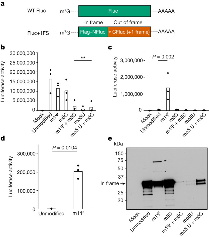
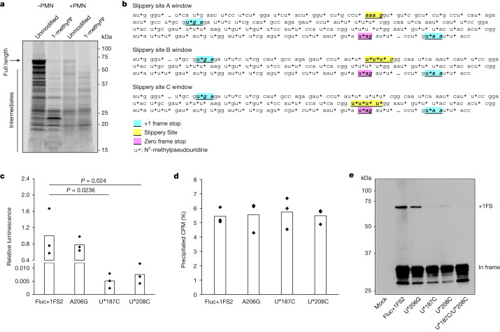
In vitro-transcribed (IVT) mRNAs are modalities that can combat human disease, exemplified by their use as vaccines for severe acute respiratory syndrome coronavirus 2 (SARS-CoV-2). IVT mRNAs are transfected into target cells, where they are translated into recombinant protein, and the biological activity or immunogenicity of the encoded protein exerts an intended therapeutic effect1,2. Modified ribonucleotides are commonly incorporated into therapeutic IVT mRNAs to decrease their innate immunogenicity3-5, but their effects on mRNA translation fidelity have not been fully explored. Here we demonstrate that incorporation of N1-methylpseudouridine into mRNA results in +1 ribosomal frameshifting in vitro and that cellular immunity in mice and humans to +1 frameshifted products from BNT162b2 vaccine mRNA translation occurs after vaccination. The +1 ribosome frameshifting observed is probably a consequence of N1-methylpseudouridine-induced ribosome stalling during IVT mRNA translation, with frameshifting occurring at ribosome slippery sequences. However, we demonstrate that synonymous targeting of such slippery sequences provides an effective strategy to reduce the production of frameshifted products. Overall, these data increase our understanding of how modified ribonucleotides affect the fidelity of mRNA translation, and although there are no adverse outcomes reported from mistranslation of mRNA-based SARS-CoV-2 vaccines in humans, these data highlight potential off-target effects for future mRNA-based therapeutics and demonstrate the requirement for sequence optimization.


Conflict of interest statement

T.E.M. and A.E.W. are inventors on a pending patent application (2305297.0) related to mRNA technology.

Figures

Fig. 1
Fig. 1. Translation of 1-methylΨ-modified mRNA produces +1 frameshifted polypeptides.
a, Structures of IVT mRNA transcripts used to probe protein synthesis fidelity. WT Fluc contains only (in-frame) Fluc coding sequence. For Fluc+1FS, the green segment represents in-frame N-terminal Fluc coding sequence (NFluc), and the orange segment represents +1 frameshifted C-terminal Fluc coding sequence (CFluc). Asterisk represents a premature stop codon. b, Luciferase activity produced by translation of WT Fluc mRNAs, either unmodified control (canonical nucleotides), or containing 1-methylΨ (m1Ψ), 5-methylC (m5C), 5-methoxyU (mo5U) or the combinations indicated. **P < 0.01 (1-methylΨ + 5-methylC, P = 0.0051; 5-methoxyU, P = 0.0023; 5-methoxyU + 5-methylC, P = 0.0042; one-way analysis of variance (ANOVA) with Dunnett’s test). c, Luciferase activity produced by translation of modified Fluc+1FS mRNAs and unmodified control. 1-methylΨ, P = 0.002 (one-way ANOVA with Dunnett’s test). d, Luciferase activity in lysates produced by transfection of HeLa cells with unmodified or 1-methylΨ Fluc+1FS mRNA for 8 h. P = 0.0104 (Welch’s one-tailed t-test). e, Western blot analysis (anti-Flag epitope) of polypeptides produced by translation of mRNAs in c. All data are obtained from n = 3 replicated experiments. e shows a single blot from n = 3 replicated experiments. Asterisks represent bands at higher molecular weight. For gel source data, see Supplementary Fig. 2.
Fig. 2
Fig. 2. +1 frameshifted products elicit off-target cellular immune responses following modified mRNA vaccination.
a, Depiction of spike and +1 frameshifted (+1FS) products produced by 1-methylΨ-modified spike mRNA translation. CDS, coding sequence. b, Splenocyte IFNγ ELISpot responses from untreated, ChAdOx1 nCoV-19-vaccinated or BNT162b2-vaccinated mice stimulated with +1FS spike peptides. IFNγ ELISpot response from BNT162b2-vaccinated mice stimulated with SARS-CoV-2 M peptides (unrelated control antigen) is included for additional comparison. SFU, spot-forming units. Each group n = 8. Untreated versus ChAdOx1 nCoV-19, P = 0.963; untreated versus BNT162b2, P = 0.0005; ChAdOx1 nCoV-19 versus BNT162b2, P = 0.001. c, Splenocyte IFNγ ELISpot responses from mice in b stimulated with spike peptides. Untreated versus ChAdOx1 nCoV-19, P = 2.05 × 10−9; untreated versus BNT162b2, P = 4.5 × 10−14; ChAdOx1 nCoV-19 versus BNT162b2, P = 1.88 × 10−13. d, Peripheral blood mononuclear cells (PBMC) IFNγ ELISpot responses from donors vaccinated with ChAdOx1 nCoV-19 (n = 20) or BNT162b2 (n = 21) stimulated with +1FS spike peptides. P = 0.0233 (Welch’s one-tailed t-test). e, PBMC IFNγ ELISpot responses from donors in c stimulated with in-frame spike peptides: total spike pool or spike S1 + S2 subpools. ChAdOx1 nCoV-19 spike versus BNT162b2 spike, P = 0.371; ChAdOx1 nCoV-19 spike versus BNT162b2 S1 + S2, P = 0.0845; BNT162b2 spike versus BNT162b2 S1 + S2, P = 0.686. f, Representative images of PBMC IFNγ ELISpot response wells for two individuals vaccinated with either BNT162b2 responder (top) or ChAdOx1 nCoV-19 (bottom). Left to right: in-frame spike response (spike peptides); +1FS spike response (+1FS spike peptides); no peptide control. P values in b,c,e were determined by one-way ANOVA and Tukey’s test. Source Data
Fig. 3
Fig. 3. Mistranslation of 1-methylΨ mRNA is due to +1 ribosomal frameshifting and not transcriptional errors.
a, Tryptic peptide coverage plot of the purified high molecular weight polypeptide produced by translation of 1-methylΨ Fluc+1FS mRNA, showing in-frame residues (top) and +1 frameshifted residues (bottom). −log10[PEP] is the mass spectrum percolator score (only high-quality peptides are shown). IP, immunoprecipitate. The structure of Fluc+1FS mRNA from Fig. 1 is re-displayed and a western blot of the translation reaction before immunoprecipitation is displayed. For gel source data, see Supplementary Fig. 3. b, Junction peptide derived from +1 ribosomal frameshifting and the originating mRNA sequence. c, Nucleotide deletions in unmodified (top) and 1-methylΨ (bottom) Fluc+1FS mRNA, quantified by n = 3 RNA-sequencing analyses. d, Nucleotide insertions in unmodified (top) and 1-methylΨ (bottom) Fluc+1FS mRNA.
Fig. 4
Fig. 4. +1 ribosomal frameshifting is dependent on mRNA slippery sequences and associated with ribosome stalling during 1-methylΨ mRNA translation.
a, SDS–polyacrylamide gel electrophoresis autoradiograph of [35S]methionine-labelled polypeptides produced by translation of unmodified or 1-methylΨ Fluc mRNA for 30 min, including or omitting 100 μM paromomycin (+PMN and −PMN, respectively). b, Diagram showing putative mRNA slippery sequences and stop-codon-flanked windows. c, Activity of +1 frameshifted products after translation of 1-methylΨ mutant mRNAs, or 1-methylΨ Fluc+1FS2 control mRNA, for 2 h. Fluc+1FS2 versus U*187C, P = 0.024; Fluc+1FS2 versus U*208C, P = 0.0236 (one-way ANOVA with Dunnett’s test). d, Total mRNA translation over 2 h for each of Fluc+1FS2 mRNA or mutant mRNAs, quantified by [35S]methionine incorporation. CPM, counts per minute. e, Western blot analysis (anti-Flag epitope) of polypeptides produced by translation of mRNAs in c, and U*187C/U*208C double-mutant 1-methylΨ mRNA. Data are from n = 3 replicated experiments. a and e show representative images from n = 3 replicated experiments. For gel source data, see Supplementary Figs. 4 and 5.
Extended Data Fig. 1
Extended Data Fig. 1. Validation of WT Fluc, Fluc + 1FS, and Fluc-1FS mRNAs.
a, UV photograph of WTFluc and Fluc+1FS mRNA transcripts analysed by agarose gel electrophoresis. b, Immunoblot of luciferase protein produced by translation of WTFluc and Fluc+1FS mRNAs using anti-FLAG antibody. c, Relative luciferase expression produced by translation of WTFluc and Fluc+1FS mRNAs. Data were obtained from n = 1 (non-replicated) analyses. For gel source data, see Supplementary Fig. 6.
Extended Data Fig. 2
Extended Data Fig. 2. Visualisation of HLA genotypes in BNT162b2-vaccinated individuals.
Major allele groups in HLA-A, -B and -C genes for n = 40 donors. The Sample ID contains 3 fields separated by underline: (i) 1 or 0, with 1 denoting an ELISpot count >40 following stimulation with the +1 FS peptide pool; (ii) donor identifier; (iii) ELISpot count if >40. SFU/million. Allele group frequency indicated by black tile, vertical dashed lines: HLA genes, horizontal dashed line: indicates the cut-off (ELISpot count >40).
Extended Data Fig. 3
Extended Data Fig. 3. HLA allele frequency distributions.
Frequencies of each major allele group in HLA-A, -B and -C genes for n = 40 donors.
Extended Data Fig. 4
Extended Data Fig. 4. NCBI BLASTP alignment of + 1 translated products.
Protein BLAST alignment of polypeptides predicted by +1 frame translation of either BNT162b2 mRNA or Wuhan SARS-CoV-2 Spike mRNA (from NC_045512.2).
Extended Data Fig. 5
Extended Data Fig. 5. Translation of 1-methylΨ-modified Fluc mRNA produces multiple polypeptides.
a, Diagram of NFLAG-WTFluc mRNA. b, Western blot analysis (anti-FLAG epitope) of polypeptides produced by translation of unmodified, or 1-methylΨ, NFLAG-WTFluc mRNA. In-frame firefly luciferase is indicated by arrow. Low molecular weight polypeptides are indicated by asterisk (*). A single blot from n = 3 replicated experiments is displayed. For gel source data, see Supplementary Fig. 7.
Extended Data Fig. 6
Extended Data Fig. 6. Annotated BNT162b2 Spike mRNA CDS putative slippery sites.
Putative ribosome slippery sites in BNT162b2 which were identified by the following formula: m1Ψm1Ψm1Ψ X, where m1Ψ is (N)1-methylpseudouridine and X is (N)1-methylpseudouridine or cytidine in the first nucleotide of the immediate downstream codon.

Comment in

References

    1. Hogan MJ, Pardi N. mRNA vaccines in the COVID-19 pandemic and beyond. Annu. Rev. Med. 2022;73:17–39. doi: 10.1146/annurev-med-042420-112725. - DOI - PubMed
    1. Chaudhary N, Weissman D, Whitehead KA. mRNA vaccines for infectious diseases: principles, delivery and clinical translation. Nat. Rev. Drug Discov. 2021;20:817–838. doi: 10.1038/s41573-021-00283-5. - DOI - PMC - PubMed
    1. Anderson BR, et al. Incorporation of pseudouridine into mRNA enhances translation by diminishing PKR activation. Nucleic Acids Res. 2010;38:5884–5892. doi: 10.1093/nar/gkq347. - DOI - PMC - PubMed
    1. Holtkamp S, et al. Modification of antigen-encoding RNA increases stability, translational efficacy, and T-cell stimulatory capacity of dendritic cells. Blood. 2006;108:4009–4017. doi: 10.1182/blood-2006-04-015024. - DOI - PubMed
    1. Andries O, et al. N1-methylpseudouridine-incorporated mRNA outperforms pseudouridine-incorporated mRNA by providing enhanced protein expression and reduced immunogenicity in mammalian cell lines and mice. J. Control. Release. 2015;217:337–344. doi: 10.1016/j.jconrel.2015.08.051. - DOI - PubMed