. 2024 Dec;39(1):2287420.
doi: 10.1080/14756366.2023.2287420. Epub 2023 Dec 7.

Suppression of lipopolysaccharide-induced COX-2 expression via p38MAPK, JNK, and C/EBPβ phosphorylation inhibition by furomagydarin A, a benzofuran glycoside from Magydaris pastinacea

Affiliations

Suppression of lipopolysaccharide-induced COX-2 expression via p38MAPK, JNK, and C/EBPβ phosphorylation inhibition by furomagydarin A, a benzofuran glycoside from Magydaris pastinacea

Shiu-Wen Huang et al. J Enzyme Inhib Med Chem. 2024 Dec.

Abstract

The phytochemical investigation of the methanol extract of the seeds of Magydaris pastinacea afforded two undescribed benzofuran glycosides, furomagydarins A-B (1, 2), together with three known coumarins. The structures of the new isolates were elucidated after extensive 1D and 2D NMR experiments as well as HR MS. Compound 1 was able to inhibit the COX-2 expression in RAW264.7 macrophages exposed to lipopolysaccharide, a pro-inflammatory stimulus. RT-qPCR and luciferase reporter assays suggested that compound 1 reduces COX-2 expression at the transcriptional level. Further studies highlighted the capability of compound 1 to suppress the LPS-induced p38MAPK, JNK, and C/EBPβ phosphorylation, leading to COX-2 down-regulation in RAW264.7 macrophages.

Keywords: C/EBPβ; COX-2; JNK; Magydaris pastinacea; benzofurans; p38MAPK.


Conflict of interest statement

No potential conflict of interest was reported by the authors.

Figures

Figure 1.
Figure 1.
Chemical structures of the isolated compounds.
Figure 2.
Figure 2.
Main HMBC and DQF-COSY correlations of compounds 12.
Figure 3.
Figure 3.
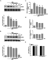
Furomagydarin A reduced COX-2 expression in LPS-stimulated RAW264.7 macrophages. (A) Cells were treated with vehicle or indicated concentrations of furomagydarin A for 30 min, followed by the treatment with LPS (100 ng/ml) for another 24 h. The COX-2 level was determined by immunoblotting. Each column represents the mean ± SEM of seven independent experiments. (B) Cells were treated with vehicle or indicated concentrations of furomagydarin B for 30 min, followed by the treatment with LPS (100 ng/ml) for another 24 h. The COX-2 level was determined by immunoblotting. Each column represents the mean ± SEM of six independent experiments. (C) Cells were treated with furomagydarin A (1–10 μM) for 30 min, followed by the treatment with LPS (100 ng/ml) for another 6 h. The extent of COX-2 mRNA was determined by an RT-qPCR assay as described in the ‘Materials and methods’ section. Each column represents the mean ± SEM of eight independent experiments. (D) Cells were transiently transfected with COX-2-luc or COX-2–3’UTR-luc and renilla-luc for 24 h. Luciferase activity was determined after treatment with LPS (100 ng/ml) for another 24 h. Data represent the mean ± SEM of eight independent experiments performed in duplicate. *P < 0.05, compared with the control group; #P < 0.05, compared with the group treated with LPS alone. (E) Cells were treated with furomagydarin A (10 μM) for 24 h. Cell viability was then determined using MTT and trypan blue exclusion assays. Data represent the mean ± SEM of four independent experiments performed in duplicate.
Figure 4.
Figure 4.
Furomagydarin A reduced LPS-induced C/EBP, p38MAPK or JNK phosphorylation in RAW264.7 macrophages. Cells were treated with furomagydarin A for 30 min, followed by the treatment with LPS (100 ng/ml) for another 30 min. The extent of C/EBP (A), p38MAPK (B) or JNK (C) phosphorylation was determined by immunoblotting. Each column represents the mean ± SEM of six independent experiments. *p < 0.05, compared with the control group; #p < 0.05, compared with the group treated with LPS alone.

References

    1. Jou IM, Lin CF, Tsai KJ, Wei SJ.. Macrophage-mediated inflammatory disorders. Mediators Inflamm. 2013;2013:316482–316483. - PMC - PubMed
    1. Sakr Y, Jaschinski U, Wittebole X, Szakmany T, Lipman J, Namendys-Silva SA, Martin-Loeches I, Leone M, Lupu MN, Vincent JL, Sepsis in intensive care unit patients: worldwide data from the intensive care over nations audit. Open Forum Infect Di. 2018;5(12):ofy313. - PMC - PubMed
    1. Roger T, Froidevaux C, Le Roy D, Reymond MK, Chanson AL, Mauri D, Burns K, Riederer BM, Akira S, Calandra T.. Protection from lethal gram-negative bacterial sepsis by targeting toll-like receptor 4. Proc Natl Acad Sci U S A. 2009;106(7):2348–2352. - PMC - PubMed
    1. Gorina R, Font-Nieves M, Marquez-Kisinousky L, Santalucia T, Planas AM.. Astrocyte TLR4 activation induces a proinflammatory environment through the interplay between MyD88-dependent NF kappa B signaling, MAPK, and Jak1/Stat1 pathways. Glia. 2011;59(2):242–255. - PubMed
    1. Chow JC, Young DW, Golenbock DT, Christ WJ, Gusovsky F.. Toll-like receptor-4 mediates lipopolysaccharide-induced signal transduction. J Biol Chem. 1999;274(16):10689–10692. - PubMed

MeSH terms

LinkOut - more resources