. 2023 Nov 26;15(11):999-1016.
doi: 10.4252/wjsc.v15.i11.999.

Hypoxia and inflammatory factor preconditioning enhances the immunosuppressive properties of human umbilical cord mesenchymal stem cells

Affiliations

Hypoxia and inflammatory factor preconditioning enhances the immunosuppressive properties of human umbilical cord mesenchymal stem cells

Hang Li et al. World J Stem Cells. .

Abstract

Background: Mesenchymal stem cells (MSCs) have great potential for the treatment of various immune diseases due to their unique immunomodulatory properties. However, MSCs exposed to the harsh inflammatory environment of damaged tissue after intravenous transplantation cannot exert their biological effects, and therefore, their therapeutic efficacy is reduced. In this challenging context, an in vitro preconditioning method is necessary for the development of MSC-based therapies with increased immunomodulatory capacity and transplantation efficacy.

Aim: To determine whether hypoxia and inflammatory factor preconditioning increases the immunosuppressive properties of MSCs without affecting their biological characteristics.

Methods: Umbilical cord MSCs (UC-MSCs) were pretreated with hypoxia (2% O2) exposure and inflammatory factors (interleukin-1β, tumor necrosis factor-α, interferon-γ) for 24 h. Flow cytometry, polymerase chain reaction, enzyme-linked immunosorbent assay and other experimental methods were used to evaluate the biological characteristics of pretreated UC-MSCs and to determine whether pretreatment affected the immunosuppressive ability of UC-MSCs in coculture with immune cells.

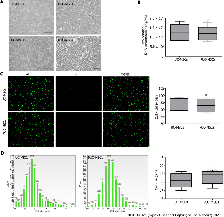
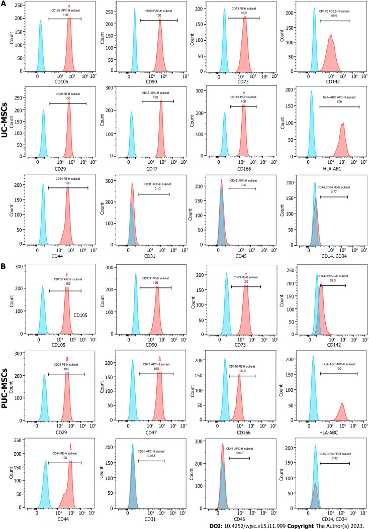
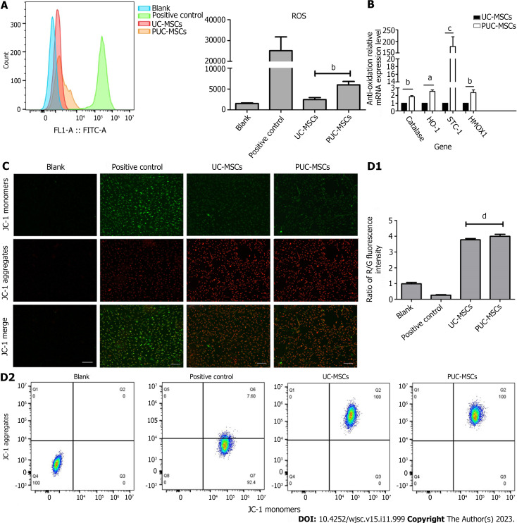
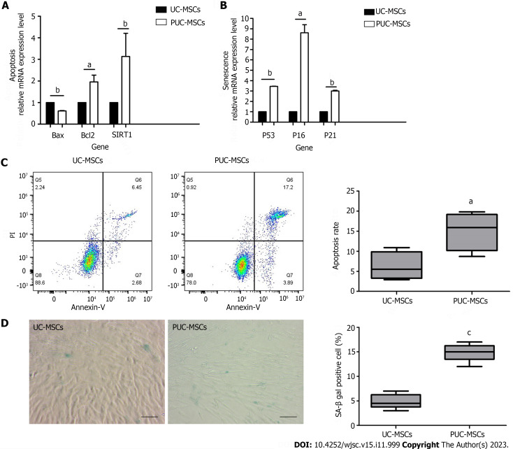
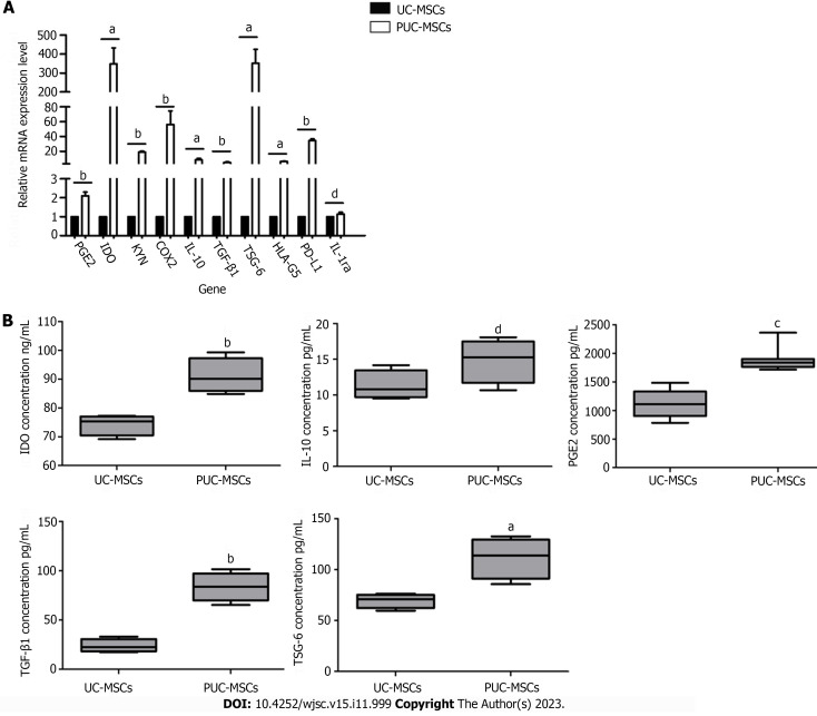
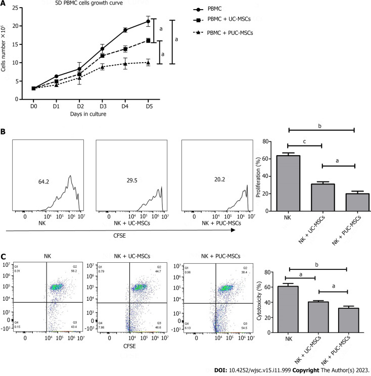
Results: Pretreatment with hypoxia and inflammatory factors caused UC-MSCs to be elongated but did not affect their viability, proliferation or size. In addition, pretreatment significantly decreased the expression of coagulation-related tissue factors but did not affect the expression of other surface markers. Similarly, mitochondrial function and integrity were retained. Although pretreatment promoted UC-MSC apoptosis and senescence, it increased the expression of genes and proteins related to immune regulation. Pretreatment increased peripheral blood mononuclear cell and natural killer (NK) cell proliferation rates and inhibited NK cell-induced toxicity to varying degrees.

Conclusion: In summary, hypoxia and inflammatory factor preconditioning led to higher immunosuppressive effects of MSCs without damaging their biological characteristics.

Keywords: Hypoxia, Inflammatory factors; Immune regulation; Mesenchymal stem cells; Preconditioning; Umbilical cord.


Conflict of interest statement

Conflict-of-interest statement: The authors report no relevant conflicts of interest for this article.

Figures

Figure 1
Figure 1
Morphology, viability, proliferation, and size of mesenchymal stem cells. A: Representative micrographs showing umbilical cord mesenchymal stem cells (UC-MSCs) and primed umbilical cord mesenchymal stem cells (PUC-MSCs). Microscopy observation of cell morphology revealed that UC-MSCs were elongated after hypoxia and inflammatory factor pretreatment (Bar = 200 μm); B: The DNA concentration was measured by an ultramicro nucleic acid protein detector. The DNA concentration of PUC-MSCs was not significantly different from that of UC-MSCs, and pretreatment had no effect on their proliferation; C: Cell viability was determined by an acridine orange (AO) propidium iodide (PI) staining cell image analyzer, and no significant difference between UC-MSCs and PUC-MSCs was observed; D: Cell size was determined with a cell imaging analyzer, and the sizes of the UC-MSCs and PUC-MSCs were comparable. dP > 0.05.
Figure 2
Figure 2
Phenotypes of mesenchymal stem cells were detected by flow cytometry. A: Cell surface markers of umbilical cord mesenchymal stem cells (UC-MSCs); B: Cell surface markers of primed umbilical cord mesenchymal stem cells (PUC-MSCs). The signals from the unstained control cells are shown in blue in the histogram, and the signals from stained cells are shown in red in the histogram. HLA: Human leukocyte antigen.
Figure 3
Figure 3
Mitochondrial function. A: Reactive oxygen species (ROS) levels in umbilical cord mesenchymal stem cells (UC-MSCs) and primed UC-MSCs (PUC-MSCs) were analyzed by flow cytometry. The fluorescence intensity of 2,7-dichlorodihydrofluorescein (DCFH-DA) in PUC-MSCs increased by 3-fold compared to UC-MSCs; B: The expression of the antioxidant-related genes catalase, stanniocalcin-1 (STC1), and hemeoxygenase-1 (HOMX1) was increased; C: Representative image showing the mitochondrial membrane potential (MMP) of mesenchymal stem cells stained with JC-1. The red fluorescence of potential-dependent JC-1 aggregation in each group and the green fluorescence of the JC-1 monomer in the cytoplasm after depolarization of the mitochondrial membrane were observed. Fluorescence microscopy showed no significant difference in red and green fluorescence between UC-MSCs and PU-MSCs; Bar = 200 μm; D: Flow cytometry was used to detect the red and green fluorescence of the JC-1 probe in UC-MSCs and PUC-MSCs. There was no difference in the ratios of red and green fluorescence. aP < 0.05; bP < 0.01; cP < 0.001; dP > 0.05.
Figure 4
Figure 4
Effects of pretreatment on apoptosis and senescence in mesenchymal stem cells. A: Apoptosis-related gene analysis showed that the expression of B-cell lymphoma 2 (BCL-2)-associated protein (Bax) was decreased and the expression of Bcl2 and silencing information regulator 2-related enzyme 1 (SIRT1) was increased after hypoxia and inflammatory factor pretreatment; B: Expression of P53, P16 and P21 was upregulated after hypoxia and inflammatory factor pretreatment; C: Umbilical cord mesenchymal stem cells (UC-MSCs) and primed UC-MSCs (PUC-MSCs) were stained with Annexin V-FITC apoptosis assay kit reagents and apoptotic cells were detected by flow cytometry. The results showed that the apoptosis index of PUC-MSCs increased; D: Representative images of β-galactosidase (SA-β-gal) staining and quantitative analysis of positive SA-β-gal staining. Compared with that of the UC-MSCs, the number of SA-β-gal-positive PUC-MSCs was significantly increased; Bar = 100 μm. aP < 0.05; bP < 0.01; cP < 0.001.
Figure 5
Figure 5
Relative expression of immunomodulatory genes and proteins in umbilical cord mesenchymal stem cells after pretreatment. A: Gene expression levels of umbilical cord mesenchymal stem cells (UC-MSCs) and primed UC-MSCs (PUC-MSCs) were analyzed by quantitative real-time polymerase chain reaction. Expression levels of prostaglandin E2 (PGE2), kynurenine, idoleamine-2,3-dioxygenase (IDO), cyclooxygenaese-2 (COX2), interleukin-10 (IL-10), transforming growth factor-beta 1 (TGF-β1), human leukocyte antigen-G5 (HLA-G5), tumor necrosis factor-α-induced protein-6 (TSG-6) and ligands for programmed cell death 1 (PD-L1) were all increased after hypoxia and inflammatory factor preconditioning. IL-1 receptor antagonist (IL-1ra) was not significantly different between the two groups; B: Levels of immunoregulatory proteins in UC-MSCs and PUC-MSCs were detected by enzyme-linked immunosorbent assay. After hypoxia and inflammatory factor pretreatment, the protein levels of IDO, PGE2, TGF-β1 and TSG-6 were increased, while the expression of IL-10 was not significantly different, as determined via three independent experiments. aP < 0.05; bP < 0.01; cP < 0.001; dP > 0.05.
Figure 6
Figure 6
Detection of the immunomodulatory function of umbilical cord mesenchymal stem cells after preconditioning. A: Peripheral blood mononuclear cells (PBMCs) alone or directly exposed to umbilical cord mesenchymal stem cells (UC-MSCs/primed UC-MSCs) for 5 d in a 3:1 ratio. The number of PBMCs in three groups was measured daily and the growth curve was plotted; B: Carboxyfluorescein diacetate succinimidyl ester (CFSE)-stained natural killer (NK) cells were cultured alone or cocultured with UC-MSCs/PUC-MSCs at a 3:1 ratio for 72 h. NK cells were harvested, and the fluorescence intensity of CFSE was measured by flow cytometry to measure the proliferation rate of NK cells; C: NK cells were collected and incubated with CFSE-stained K562 target cells for 4 h at an effector ratio (E:T = 1:5). Effector cell (NK cell)-mediated cytotoxicity of K562 cells was analyzed by flow cytometry. aP < 0.05; bP < 0.01; cP < 0.001; dP > 0.05.

References

    1. Yu M, Ma L, Yuan Y, Ye X, Montagne A, He J, Ho TV, Wu Y, Zhao Z, Sta Maria N, Jacobs R, Urata M, Wang H, Zlokovic BV, Chen JF, Chai Y. Cranial Suture Regeneration Mitigates Skull and Neurocognitive Defects in Craniosynostosis. Cell. 2021;184:243–256.e18. - PMC - PubMed
    1. Berebichez-Fridman R, Montero-Olvera PR. Sources and Clinical Applications of Mesenchymal Stem Cells: State-of-the-art review. Sultan Qaboos Univ Med J. 2018;18:e264–e277. - PMC - PubMed
    1. Stefańska K, Nemcova L, Blatkiewicz M, Pieńkowski W, Ruciński M, Zabel M, Mozdziak P, Podhorska-Okołów M, Dzięgiel P, Kempisty B. Apoptosis Related Human Wharton's Jelly-Derived Stem Cells Differentiation into Osteoblasts, Chondrocytes, Adipocytes and Neural-like Cells-Complete Transcriptomic Assays. Int J Mol Sci. 2023;24 - PMC - PubMed
    1. Zhou T, Yuan Z, Weng J, Pei D, Du X, He C, Lai P. Challenges and advances in clinical applications of mesenchymal stromal cells. J Hematol Oncol. 2021;14:24. - PMC - PubMed
    1. Gao F, Chiu SM, Motan DA, Zhang Z, Chen L, Ji HL, Tse HF, Fu QL, Lian Q. Mesenchymal stem cells and immunomodulation: current status and future prospects. Cell Death Dis. 2016;7:e2062. - PMC - PubMed